International Chemistry Olympiad
IChO 2001
Mumbai, India · 7 theory problems with complete solutions
Problem 1: Hydrogen Atom and Hydrogen Molecule
Niels Bohr (1885-1962)
The observed wavelengths in the line spectrum of hydrogen atom were first expressed in terms of a series by Johann Jakob Balmer, a Swiss teacher. Balmer’s empirical formula is
\[ \frac{1}{\lambda} = R_{\mathrm H}\left(\frac{1}{2^2}-\frac{1}{n^2}\right); \qquad n=3,4,5,\ldots \]
Here,
\[ R_{\mathrm H} = \frac{m_e e^4}{8\varepsilon_0^2h^3c} = 109\,678\ \mathrm{cm^{-1}} \]
is the Rydberg konstant, \(m_e\) is the mass of an electron. Niels Bohr derived this expression theoretically in 1913. The formula is easily generalized to any one electron atom/ion.
1.1 Calculate the longest wavelength in Å (\(1\ \text{Å}=10^{-10}\ \mathrm m\)) in the ‘Balmer series’ of singly ionized helium (\(\ce{He+}\)). Ignore nuclear motion in your calculation.
1.2 A formula analogous to Balmer’s formula applies to the series of spectral lines which arise from transitions from higher energy levels to the lowest energy level of hydrogen atom. Write this formula and use it to determine the ground state energy of a hydrogen atom in eV.
A ‘muonic hydrogen atom’ is like a hydrogen atom in which the electron is replaced by a heavier particle, the muon. The mass of a muon is about 207 times the mass of an electron, while its charge is the same as that of an electron. A muon has a very short lifetime, but we ignore its unstable nature here.
1.3 Determine the lowest energy and the radius of the first Bohr orbit of the muonic hydrogen atom. Ignore the motion of the nucleus in your calculation. The radius of the first Bohr orbit of a hydrogen atom (called the Bohr radius, \(a_0=\dfrac{\varepsilon_0h^2}{m_e e^2\pi}\)) is \(0.53\ \text{Å}\).
The classical picture of an "orbit" in Bohr’s theory has now been replaced by the quantum mechanical notion of an ‘orbital’. The orbital \(\psi1\sigma_{1s}(r)\) for the ground state of a hydrogen atom is given by
\[ \Psi_{1s}(r) = \frac{1}{\sqrt{\pi a_0^3}}e^{-r/a_0} \]
where \(r\) is the distance of the electron from the nucleus and \(a_0\) is the Bohr radius.
1.4 Consider a spherical shell of radius \(a_0\) and thickness \(0.001a_0\). Estimate the probability of finding the electron in this shell. Volume of a spherical shell of inner radius \(r\) and small thickness \(\Delta r\) equals \(4\pi r^2\Delta r\).
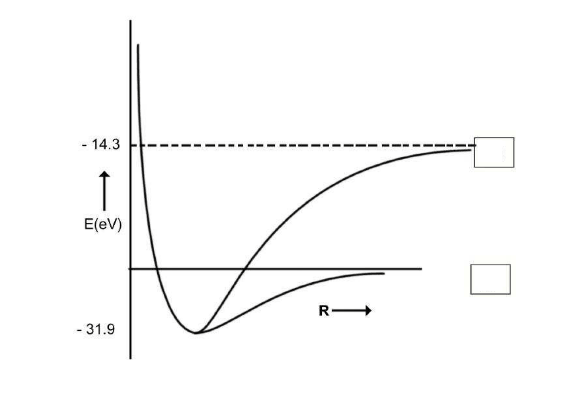
The \(\ce{H2}\) molecule can dissociate through two different channels:

The graph of energy (\(E\)) vs internuclear distance (\(R\)) for \(\ce{H2}\) is shown schematically in the figure. The atomic and molecular energies are given in the same scale.
1.5 Put appropriate channel labels (i) or (ii) in the boxes below.
1.6 Determine the values of the dissociation energies (\(D_e\) in eV) of the \(\ce{H2}\) molecule corresponding to
channel (i) \(\boxed{\phantom{000000}}\)
channel (ii) \(\boxed{\phantom{000000}}\)
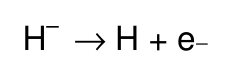
1.7 From the given data, calculate the energy change for the process
1.8 \(\ce{H-}\) is a two-electron atomic system. Assuming that the Bohr energy formula is valid for each electron with nuclear charge \(Z\) replaced by \(Z_{\mathrm{eff}}\), calculate \(Z_{\mathrm{eff}}\) for \(\ce{H-}\).
Official English text of the problem.
Problem 2: Phosphoric Acid
Phosphoric acid is of a great importance in fertilizer industry. Besides, phosphoric acid and its various salts have a n umber of applications in metal treatment, food, detergent and toothpaste industries.
2.1 The pK values of the three successive dissociations of phosphoric acid at 25 °C are:
\[ pK_{1a}=2.12 \]
\[ pK_{2a}=7.21 \]
\[ pK_{3a}=12.32 \]
Write down the conjugate base of dihydrogen phosphate ion and determine its \(pK_b\) value.
Small quantities of phosphoric acid are extensively used to impart the sour or tart taste to many soft drinks such as colas and root beers. A cola having a density of \(1.00\ \mathrm{g\,cm^{-3}}\) contains 0.05 % by weight of phosphoric acid.
2.2 Determine the pH of the cola (ignoring the second and the third dissociation steps for phosphoric acid). Assume that the acidity of the cola arises only from phosphoric acid.
2.3 Phosphoric acid is used as a fertiliser for agriculture. \(1.00\times10^{-3}\ \mathrm{M}\) phosphoric acid is added to an aqueous soil suspension and the pH is found to be 7.00.
Determine the fractional concentrations of all the different phosphate species present in the solution. Assume that no component of the soil interacts with any phosphate species.
2.4 Zinc is an essential micronutrient for plant growth. Plant can absorb zinc in water soluble form only. In a given soil water with \(pH=7.0\), zinc phosphate was found to be the only source of zinc and phosphate. Calculate the concentration of \([\ce{Zn^2+}]\) and \([\ce{PO4^3-}]\) ions in the solution. \(K_{\mathrm{sp}}\) for zinc phosphate is \(9.1\times10^{-33}\).
Official English text of the problem.
Problem 3: Second Law of Thermodynamics
J.W.Gibbs (1839 –1903)
The second law of thermodynamics is a fundamental law of science. In this problem we consider the thermodynamics of an ideal gas, phase transitions and chemical equilibrium.
3.00 mol of \(\ce{CO2}\) gas expands isothermically (in thermal contact with the surroundings; temperature = 15 °C) against a fixed external pressure of 1.00 bar. The initial and final volumes of the gas are 10.0 dm³ and 30.0 dm³, respectively.
3.1 Choose the correct option for change in the entropy of the system \((\Delta S_{\mathrm{sys}})\) and of the surroundings \((\Delta S_{\mathrm{sur}})\):
\(\Delta S_{\mathrm{sys}}\) \(\Delta S_{\mathrm{sur}}\) (a) \(>0\) \(=0\) ☐ (b) \(<0\) \(>0\) ☐ (c) \(>0\) \(<0\) ☐ d) \(=0\) \(=0\) ☐ (Mark X in the correct box.)
3.2 Calculate \(\Delta S_{\mathrm{sys}}\) assuming \(\ce{CO2}\) to be an ideal gas.
3.3 Calculate \(\Delta S_{\mathrm{sur}}\).
3.4 Calculate the change in entropy of the universe.
Does your answer agree with the Second Law of Thermodynamics?
(Mark X in the correct box.)
☐ Yes ☐ No
The pressure – temperature phase diagram of \(\ce{CO2}\) is given below schematically. The diagram is not to scale.
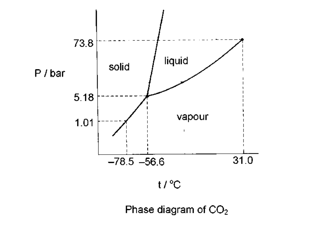
Phase diagram of \(\ce{CO2}\)
3.5 \(\ce{CO2}\) gas, initially at a pressure of 4.0 bar and temperature of 10.0 °C is cooled at constant pressure. In this process,
(a) it goes first to the liquid phase and then to the solid phase. ☐
(b) it goes to the solid phase without going through the liquid phase. ☐
3.6 Starting with the same pressure and temperature as above (in 3.5), \(\ce{CO2}\) is compressed isothermatically. In this process,
(a) it goes first to the liquid phase and then to the solid phase. ☐
(b) it goes to the solid phase without going through the liquid phase. ☐
3.7 From the data given in the phase diagram, calculate the molar enthalpy change of sublimation of \(\ce{CO2}\). Write down the formula used.
3.8 \(\ce{CO}\) gas, used extensively in organic synthesis, can be obtained by reacting \(\ce{CO2}\) gas with graphite. Use the data given below to show that the equilibrium constant at 298.15 K is less than unity.
At 298.15 K
\(\Delta H_f^0\ /\ \mathrm{kJ\,mol^{-1}}\) \(S^0\ /\ \mathrm{J\,K^{-1}\,mol^{-1}}\) \(\ce{CO2(g)}\) −393.51 213.79 \(\ce{CO(g)}\) −110.53 197.66 \(\ce{C(graphite)}\) 5.74 3.9 Estimate the temperature at which the reaction would have an equilibrium constant equal to 1. Ignore slight variations in the thermodynamic data with temperature.
3.10 The reaction above (in 3.8) is carried out between \(\ce{CO2}\) and excess hot graphite in a reactor maintained at about 800 °C and a total pressure of 5.0 bar. The equilibrium constant \(K_p\) under these conditions is 10.0. Calculate the partial pressure of \(\ce{CO}\) at equilibrium.
Official English text of the problem.
Problem 4: Thorium, uranium and lead in monazite
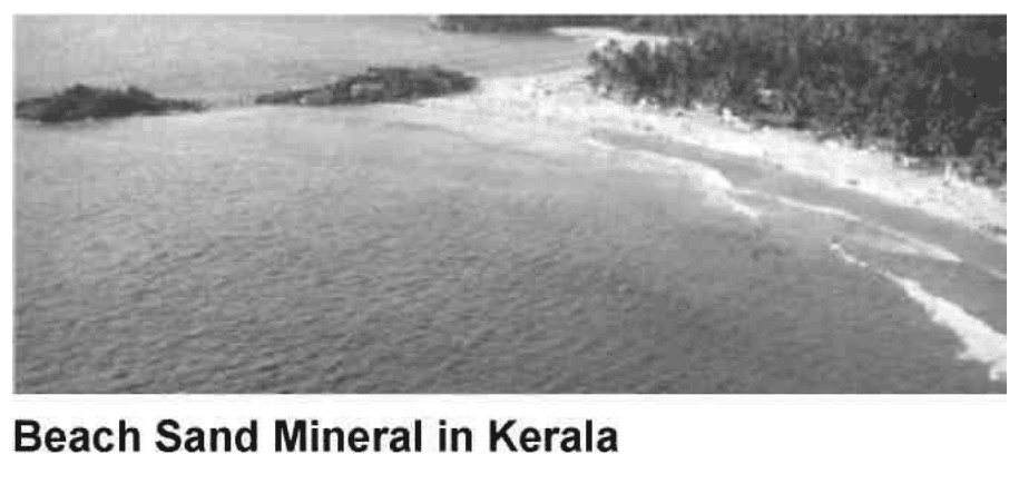
Beach Sand Mineral in Kerala
Beach sand mineral, monazite, is a rich source of thorium, available in large quantities in the state of Kerala in India. A typical monazite sample contains about 9 % \(\ce{ThO2}\) and 0.35 % \(\ce{U3O8}\). \(\ce{^{208}Pb}\) a \(\ce{^{206}Pb}\) are the stable end-products in the radioactive decay series of \(\ce{^{232}Th}\) and \(\ce{^{238}U}\), respectively. All the lead (\(\ce{Pb}\)) found in monazite is of radiogenic origin.
The isotopic atom ratio \(\ce{^{208}Pb}/\ce{^{232}Th}\), measured mass spectrometrically, in a monazite sample was found to be 0.104. The half-lives of \(\ce{^{232}Th}\) and \(\ce{^{238}U}\) \(1.41\times10^{10}\) years and \(4.47\times10^9\) years, respectively. Assume that \(\ce{^{208}Pb}\), \(\ce{^{206}Pb}\), \(\ce{^{232}Th}\) and \(\ce{^{238}U}\) remained entirely in the monazite sample since the formation of monazite mineral.
4.1 Calculate the age (time elapsed since its formation) of the monazite sample.
4.2 Estimate the isotopic atom ratio \(\ce{^{206}Pb}/\ce{^{238}U}\) in the monazite sample.
4.3 Thorium-232 is a fertile material for nuclear energy. In thermal neutron irradiation, it absorbs a neutron and the resulting isotope forms \(\ce{^{233}U}\) by successive \(\beta^-\) decays. Write the nuclear reactions for the formation of \(\ce{^{233}U}\) from \(\ce{^{232}Th}\).
In nuclear fission of \(\ce{^{233}U}\) a complex mixture of radioactive fission products is formed. The fission product \(\ce{^{101}Mo}\) initially undergoes radioactive decay as shown below:

4.4 A freshly prepared radiochemically pure sample of \(\ce{^{101}Mo}\) contains 5000 atoms of \(\ce{^{101}Mo}\) initially. How many atoms of
i) \(\ce{^{101}Mo}\)
ii) \(\ce{^{101}Tc}\)
iii) \(\ce{^{101}Ru}\)
will be present in the sample after 14.6 min?
Official English text of the problem.
Problem 5: Halogen Chemistry
Halogens in their reactions among themselves and with a variety of other elements give rise to a large number of compounds with diverse structure, bonding and chemical behaviour. Metal halides, halogen derivatives and interhalogens represent major types of halogen compounds.
(A) Photography
A "black and white" photographic film contains a coating of silver bromide on a support such as cellulose acetate.
5.1 Write the photochemical reaction that occurs when light falls on \(\ce{AgBr(s)}\) coated on a film.
5.2 During the developing process, unexposed \(\ce{AgBr}\) is washed away by complexation of Ag(I) by sodium thiosulphate solution. Write down this chemical reaction.
5.3 These washings are often disposed of as waste. However, metallic silver can be recovered from them by adding cyanide, followed by zinc. Write down the reactions involved.
(B) Shapes, spectra and reactivity
The most reactive halogen, fluorine, reacts with other halogens \(\ce{Cl2}\), \(\ce{Br2}\) and \(\ce{I2}\) under controlled conditions giving a tetra-atomic, hexa-atomic and an octa-atomic molecule, respectively.
5.4 Write the formulae and 3-dimensional structures of these interhalogen molecules on the basis of VSEPR theory. Show the disposition of the lone pairs on the central atom, where appropriate.
A mixture of iodine vapour and chlorine gas when fed into a mass spectrometer gave two sets (A and B) of mass spectral peaks corresponding to molecular ions of two chemical species at \(m/z\):
A: 162, 164
B: 464, 466, 468, 470, 472, 474, 476
5.5 Identify the molecular species corresponding to \(m/z = 162,\ 164,\ 466\) and \(476\). Draw the structure of the heaviest species (\(m/z = 476\)) indicating clearly the lone pairs on atom(s) of I (iodine). Show the isotopic composition of each species.
In aqueous medium chlorine gas oxidises sodium thiosulphate to an ion containing the highest oxidation state of sulphur.
5.6 Write down the chemical equation for this reaction.
5.7 Write down the Lewis dot structure of the thiosulphate ion. Circle the sulphur atom that has the lower oxidation state.
5.8 Chlorine dioxide reacts with sodium hydroxide as shown below. Identify the products X and Y (both containing chlorine) and balance the equation.
5.9 Reaction of chlorine an alkali is used by manufacturing bleach. Write the chemical reaction for its formation.
5.10 Write the oxidation state(s) of chlorine in bleach.
(C) Alkali metal halides and X-ray crystallography
X-ray crystallography reveals many aspects of the structure of metal halides. The radius ratio \((r_+/r_-)\) is a useful parameter to rationalise their structure and stability. A table of radius ratio \((r_+/r_-)\) for some alkali halides with radius ratio (\(r_-\) kept constant) is shown schematically for \(\ce{NaCl}\)-type and \(\ce{CsCl}\)-type crystal structures.
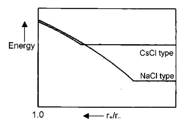
\(\ce{Li+}\) \(\ce{Na+}\) \(\ce{K+}\) \(\ce{Rb+}\) \(\ce{Cs+}\) \(\ce{Cl-}\) 0.33 0.52 0.74 0.82 0.93 \(\ce{Br-}\) 0.31 0.49 0.68 0.76 0.87 \(\ce{I-}\) 0.28 0.44 0.62 0.69 0.78 5.11 For a given anion, the graph for \(\ce{NaCl}\)-type structure levels off at low \(r_+/r_-\) values because of
(a) cation-cation contact along the face diagonal. \(\square\)
(b) anion-anion contact along the face diagonal. \(\square\)
(c) cation-anion contact along the cell edge. \(\square\)
(Mark X in the correct box.)
5.12 Which among the halides \(\ce{LiBr}\), \(\ce{NaBr}\) and \(\ce{RbBr}\) is likely to undergo phase transition from \(\ce{NaCl}\)-type to \(\ce{CsCl}\)-type structure with change of temperature and / or pressure?
5.13 Show by calculation the radius ratio \((r_+/r_-)\) at which the energy of \(\ce{CsCl}\)-type structure levels off.
5.14 Using CuKα X-rays (\(\lambda = 154\ \mathrm{nm}\)), diffraction by a \(\ce{KCl}\) crystal (fcc structure) is observed at an angle (\(\theta\)) of \(14.2^\circ\). Given that (i) diffraction takes place from the planes with \(h^2+k^2+l^2=4\), (ii) in a cubic crystal \(d_{hkl}=a/(h^2+k^2+l^2)^{1/2}\), where "d" is the distance between adjacent hkl planes and "a" is a lattice parameter, and (iii) reflections in an fcc structure can occur only from planes with "all odd" or "all even" hkl (Miller) indices, calculate the lattice parameter "a" for \(\ce{KCl}\).
5.15 Indicate in the table given below the required information for the \(2^{\mathrm{nd}}\) and \(3^{\mathrm{rd}}\) nearest neighbours of a \(\ce{K+}\) ion in the \(\ce{KCl}\) lattice.
\(2^{\mathrm{nd}}\) nearest neighbours \(3^{\mathrm{rd}}\) nearest neighbours number sign of the charge distance (pm) number sign of the charge distance (pm) 5.16 Determine the lowest value of diffraction angle \(\theta\) possible for the \(\ce{KCl}\) structure.
Official English text of the problem.
Problem 6: Organic Chemistry of Indian Spices
The rhizomes of ginger (Zingiber officinale) are well known for their medicinal and flavouring properties. In Ayurveda (the traditional system of medicine in India) different formulations of ginger are used for the treatment of gastrointestinal problems, common cold and other ailments. Several compounds are responsible for the pungency of ginger. Many are simple substituted aromatic compounds with different side chains. Three of them, Zingerone, (+)[6] Gingerol (to be referred hereafter as Gingerol only), and Shogaol are particularly important.
Zingerone: \(\ce{C11H14O3}\)
Gingerol: \(\ce{C17H14O4}\)
Shogaol: \(\ce{C17H24O3}\)6.1 Zingerone gives positive \(\ce{FeCl3}\) and 2,4-DNP (2,4-dinitrophenylhydrazine) tests. It does not react with Tollen's reagent. Therefore, Zingerone contains the following functional groups: (Mark X in the correct boxes.)
(a) alcoholic hydroxyl \(\square\) (e) ester \(\square\) (b) aldehydic carbonyl \(\square\) (f) alkoxyl \(\square\) (c) ketonic carbonyl \(\square\) (g) unsaturation \(\square\) (d) phenolic hydroxyl \(\square\) The data obtained from the \(\ce{^1H}\) NMR spectrum of Zingerone are shown in Table 1. Some other relevant information is given in Table 2.
Table 1: \(\ce{^1H}\) NMR spectral data on Zingerone
Chemical shifts \((\delta)\) Multiplicity Relative intensity 2.04 singlet 3 2.69, 2.71 two (closely spaced) triplets of equal intensity 4 3.81 singlet 3 5.90 broad singlet (\(\ce{D2O}\) exchangeable) 1 6.4 – 6.8 two doublets with similar chemical shifts and one singlet 3 *) For clarity, some of the data have been altered slightly.)
Table 2: Approximate \(\ce{^1H}\) chemical shifts \((\delta)\) and spin-spin coupling constants \((J)\) of some protons
\(\ce{^1H}\) Chemical shifts
alkyl - H 0.9 - 1.5 [S1] 3.4 - 3.6 [S2] 2.0 - 2.7 [S3] 4.0 - 7.0 [S4] 2.3 - 2.9 [S5] 6.0 - 8.0 [S6] 3.3 - 3.9 [S7] 4.0 - 11.0 [S1]
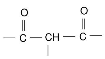
[S2]
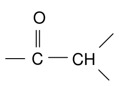
[S3]
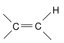
[S4]
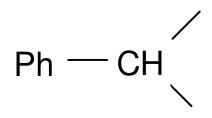
[S5]
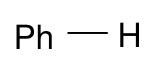
[S6]
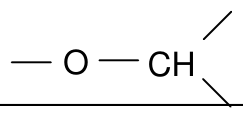
[S7]
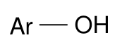
Spin–spin coupling constants \((J)\)
Alkenes cis 5 – 14 Hz (commonly around 6 – 8 Hz) trans 11 – 19 Hz (commonly around 14 – 16 Hz) Zingerone on bromination with bromine water gives only one nuclear mono brominated product. The IR spectrum of Zingerone indicates the presence of a week intramolecular hydrogen bond. The same is present even after Clemmensen reduction (\(\ce{Zn}\) – \(\ce{Hg}/\ce{HCl}\)) of Zingerone.
6.2 From the information above deduce the following:
i) side chain in Zingerone
ii) substituent on the aromatic ring
iii) relative positions of the substituents on the ring
6.3 Draw a possible structure of Zingerone based on the above inferences.
6.4 Complete the following reaction sequence for the synthesis of Zingerone.
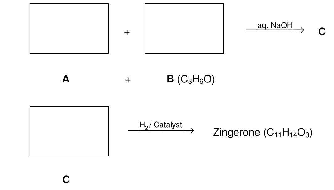
6.5 Zingerone can be easily converted into Gingerol by the following reaction sequence:

Notes: (1) \(\ce{Me3SiCl}/\ce{(Me3Si)2NH}\) is used to convert OH into \(\ce{-OSiMe3}\); the group \(\ce{-SiMe3}\) can be removed by acid hydrolysis.
(2) LDA is lithium disopropylamine, a strong, very hindered, non.nucleophilic base.
i) Draw the structure of D.
ii) Draw the structure of Gingerol.
iii) Complete the Fischer projection of the \(R\)-enantiomer of Gingerol.
iv) In the above reaction sequence (6.5), about 2 – 3 % another constitutional isomer (E) of Gingerol is obtained. Draw the likely structure of E.
v) Will the compound E be formed as
(a) a pair of enatiomers? \(\square\)
(b) a mixture of diastereomers? \(\square\)
(c) a mixture of an enantiomeric pair and a meso isomer? \(\square\)
(Mark X in the correct box.)
vi) Gingerol (\(\ce{C17H26O4}\)) when heated with a mild acid (such as \(\ce{KHSO4}\)) gives Shogaol (\(\ce{C17H24O3}\)). Draw the structure of Shogaol.
6.6 Turmeric (Curcuma longa) is a commonly used spice in Indian food. It is also used in Ayurvedic medicinal formulations. Curmunin (\(\ce{C21H20O6}\)), an active ingredient of turmeric, is structurally related to Gingerol. It exhibits keto-enol tautomerism. Curcumin is responsible for the yellow colour of turmeric and probably also for the pungent taste.
The \(\ce{^1H}\) NMR spectrum of the keto form of Curcumin shows aromatic signal similar to that of Gingerol. It also shows a singlet around \(\delta\) 3.5 (2H) and two doublets (2H each) in the region \(\delta\) 6 – 7 with \(J=16\ \mathrm{Hz}\). It can be synthesized by condensing TWO moles of A (refer to 6.4) with one mole of pentan-2,4-dione.
i) Draw the stereochemical structure of Curcumin.
ii) Draw the structure of the enol form of Curcumin.
iii) Curcumin is yellow in colour because it has
(a) a phenyl ring \(\square\)
(b) a carbonyl group \(\square\)
(c) an extended conjugation \(\square\)
(d) a hydroxyl group \(\square\)
(Mark X in the correct box.)
Official English text of the problem.
Problem 7: Peptides and Proteins
[In the amino-acid tables, S1–S8 refer to the structure figures reproduced immediately below each table.]
PROBLEM 7
Peptides and Proteins
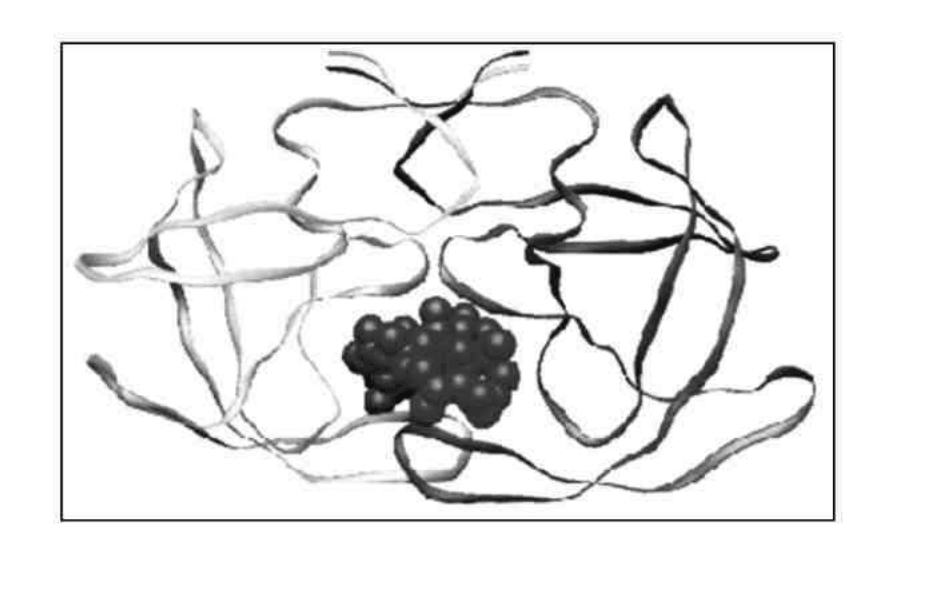
Protein folding
Proteins (polypeptides) are known to assume a variety of backbone conformations. In one conformation, the backbone is almost fully extended arrangement (as in a parallel or anti-parallel β-sheet), and in another conformation it is in a fully folded arrangement (as in an α-helix).
7.1 The end-to-end distance in a hexapeptide when it is in a fully extended conformation is approximately:
(a) \(10\ \text{Å}\) ☐
(b) \(15\ \text{Å}\) ☐
(c) \(20\ \text{Å}\) ☐
(d) \(25\ \text{Å}\) ☐(Mark X in the correct box.)
7.2 Assuming that the following hexapeptide is a α-helix conformation, draw a connecting arrow between an oxygen atom and an amide \(\ce{NH}\) with which it is hydrogen bonded.
7.3 The following hexapeptides (A and B) display contrasting conformations in water at pH 7.0, especially when their serine hydroxyls are phosphorylated. A is moderately helical and it becomes a stronger helix on serine phosphorylation. B is weakly helical and it is completely disorded on serine phosphorylation. Draw connecting arrows to indicate the interactions between the residues that are responsible for this differing behaviour.
Consider the following process of folding/unfolding of a tripeptide segment in a large protein. The free energy change (\(\Delta G\)) will depend on the interaction of the unfolded tripeptide with the solvent (water) and with the rest of the protein in the folded state (see below). Assume that the tripeptide is made up of one non-polar (hydrophobic; shaded) and two polar (hydrophilic; unshaded) residues. Assume the following approximate changes in the interaction free energies :
(a) a non-polar residue and the solvent (water): \(\Delta G=+8\ \mathrm{kJ\,mol^{-1}}\)
(b) a non-polar residue and the rest of the protein: \(\Delta G=-4\ \mathrm{kJ\,mol^{-1}}\)
(c) a polar residue and the solvent (water): \(\Delta G=-16\ \mathrm{kJ\,mol^{-1}}\)
(d) a polar residue and the rest of the protein: \(\Delta G=-14\ \mathrm{kJ\,mol^{-1}}\)
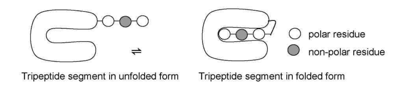
7.4 Calculate \(\Delta G\) for the folding of the tripeptide segment.
7.5 Calculate \(\Delta G\) for the folding of the tripeptide segment if all the three residues are polar.
β - lactamase and drug resistance
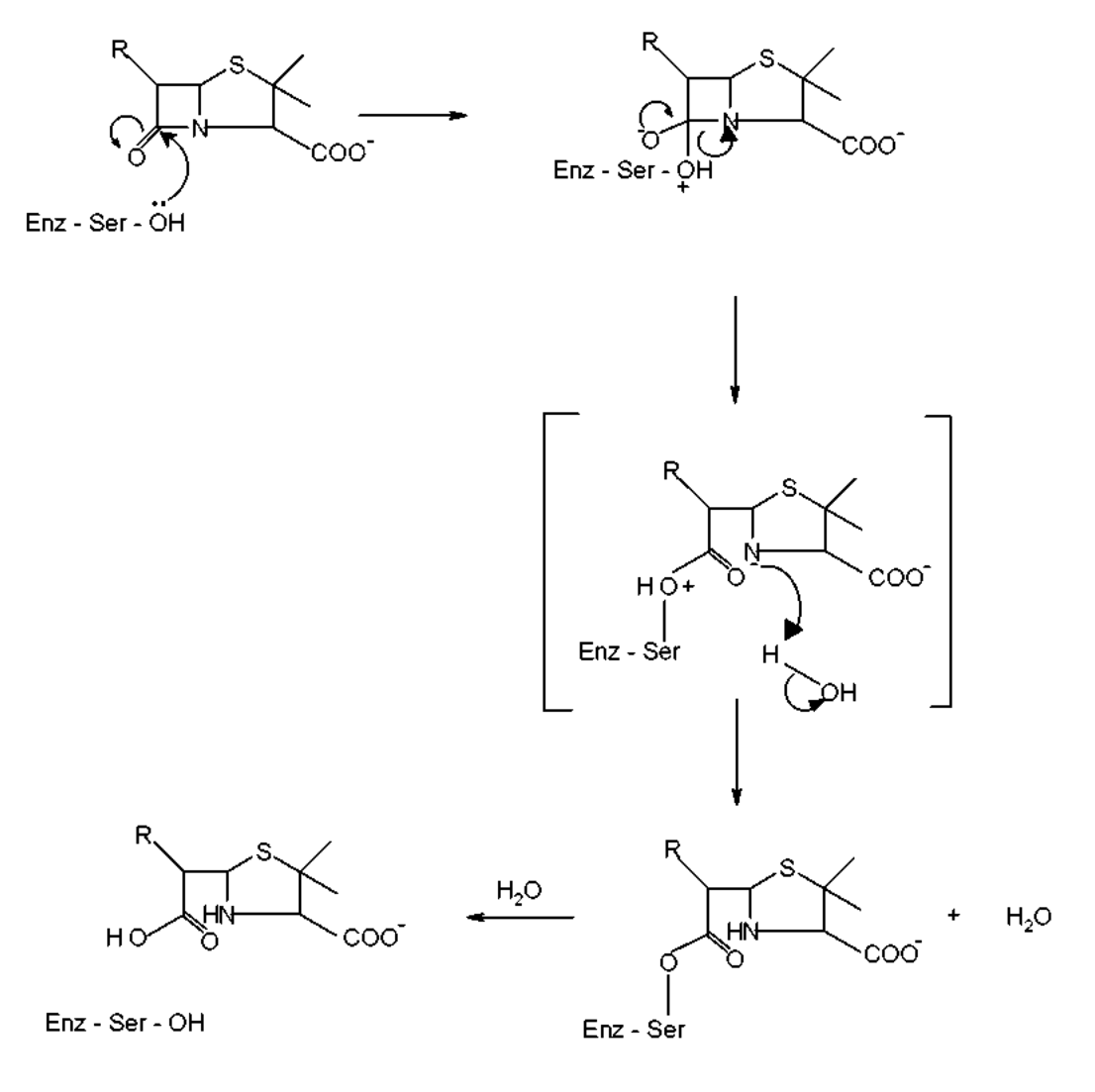
Penicillins are effective drugs in combating bacterial infections. An alarming development over the years has, however, been the emergence of drug resistant bacteria. Penicillin resistance arises due to the secretion of an enzyme called (3-lactamase (also known as penicillinase), which inactivates penicillin by opening its (3-lactam ring. The mechanism for this (3-lactam ring opening involves the nucleophilic attack by serine-\(\ce{OH}\) at the active site of the enzyme as shown below.
An attempt was made to characterize the b–lactamase from Staphylococcus aureus. When the pure enzyme was treated with a radiolabelled [\(\ce{^{32}P}\)] phosphorylating agent, only the active site serine got labelled. On analysis, this serine {molecular mass = 105 mass units (Da)} constituted 0.35% by weight of the b–lactamase.
7.6 Estimate the minimal molecular mass of this b–lactamase.
7.7 The approximate number of amino acid residues present in a protein of this size is :
(a) 100 ☐
(b) 150 ☐
(c) 275 ☐
(d) 375 ☐[Mark X in the correct box.]
To map its active site, the b–lactamase was hydrolysed using trypsin, a specific enzyme. This resulted in a hexapeptide P1 containing the active site serine. Amino acid analysis revealed the following in equimolar proportion : Glu, Leu, Lys, Met, Phe and Ser. Treatment of P1 with Edman’s reagent (phenyl isothiocyanate) yielded phenyl thiohydantoin (PTH) derivative of phenyalanine and a peptide P2.
Treatment of P1 with cyanogen bromide (\(\ce{CNBr}\)) gave an acidic tetrapeptide P3 and a dipeptide P4.
Treatment of P2 with 1–fluoro–2,4–dinitrobenzene, followed by complete hydrolysis, yielded N–2,4–dinitrophenyl–Glu. P1, P2, and P3 contain the active site serine.
7.8 From the above information, deduce the amino acid sequence of P1, P2, P3 and P4.
7.9 Calculate the molecular mass of P3 in mass units (Da) from the information given in the attached Table.
The β–lactamase active site provides a unique microenvironment that makes the catalytic serine–\(\ce{OH}\) an unusually reactive nucleophile. The first order rate constant for β–lactamase catalysed reaction is \(350\ \mathrm{s^{-1}}\). For penicillin hydrolysis by free serine–\(\ce{OH}\) (at \(1\ \mathrm{M}\)) in solution, the pseudo first order rate constant is \(0.5\ \mathrm{s^{-1}}\).
7.10 From the information above, calculate the effective concentration of this nucleophile at the enzyme active site?
A molecule competing with penicillin for binding to the b–lactamase active site can inhibit the enzyme. Dissociation constants (\(K_D\)) for the inhibitor–lactamase complex for three different inhibitors are given below :
Inhibitor Dissociation constant (\(K_D\)) A \(2.0\times10^{-3}\) B \(1.0\times10^{-6}\) C \(5.0\times10^{-9}\) 7.11 Indicate which of these inhibitors is most effective in protecting penicillin against β–lactamase. [Mark X in the correct box]
A ☐
B ☐
C ☐A β–lactamase inhibitor was designed rationally. On binding to the enzyme active site, a nucleophilic attack by the \(\ce{OH}\) group of serine resulted in the opening of the β–lactam moiety of the inhibitor and elimination of \(\ce{Br^-}\). A reactive electrophile is generated as a result and it captures an active site residue X, inactivating the enzyme.
7.12 Based on the above information, identify the electrophile (A) generated and the final product (B) formed in the enzyme inactivation by the inhibitor shown.
Table
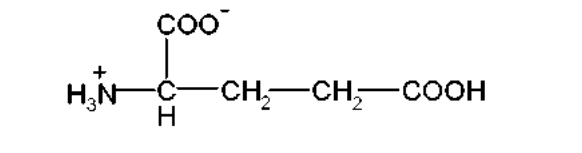
Amino Acid Structure Molecular mass (Da) Glu – Glutamic acid S1 147 Hms – Homoserine S2 119 Leu – Leucine S3 131 Met – Methionine S4 149 Lys – Lysine S5 146 Phe – Phenylalanine S6 165 S1:
S2:
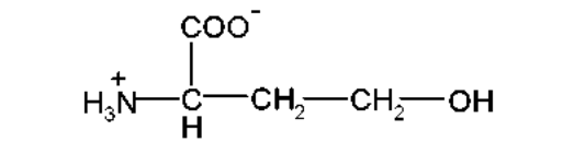
S3:
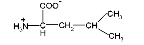
S4:
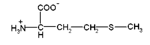
S5:
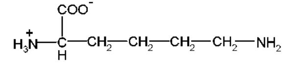
S6:
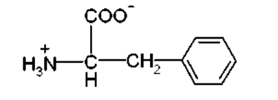
Table (continued)
Amino Acid Structure Molecular mass (Da) Arg – Arginine S7 174 Ser – Serine S8 105 S7:
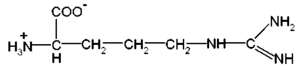
S8:
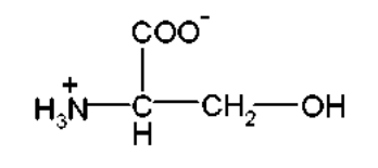
Official English text of the problem.
Internationale Chemie-Olympiade
IChO 2001
Mumbai, Indien · 7 Theorieaufgaben mit vollständigen Lösungen
Aufgabe 1: Wasserstoffatom und Wasserstoffmolekül
Niels Bohr (1885-1962)
Die im Linienspektrum des Wasserstoffatoms beobachteten Wellenlängen wurden erstmals von Johann Jakob Balmer, einem Schweizer Lehrer, durch eine Serienformel beschrieben. Balmers empirische Formel lautet
\[ \frac{1}{\lambda} = R_{\mathrm H}\left(\frac{1}{2^2}-\frac{1}{n^2}\right); \qquad n=3,4,5,\ldots \]
Dabei ist
\[ R_{\mathrm H} = \frac{m_e e^4}{8\varepsilon_0^2h^3c} = 109\,678\ \mathrm{cm^{-1}} \]
die Rydberg-Konstante, und \(m_e\) ist die Masse eines Elektrons. Niels Bohr leitete diesen Ausdruck 1913 theoretisch her. Die Formel lässt sich leicht auf jedes Atom bzw. Ion mit einem Elektron verallgemeinern.
1.1 Berechnen Sie die längste Wellenlänge in Å (\(1\ \text{Å}=10^{-10}\ \mathrm m\)) in der „Balmer-Serie“ des einfach ionisierten Heliums (\(\ce{He+}\)). Vernachlässigen Sie bei Ihrer Berechnung die Kernbewegung.
1.2 Eine zur Balmer-Formel analoge Formel gilt für die Serie von Spektrallinien, die bei Übergängen von höheren Energieniveaus zum niedrigsten Energieniveau des Wasserstoffatoms entstehen. Geben Sie diese Formel an und bestimmen Sie damit die Grundzustandsenergie eines Wasserstoffatoms in eV.
Ein „myonisches Wasserstoffatom“ gleicht einem Wasserstoffatom, in dem das Elektron durch ein schwereres Teilchen, das Myon, ersetzt ist. Die Masse eines Myons beträgt etwa das 207-Fache der Masse eines Elektrons, während seine Ladung der eines Elektrons entspricht. Ein Myon hat eine sehr kurze Lebensdauer, doch seine Instabilität wird hier vernachlässigt.
1.3 Bestimmen Sie die niedrigste Energie und den Radius der ersten Bohr'schen Bahn des myonischen Wasserstoffatoms. Vernachlässigen Sie bei Ihrer Berechnung die Bewegung des Kerns. Der Radius der ersten Bohr'schen Bahn eines Wasserstoffatoms (als Bohr-Radius bezeichnet, \(a_0=\dfrac{\varepsilon_0h^2}{m_e e^2\pi}\)) beträgt \(0.53\ \text{Å}\).
Das klassische Bild einer „Bahn“ in Bohrs Theorie ist inzwischen durch den quantenmechanischen Begriff des „Orbitals“ ersetzt worden. Das Orbital \(\psi1\sigma_{1s}(r)\) für den Grundzustand eines Wasserstoffatoms ist gegeben durch
\[ \Psi_{1s}(r) = \frac{1}{\sqrt{\pi a_0^3}}e^{-r/a_0} \]
wobei \(r\) der Abstand des Elektrons vom Kern und \(a_0\) der Bohr-Radius ist.
1.4 Betrachten Sie eine Kugelschale mit dem Radius \(a_0\) und der Dicke \(0.001a_0\). Schätzen Sie die Wahrscheinlichkeit ab, das Elektron in dieser Schale anzutreffen. Das Volumen einer Kugelschale mit dem Innenradius \(r\) und einer kleinen Dicke \(\Delta r\) beträgt \(4\pi r^2\Delta r\).
Das \(\ce{H2}\)-Molekül kann über zwei verschiedene Kanäle dissoziieren:
Der Verlauf der Energie (\(E\)) in Abhängigkeit vom Kernabstand (\(R\)) für \(\ce{H2}\) ist in der Abbildung schematisch dargestellt. Die atomaren und molekularen Energien sind auf derselben Skala angegeben.
1.5 Tragen Sie die passenden Kanalbezeichnungen (i) oder (ii) in die unten stehenden Kästchen ein.
1.6 Bestimmen Sie die Werte der Dissoziationsenergien (\(D_e\) in eV) des \(\ce{H2}\)-Moleküls für
Kanal (i) \(\boxed{\phantom{000000}}\)
Kanal (ii) \(\boxed{\phantom{000000}}\)
1.7 Berechnen Sie anhand der gegebenen Daten die Energieänderung für den Vorgang
1.8 \(\ce{H-}\) ist ein atomares Zweielektronensystem. Berechnen Sie \(Z_{\mathrm{eff}}\) für \(\ce{H-}\) unter der Annahme, dass die Bohr'sche Energieformel für jedes Elektron gilt, wobei die Kernladungszahl \(Z\) durch \(Z_{\mathrm{eff}}\) ersetzt wird.
Aufgabentext aus dem offiziellen englischen Text übersetzt.
Aufgabe 2: Phosphorsäure
Phosphorsäure ist von großer Bedeutung für die Düngemittelindustrie. Darüber hinaus finden Phosphorsäure und ihre verschiedenen Salze zahlreiche Anwendungen in der Metallbehandlung sowie in der Lebensmittel-, Waschmittel- und Zahnpastaindustrie.
2.1 Die pK-Werte der drei aufeinanderfolgenden Dissoziationsstufen der Phosphorsäure bei 25 °C betragen:
\[ pK_{1a}=2.12 \]
\[ pK_{2a}=7.21 \]
\[ pK_{3a}=12.32 \]
Geben Sie die konjugierte Base des Dihydrogenphosphat-Ions an und bestimmen Sie ihren \(pK_b\)-Wert.
Kleine Mengen Phosphorsäure werden häufig verwendet, um vielen Erfrischungsgetränken wie Cola und Root Beer einen sauren oder herben Geschmack zu verleihen. Eine Cola mit einer Dichte von \(1.00\ \mathrm{g\,cm^{-3}}\) enthält 0.05 Massen-% Phosphorsäure.
2.2 Bestimmen Sie den pH-Wert der Cola (unter Vernachlässigung der zweiten und dritten Dissoziationsstufe der Phosphorsäure). Nehmen Sie an, dass der Säuregehalt der Cola ausschließlich auf Phosphorsäure zurückzuführen ist.
2.3 Phosphorsäure wird als Düngemittel in der Landwirtschaft eingesetzt. Einer wässrigen Bodensuspension wird \(1.00\times10^{-3}\ \mathrm{M}\) Phosphorsäure zugesetzt, und es wird ein pH-Wert von 7.00 gemessen.
Bestimmen Sie die Konzentrationsanteile aller verschiedenen in der Lösung vorliegenden Phosphatspezies. Nehmen Sie an, dass kein Bestandteil des Bodens mit einer Phosphatspezies wechselwirkt.
2.4 Zink ist ein essenzieller Mikronährstoff für das Pflanzenwachstum. Pflanzen können Zink nur in wasserlöslicher Form aufnehmen. In einem gegebenen Bodenwasser mit \(pH=7.0\) wurde festgestellt, dass Zinkphosphat die einzige Quelle für Zink und Phosphat ist. Berechnen Sie die Konzentrationen der Ionen \([\ce{Zn^2+}]\) und \([\ce{PO4^3-}]\) in der Lösung. \(K_{\mathrm{sp}}\) für Zinkphosphat beträgt \(9.1\times10^{-33}\).
Aufgabentext aus dem offiziellen englischen Text übersetzt.
Aufgabe 3: Zweiter Hauptsatz der Thermodynamik
J.W.Gibbs (1839 –1903)
Der zweite Hauptsatz der Thermodynamik ist ein grundlegendes Naturgesetz. In dieser Aufgabe betrachten wir die Thermodynamik eines idealen Gases, Phasenübergänge und das chemische Gleichgewicht.
3.00 mol gasförmiges \(\ce{CO2}\) expandieren isotherm (im thermischen Kontakt mit der Umgebung; Temperatur = 15 °C) gegen einen konstanten äußeren Druck von 1.00 bar. Das Anfangs- und Endvolumen des Gases betragen 10.0 dm³ bzw. 30.0 dm³.
3.1 Wählen Sie die richtige Option für die Entropieänderung des Systems \((\Delta S_{\mathrm{sys}})\) und der Umgebung \((\Delta S_{\mathrm{sur}})\):
\(\Delta S_{\mathrm{sys}}\) \(\Delta S_{\mathrm{sur}}\) (a) \(>0\) \(=0\) ☐ (b) \(<0\) \(>0\) ☐ (c) \(>0\) \(<0\) ☐ d) \(=0\) \(=0\) ☐ (Markieren Sie das richtige Kästchen mit X.)
3.2 Berechnen Sie \(\Delta S_{\mathrm{sys}}\) unter der Annahme, dass \(\ce{CO2}\) ein ideales Gas ist.
3.3 Berechnen Sie \(\Delta S_{\mathrm{sur}}\).
3.4 Berechnen Sie die Entropieänderung des Universums.
Stimmt Ihre Antwort mit dem zweiten Hauptsatz der Thermodynamik überein?
(Markieren Sie das richtige Kästchen mit X.)
☐ Ja ☐ Nein
Das Druck-Temperatur-Phasendiagramm von \(\ce{CO2}\) ist unten schematisch dargestellt. Das Diagramm ist nicht maßstabsgetreu.
Phasendiagramm von \(\ce{CO2}\)
3.5 Gasförmiges \(\ce{CO2}\), das sich anfangs bei einem Druck von 4.0 bar und einer Temperatur von 10.0 °C befindet, wird bei konstantem Druck abgekühlt. Dabei
(a) geht es zunächst in die flüssige Phase und anschließend in die feste Phase über. ☐
(b) geht es in die feste Phase über, ohne die flüssige Phase zu durchlaufen. ☐
3.6 Ausgehend von demselben Druck und derselben Temperatur wie oben (in 3.5) wird \(\ce{CO2}\) isotherm komprimiert. Dabei
(a) geht es zunächst in die flüssige Phase und anschließend in die feste Phase über. ☐
(b) geht es in die feste Phase über, ohne die flüssige Phase zu durchlaufen. ☐
3.7 Berechnen Sie anhand der im Phasendiagramm angegebenen Daten die molare Sublimationsenthalpie von \(\ce{CO2}\). Geben Sie die verwendete Formel an.
3.8 Gasförmiges \(\ce{CO}\), das in großem Umfang in der organischen Synthese eingesetzt wird, kann durch die Reaktion von gasförmigem \(\ce{CO2}\) mit Graphit gewonnen werden. Zeigen Sie anhand der unten angegebenen Daten, dass die Gleichgewichtskonstante bei 298.15 K kleiner als eins ist.
Bei 298.15 K
\(\Delta H_f^0\ /\ \mathrm{kJ\,mol^{-1}}\) \(S^0\ /\ \mathrm{J\,K^{-1}\,mol^{-1}}\) \(\ce{CO2(g)}\) −393.51 213.79 \(\ce{CO(g)}\) −110.53 197.66 \(\ce{C(graphite)}\) 5.74 3.9 Schätzen Sie die Temperatur ab, bei der die Gleichgewichtskonstante der Reaktion gleich 1 wäre. Vernachlässigen Sie geringfügige Änderungen der thermodynamischen Daten mit der Temperatur.
3.10 Die obige Reaktion (in 3.8) wird zwischen \(\ce{CO2}\) und überschüssigem heißem Graphit in einem Reaktor durchgeführt, der bei etwa 800 °C und einem Gesamtdruck von 5.0 bar gehalten wird. Die Gleichgewichtskonstante \(K_p\) beträgt unter diesen Bedingungen 10.0. Berechnen Sie den Partialdruck von \(\ce{CO}\) im Gleichgewicht.
Aufgabentext aus dem offiziellen englischen Text übersetzt.
Aufgabe 4: Thorium, Uran und Blei in Monazit
Mineral im Strandsand von Kerala
Das im Strandsand vorkommende Mineral Monazit ist eine ergiebige Thoriumquelle und kommt im indischen Bundesstaat Kerala in großen Mengen vor. Eine typische Monazitprobe enthält etwa 9 % \(\ce{ThO2}\) und 0.35 % \(\ce{U3O8}\). \(\ce{^{208}Pb}\) und \(\ce{^{206}Pb}\) sind die stabilen Endprodukte der radioaktiven Zerfallsreihen von \(\ce{^{232}Th}\) beziehungsweise \(\ce{^{238}U}\). Das gesamte im Monazit enthaltene Blei (\(\ce{Pb}\)) ist radiogenen Ursprungs.
Das massenspektrometrisch gemessene Isotopen-Atomzahlverhältnis \(\ce{^{208}Pb}/\ce{^{232}Th}\) in einer Monazitprobe beträgt 0.104. Die Halbwertszeiten von \(\ce{^{232}Th}\) und \(\ce{^{238}U}\) betragen \(1.41\times10^{10}\) Jahre beziehungsweise \(4.47\times10^9\) Jahre. Nehmen Sie an, dass \(\ce{^{208}Pb}\), \(\ce{^{206}Pb}\), \(\ce{^{232}Th}\) und \(\ce{^{238}U}\) seit der Bildung des Minerals Monazit vollständig in der Monazitprobe verblieben sind.
4.1 Berechnen Sie das Alter (die seit ihrer Bildung verstrichene Zeit) der Monazitprobe.
4.2 Schätzen Sie das Isotopen-Atomzahlverhältnis \(\ce{^{206}Pb}/\ce{^{238}U}\) in der Monazitprobe ab.
4.3 Thorium-232 ist ein Brutstoff zur Kernenergiegewinnung. Bei der Bestrahlung mit thermischen Neutronen absorbiert es ein Neutron, und das entstehende Isotop bildet durch aufeinanderfolgende \(\beta^-\)-Zerfälle \(\ce{^{233}U}\). Geben Sie die Kernreaktionen für die Bildung von \(\ce{^{233}U}\) aus \(\ce{^{232}Th}\) an.
Bei der Kernspaltung von \(\ce{^{233}U}\) entsteht ein komplexes Gemisch radioaktiver Spaltprodukte. Das Spaltprodukt \(\ce{^{101}Mo}\) zerfällt zunächst radioaktiv wie unten dargestellt:
4.4 Eine frisch hergestellte, radiochemisch reine Probe von \(\ce{^{101}Mo}\) enthält anfangs 5000 Atome \(\ce{^{101}Mo}\). Wie viele Atome von
i) \(\ce{^{101}Mo}\)
ii) \(\ce{^{101}Tc}\)
iii) \(\ce{^{101}Ru}\)
sind nach 14.6 min in der Probe vorhanden?
Aufgabentext aus dem offiziellen englischen Text übersetzt.
Aufgabe 5: Halogenchemie
Halogene bilden bei ihren Reaktionen untereinander und mit einer Vielzahl anderer Elemente zahlreiche Verbindungen mit unterschiedlichen Strukturen, Bindungsverhältnissen und chemischen Eigenschaften. Metallhalogenide, Halogenderivate und Interhalogenverbindungen stellen wichtige Typen von Halogenverbindungen dar.
(A) Fotografie
Ein fotografischer Schwarzweißfilm enthält eine Schicht aus Silberbromid auf einem Trägermaterial wie Celluloseacetat.
5.1 Geben Sie die photochemische Reaktion an, die stattfindet, wenn Licht auf das auf einem Film aufgebrachte \(\ce{AgBr(s)}\) fällt.
5.2 Während des Entwicklungsprozesses wird unbelichtetes \(\ce{AgBr}\) durch Komplexierung von Ag(I) mit Natriumthiosulfatlösung ausgewaschen. Geben Sie diese chemische Reaktion an.
5.3 Diese Waschlösungen werden häufig als Abfall entsorgt. Metallisches Silber lässt sich jedoch daraus zurückgewinnen, indem zunächst Cyanid und anschließend Zink zugegeben wird. Geben Sie die dabei ablaufenden Reaktionen an.
(B) Molekülgestalten, Spektren und Reaktivität
Das reaktivste Halogen, Fluor, reagiert unter kontrollierten Bedingungen mit den anderen Halogenen \(\ce{Cl2}\), \(\ce{Br2}\) und \(\ce{I2}\) unter Bildung eines vieratomigen, eines sechsatomigen beziehungsweise eines achtatomigen Moleküls.
5.4 Geben Sie auf Grundlage der VSEPR-Theorie die Formeln und dreidimensionalen Strukturen dieser Interhalogenmoleküle an. Zeigen Sie gegebenenfalls die Anordnung der freien Elektronenpaare am Zentralatom.
Ein Gemisch aus Ioddampf und Chlorgas ergab beim Einleiten in ein Massenspektrometer zwei Gruppen (A und B) massenspektrometrischer Peaks, die den Molekülionen zweier chemischer Spezies bei folgenden \(m/z\)-Werten entsprechen:
A: 162, 164
B: 464, 466, 468, 470, 472, 474, 476
5.5 Identifizieren Sie die molekularen Spezies, die \(m/z = 162,\ 164,\ 466\) und \(476\) entsprechen. Zeichnen Sie die Struktur der schwersten Spezies (\(m/z = 476\)) und kennzeichnen Sie dabei deutlich die freien Elektronenpaare am Atom bzw. an den Atomen von I (Iod). Geben Sie die Isotopenzusammensetzung jeder Spezies an.
In wässrigem Medium oxidiert Chlorgas Natriumthiosulfat zu einem Ion, in dem Schwefel seine höchste Oxidationsstufe aufweist.
5.6 Geben Sie die chemische Gleichung für diese Reaktion an.
5.7 Zeichnen Sie die Lewis-Punktformel des Thiosulfat-Ions. Kreisen Sie das Schwefelatom mit der niedrigeren Oxidationsstufe ein.
5.8 Chlordioxid reagiert mit Natriumhydroxid wie unten gezeigt. Identifizieren Sie die Produkte X und Y (beide enthalten Chlor) und gleichen Sie die Gleichung aus.
5.9 Die Reaktion von Chlor mit einer Lauge wird zur Herstellung von Bleichlauge genutzt. Geben Sie die chemische Reaktion für deren Bildung an.
5.10 Geben Sie die Oxidationsstufe(n) von Chlor in Bleichlauge an.
(C) Alkalimetallhalogenide und Röntgenkristallographie
Die Röntgenkristallographie liefert Aufschluss über viele Aspekte der Struktur von Metallhalogeniden. Das Radienverhältnis \((r_+/r_-)\) ist ein nützlicher Parameter zur Erklärung ihrer Struktur und Stabilität. Eine Tabelle der Radienverhältnisse \((r_+/r_-)\) einiger Alkalihalogenide ist angegeben; die Abhängigkeit vom Radienverhältnis (bei konstant gehaltenem \(r_-\)) ist für Kristallstrukturen vom \(\ce{NaCl}\)-Typ und vom \(\ce{CsCl}\)-Typ schematisch dargestellt.
\(\ce{Li+}\) \(\ce{Na+}\) \(\ce{K+}\) \(\ce{Rb+}\) \(\ce{Cs+}\) \(\ce{Cl-}\) 0.33 0.52 0.74 0.82 0.93 \(\ce{Br-}\) 0.31 0.49 0.68 0.76 0.87 \(\ce{I-}\) 0.28 0.44 0.62 0.69 0.78 5.11 Für ein gegebenes Anion geht die Kurve für die Struktur vom \(\ce{NaCl}\)-Typ bei kleinen \(r_+/r_-\)-Werten in ein Plateau über, aufgrund von
(a) Kation-Kation-Kontakt entlang der Flächendiagonalen. \(\square\)
(b) Anion-Anion-Kontakt entlang der Flächendiagonalen. \(\square\)
(c) Kation-Anion-Kontakt entlang der Kante der Elementarzelle. \(\square\)
(Markieren Sie das richtige Kästchen mit X.)
5.12 Welches der Halogenide \(\ce{LiBr}\), \(\ce{NaBr}\) und \(\ce{RbBr}\) wird bei einer Änderung der Temperatur und/oder des Drucks am ehesten einen Phasenübergang von einer Struktur vom \(\ce{NaCl}\)-Typ zu einer Struktur vom \(\ce{CsCl}\)-Typ durchlaufen?
5.13 Berechnen Sie das Radienverhältnis \((r_+/r_-)\), bei dem die Energie der Struktur vom \(\ce{CsCl}\)-Typ in ein Plateau übergeht.
5.14 Bei Verwendung von CuKα-Röntgenstrahlung (\(\lambda = 154\ \mathrm{nm}\)) wird an einem \(\ce{KCl}\)-Kristall (kubisch flächenzentrierte Struktur, fcc) Beugung unter einem Winkel (\(\theta\)) von \(14.2^\circ\) beobachtet. Gegeben ist, dass (i) die Beugung an Ebenen mit \(h^2+k^2+l^2=4\) stattfindet, (ii) in einem kubischen Kristall \(d_{hkl}=a/(h^2+k^2+l^2)^{1/2}\) gilt, wobei „d“ der Abstand zwischen benachbarten hkl-Ebenen und „a“ ein Gitterparameter ist, und (iii) Reflexe in einer fcc-Struktur nur von Ebenen mit „ausschließlich ungeraden“ oder „ausschließlich geraden“ hkl-Indizes (Miller-Indizes) auftreten können. Berechnen Sie den Gitterparameter „a“ für \(\ce{KCl}\).
5.15 Tragen Sie in die folgende Tabelle die geforderten Angaben zu den \(2^{\mathrm{nd}}\) und \(3^{\mathrm{rd}}\) nächsten Nachbarn eines \(\ce{K+}\)-Ions im \(\ce{KCl}\)-Gitter ein.
\(2^{\mathrm{nd}}\) nächste Nachbarn \(3^{\mathrm{rd}}\) nächste Nachbarn Anzahl Vorzeichen der Ladung Abstand (pm) Anzahl Vorzeichen der Ladung Abstand (pm) 5.16 Bestimmen Sie den kleinsten möglichen Wert des Beugungswinkels \(\theta\) für die \(\ce{KCl}\)-Struktur.
Aufgabentext aus dem offiziellen englischen Text übersetzt.
Aufgabe 6: Organische Chemie indischer Gewürze
Die Rhizome des Ingwers (Zingiber officinale) sind für ihre heilenden und würzenden Eigenschaften bekannt. Im Ayurveda (dem traditionellen Medizinsystem Indiens) werden verschiedene Ingwerzubereitungen zur Behandlung von Magen-Darm-Beschwerden, Erkältungen und anderen Erkrankungen verwendet. Mehrere Verbindungen sind für die Schärfe des Ingwers verantwortlich. Viele davon sind einfach substituierte aromatische Verbindungen mit unterschiedlichen Seitenketten. Drei von ihnen, Zingeron, (+)[6]-Gingerol (im Folgenden nur als Gingerol bezeichnet) und Shogaol, sind besonders wichtig.
Zingeron: \(\ce{C11H14O3}\)
Gingerol: \(\ce{C17H14O4}\)
Shogaol: \(\ce{C17H24O3}\)6.1 Zingeron ergibt positive Nachweise mit \(\ce{FeCl3}\) und 2,4-DNP (2,4-Dinitrophenylhydrazin). Es reagiert nicht mit Tollens-Reagenz. Daher enthält Zingeron die folgenden funktionellen Gruppen: (Markieren Sie die richtigen Kästchen mit X.)
(a) alkoholische Hydroxygruppe \(\square\) (e) Estergruppe \(\square\) (b) aldehydische Carbonylgruppe \(\square\) (f) Alkoxygruppe \(\square\) (c) ketonische Carbonylgruppe \(\square\) (g) Ungesättigtheit \(\square\) (d) phenolische Hydroxygruppe \(\square\) Die aus dem \(\ce{^1H}\)-NMR-Spektrum von Zingeron erhaltenen Daten sind in Tabelle 1 aufgeführt. Einige weitere relevante Informationen sind in Tabelle 2 angegeben.
Tabelle 1: \(\ce{^1H}\)-NMR-spektroskopische Daten von Zingeron
Chemische Verschiebungen \((\delta)\) Multiplizität Relative Intensität 2.04 Singulett 3 2.69, 2.71 zwei (eng benachbarte) Tripletts gleicher Intensität 4 3.81 Singulett 3 5.90 breites Singulett (mit \(\ce{D2O}\) austauschbar) 1 6.4 – 6.8 zwei Dubletts mit ähnlichen chemischen Verschiebungen und ein Singulett 3 *) Zur besseren Übersicht wurden einige Daten geringfügig verändert.)
Tabelle 2: Ungefähre chemische \(\ce{^1H}\)-Verschiebungen \((\delta)\) und Spin-Spin-Kopplungskonstanten \((J)\) einiger Protonen
Chemische \(\ce{^1H}\)-Verschiebungen
Alkyl - H 0.9 - 1.5 [S1] 3.4 - 3.6 [S2] 2.0 - 2.7 [S3] 4.0 - 7.0 [S4] 2.3 - 2.9 [S5] 6.0 - 8.0 [S6] 3.3 - 3.9 [S7] 4.0 - 11.0 [S1]
[S2]
[S3]
[S4]
[S5]
[S6]
[S7]
Spin-Spin-Kopplungskonstanten \((J)\)
Alkene cis 5 – 14 Hz (gewöhnlich etwa 6 – 8 Hz) trans 11 – 19 Hz (gewöhnlich etwa 14 – 16 Hz) Bei der Bromierung mit Bromwasser ergibt Zingeron nur ein einziges am Ring monobromiertes Produkt. Das IR-Spektrum von Zingeron weist auf eine schwache intramolekulare Wasserstoffbrückenbindung hin. Diese ist auch nach der Clemmensen-Reduktion (\(\ce{Zn}\) – \(\ce{Hg}/\ce{HCl}\)) von Zingeron noch vorhanden.
6.2 Leiten Sie aus den obigen Informationen Folgendes ab:
i) die Seitenkette in Zingeron
ii) den Substituenten am aromatischen Ring
iii) die relativen Positionen der Substituenten am Ring
6.3 Zeichnen Sie auf der Grundlage der obigen Schlussfolgerungen eine mögliche Struktur von Zingeron.
6.4 Vervollständigen Sie die folgende Reaktionsfolge zur Synthese von Zingeron.
6.5 Zingeron lässt sich durch die folgende Reaktionsfolge leicht in Gingerol überführen:
Hinweise: (1) \(\ce{Me3SiCl}/\ce{(Me3Si)2NH}\) wird verwendet, um OH in \(\ce{-OSiMe3}\) umzuwandeln; die Gruppe \(\ce{-SiMe3}\) kann durch saure Hydrolyse entfernt werden.
(2) LDA ist Lithiumdiisopropylamin, eine starke, stark sterisch gehinderte, nicht nukleophile Base.
i) Zeichnen Sie die Struktur von D.
ii) Zeichnen Sie die Struktur von Gingerol.
iii) Vervollständigen Sie die Fischer-Projektion des \(R\)-Enantiomers von Gingerol.
iv) In der obigen Reaktionsfolge (6.5) entstehen etwa 2 – 3 % eines weiteren Konstitutionsisomers (E) von Gingerol. Zeichnen Sie die wahrscheinliche Struktur von E.
v) Entsteht die Verbindung E als
(a) ein Enantiomerenpaar? \(\square\)
(b) ein Gemisch von Diastereomeren? \(\square\)
(c) ein Gemisch aus einem Enantiomerenpaar und einem meso-Isomer? \(\square\)
(Markieren Sie das richtige Kästchen mit X.)
vi) Beim Erhitzen mit einer milden Säure (wie \(\ce{KHSO4}\)) ergibt Gingerol (\(\ce{C17H26O4}\)) Shogaol (\(\ce{C17H24O3}\)). Zeichnen Sie die Struktur von Shogaol.
6.6 Kurkuma (Curcuma longa) ist ein häufig verwendetes Gewürz in der indischen Küche. Es wird auch in ayurvedischen Arzneizubereitungen verwendet. Curcumin (\(\ce{C21H20O6}\)), ein wirksamer Inhaltsstoff von Kurkuma, ist strukturell mit Gingerol verwandt. Es zeigt Keto-Enol-Tautomerie. Curcumin ist für die gelbe Farbe von Kurkuma und wahrscheinlich auch für den scharfen Geschmack verantwortlich.
Das \(\ce{^1H}\)-NMR-Spektrum der Ketoform von Curcumin zeigt ein aromatisches Signal ähnlich dem von Gingerol. Es zeigt außerdem ein Singulett bei etwa \(\delta\) 3.5 (2H) und zwei Dubletts (je 2H) im Bereich \(\delta\) 6 – 7 mit \(J=16\ \mathrm{Hz}\). Es lässt sich durch Kondensation von ZWEI Mol A (siehe 6.4) mit einem Mol Pentan-2,4-dion synthetisieren.
i) Zeichnen Sie die stereochemische Struktur von Curcumin.
ii) Zeichnen Sie die Struktur der Enolform von Curcumin.
iii) Curcumin ist gelb, weil es Folgendes besitzt:
(a) einen Phenylring \(\square\)
(b) eine Carbonylgruppe \(\square\)
(c) ein ausgedehntes konjugiertes System \(\square\)
(d) eine Hydroxygruppe \(\square\)
(Markieren Sie das richtige Kästchen mit X.)
Aufgabentext aus dem offiziellen englischen Text übersetzt.
Aufgabe 7: Peptide und Proteine
[In den Aminosäuretabellen beziehen sich S1–S8 auf die Strukturabbildungen, die unmittelbar unter der jeweiligen Tabelle wiedergegeben sind.]
AUFGABE 7
Peptide und Proteine
Proteinfaltung
Proteine (Polypeptide) können bekanntlich verschiedene Konformationen des Peptidrückgrats annehmen. In einer Konformation ist das Rückgrat nahezu vollständig gestreckt (wie in einem parallelen oder antiparallelen β-Faltblatt), in einer anderen liegt es in einer vollständig gefalteten Anordnung vor (wie in einer α-Helix).
7.1 Der Abstand zwischen den beiden Enden eines Hexapeptids in vollständig gestreckter Konformation beträgt ungefähr:
(a) \(10\ \text{Å}\) ☐
(b) \(15\ \text{Å}\) ☐
(c) \(20\ \text{Å}\) ☐
(d) \(25\ \text{Å}\) ☐(Markieren Sie das richtige Kästchen mit X.)
7.2 Nehmen Sie an, dass das folgende Hexapeptid in einer α-Helix-Konformation vorliegt. Zeichnen Sie einen Verbindungspfeil zwischen einem Sauerstoffatom und einer Amid-\(\ce{NH}\)-Gruppe, mit der es eine Wasserstoffbrücke bildet.
7.3 Die folgenden Hexapeptide (A und B) zeigen in Wasser bei pH 7.0 unterschiedliche Konformationen, insbesondere wenn ihre Serin-Hydroxygruppen phosphoryliert sind. A ist mäßig helikal und bildet nach Phosphorylierung des Serins eine stärker ausgeprägte Helix. B ist schwach helikal und nach Phosphorylierung des Serins vollständig ungeordnet. Zeichnen Sie Verbindungspfeile ein, um die Wechselwirkungen zwischen den Resten anzugeben, die für dieses unterschiedliche Verhalten verantwortlich sind.
Betrachten Sie den folgenden Vorgang der Faltung/Entfaltung eines Tripeptidsegments in einem großen Protein. Die Änderung der freien Enthalpie (\(\Delta G\)) hängt von der Wechselwirkung des entfalteten Tripeptids mit dem Lösungsmittel (Wasser) und im gefalteten Zustand mit dem übrigen Protein ab (siehe unten). Nehmen Sie an, dass das Tripeptid aus einem unpolaren (hydrophoben; schattierten) und zwei polaren (hydrophilen; nicht schattierten) Resten besteht. Nehmen Sie die folgenden ungefähren Änderungen der freien Wechselwirkungsenthalpien an:
(a) ein unpolarer Rest und das Lösungsmittel (Wasser): \(\Delta G=+8\ \mathrm{kJ\,mol^{-1}}\)
(b) ein unpolarer Rest und das übrige Protein: \(\Delta G=-4\ \mathrm{kJ\,mol^{-1}}\)
(c) ein polarer Rest und das Lösungsmittel (Wasser): \(\Delta G=-16\ \mathrm{kJ\,mol^{-1}}\)
(d) ein polarer Rest und das übrige Protein: \(\Delta G=-14\ \mathrm{kJ\,mol^{-1}}\)
7.4 Berechnen Sie \(\Delta G\) für die Faltung des Tripeptidsegments.
7.5 Berechnen Sie \(\Delta G\) für die Faltung des Tripeptidsegments, wenn alle drei Reste polar sind.
β-Lactamase und Arzneimittelresistenz
Penicilline sind wirksame Arzneimittel zur Bekämpfung bakterieller Infektionen. Eine besorgniserregende Entwicklung im Laufe der Jahre ist jedoch das Auftreten arzneimittelresistenter Bakterien. Penicillinresistenz entsteht durch die Sekretion eines Enzyms namens β-Lactamase (auch als Penicillinase bekannt), das Penicillin durch Öffnung seines β-Lactamrings inaktiviert. Der Mechanismus dieser β-Lactamringöffnung umfasst den nukleophilen Angriff durch Serin-\(\ce{OH}\) im aktiven Zentrum des Enzyms, wie unten gezeigt.
Es wurde versucht, die β-Lactamase aus Staphylococcus aureus zu charakterisieren. Bei der Behandlung des reinen Enzyms mit einem radioaktiv markierten [\(\ce{^{32}P}\)] Phosphorylierungsreagenz wurde nur das Serin im aktiven Zentrum markiert. Die Analyse ergab, dass dieses Serin {Molekülmasse = 105 Masseneinheiten (Da)} einen Massenanteil von 0.35% an der β-Lactamase ausmachte.
7.6 Schätzen Sie die minimale Molekülmasse dieser β-Lactamase ab.
7.7 Die ungefähre Anzahl der Aminosäurereste in einem Protein dieser Größe beträgt:
(a) 100 ☐
(b) 150 ☐
(c) 275 ☐
(d) 375 ☐[Markieren Sie das richtige Kästchen mit X.]
Zur Untersuchung ihres aktiven Zentrums wurde die β-Lactamase mit Trypsin, einem spezifischen Enzym, hydrolysiert. Dabei entstand ein Hexapeptid P1, das das Serin des aktiven Zentrums enthielt. Die Aminosäureanalyse ergab die folgenden Aminosäuren in äquimolaren Anteilen: Glu, Leu, Lys, Met, Phe und Ser. Die Behandlung von P1 mit dem Edman-Reagenz (Phenylisothiocyanat) lieferte das Phenylthiohydantoin-Derivat (PTH-Derivat) von Phenylalanin und ein Peptid P2.
Die Behandlung von P1 mit Bromcyan (\(\ce{CNBr}\)) ergab ein saures Tetrapeptid P3 und ein Dipeptid P4.
Die Behandlung von P2 mit 1-Fluor-2,4-dinitrobenzol mit anschließender vollständiger Hydrolyse ergab N-2,4-Dinitrophenyl-Glu. P1, P2 und P3 enthalten das Serin des aktiven Zentrums.
7.8 Leiten Sie aus den obigen Angaben die Aminosäuresequenzen von P1, P2, P3 und P4 ab.
7.9 Berechnen Sie anhand der Angaben in der beigefügten Tabelle die Molekülmasse von P3 in Masseneinheiten (Da).
Das aktive Zentrum der β-Lactamase bietet eine einzigartige Mikroumgebung, die das katalytische Serin-\(\ce{OH}\) zu einem ungewöhnlich reaktiven Nukleophil macht. Die Geschwindigkeitskonstante erster Ordnung für die durch β-Lactamase katalysierte Reaktion beträgt \(350\ \mathrm{s^{-1}}\). Für die Penicillinhydrolyse durch freies Serin-\(\ce{OH}\) (bei \(1\ \mathrm{M}\)) in Lösung beträgt die Geschwindigkeitskonstante pseudoerster Ordnung \(0.5\ \mathrm{s^{-1}}\).
7.10 Berechnen Sie anhand der obigen Angaben die effektive Konzentration dieses Nukleophils im aktiven Zentrum des Enzyms.
Ein Molekül, das mit Penicillin um die Bindung an das aktive Zentrum der β-Lactamase konkurriert, kann das Enzym hemmen. Die Dissoziationskonstanten (\(K_D\)) des Inhibitor-Lactamase-Komplexes für drei verschiedene Inhibitoren sind nachstehend angegeben:
Inhibitor Dissoziationskonstante (\(K_D\)) A \(2.0\times10^{-3}\) B \(1.0\times10^{-6}\) C \(5.0\times10^{-9}\) 7.11 Geben Sie an, welcher dieser Inhibitoren Penicillin am wirksamsten vor β-Lactamase schützt. [Markieren Sie das richtige Kästchen mit X.]
A ☐
B ☐
C ☐Ein β-Lactamase-Inhibitor wurde gezielt auf rationaler Grundlage entwickelt. Nach der Bindung an das aktive Zentrum des Enzyms führte ein nukleophiler Angriff der \(\ce{OH}\)-Gruppe des Serins zur Öffnung der β-Lactam-Einheit des Inhibitors und zur Abspaltung von \(\ce{Br^-}\). Dadurch entsteht ein reaktives Elektrophil, das einen Rest X im aktiven Zentrum bindet und so das Enzym inaktiviert.
7.12 Identifizieren Sie anhand der obigen Angaben das gebildete Elektrophil (A) und das Endprodukt (B), das bei der Inaktivierung des Enzyms durch den gezeigten Inhibitor entsteht.
Tabelle
Aminosäure Struktur Molekülmasse (Da) Glu – Glutaminsäure S1 147 Hms – Homoserin S2 119 Leu – Leucin S3 131 Met – Methionin S4 149 Lys – Lysin S5 146 Phe – Phenylalanin S6 165 S1:
S2:
S3:
S4:
S5:
S6:
Tabelle (Fortsetzung)
Aminosäure Struktur Molekülmasse (Da) Arg – Arginin S7 174 Ser – Serin S8 105 S7:
S8:
Aufgabentext aus dem offiziellen englischen Text übersetzt.
Uluslararası Kimya Olimpiyatı
IChO 2001
Mumbai, Hindistan · 7 teori sorusu, tam çözümleriyle
Soru 1: Hidrojen Atomu ve Hidrojen Molekülü
Niels Bohr (1885-1962)
Hidrojen atomunun çizgi spektrumunda gözlenen dalga boyları ilk kez İsviçreli bir öğretmen olan Johann Jakob Balmer tarafından bir seri biçiminde ifade edilmiştir. Balmer’in ampirik formülü şöyledir:
\[ \frac{1}{\lambda} = R_{\mathrm H}\left(\frac{1}{2^2}-\frac{1}{n^2}\right); \qquad n=3,4,5,\ldots \]
Burada,
\[ R_{\mathrm H} = \frac{m_e e^4}{8\varepsilon_0^2h^3c} = 109\,678\ \mathrm{cm^{-1}} \]
Rydberg sabitidir; \(m_e\) ise elektronun kütlesidir. Niels Bohr bu ifadeyi 1913 yılında kuramsal olarak türetmiştir. Formül, tek elektronlu herhangi bir atom veya iyona kolayca genellenebilir.
1.1 Bir kez iyonlaşmış helyumun (\(\ce{He+}\)) ‘Balmer serisi’ndeki en uzun dalga boyunu Å (\(1\ \text{Å}=10^{-10}\ \mathrm m\)) cinsinden hesaplayınız. Hesabınızda çekirdeğin hareketini ihmal ediniz.
1.2 Balmer formülüne benzer bir formül, hidrojen atomunun daha yüksek enerji düzeylerinden en düşük enerji düzeyine geçişlerinden kaynaklanan spektrum çizgileri serisi için de geçerlidir. Bu formülü yazınız ve bunu kullanarak hidrojen atomunun temel hâl enerjisini eV cinsinden belirleyiniz.
‘Müonik hidrojen atomu’, elektronun yerini daha ağır bir parçacık olan müonun aldığı bir hidrojen atomu gibidir. Müonun kütlesi elektronun kütlesinin yaklaşık 207 katıdır; yükü ise elektronunkiyle aynıdır. Müonun ömrü çok kısadır, ancak burada kararsız oluşunu göz ardı ediyoruz.
1.3 Müonik hidrojen atomunun en düşük enerjisini ve birinci Bohr yörüngesinin yarıçapını belirleyiniz. Hesabınızda çekirdeğin hareketini ihmal ediniz. Hidrojen atomunun birinci Bohr yörüngesinin yarıçapı (Bohr yarıçapı olarak adlandırılır, \(a_0=\dfrac{\varepsilon_0h^2}{m_e e^2\pi}\)) \(0.53\ \text{Å}\)’dur.
Bohr kuramındaki klasik "yörünge" tasvirinin yerini artık kuantum mekaniğindeki ‘orbital’ kavramı almıştır. Hidrojen atomunun temel hâline ait \(\psi1\sigma_{1s}(r)\) orbitali şu şekilde verilir:
\[ \Psi_{1s}(r) = \frac{1}{\sqrt{\pi a_0^3}}e^{-r/a_0} \]
Burada \(r\), elektronun çekirdeğe olan uzaklığı, \(a_0\) ise Bohr yarıçapıdır.
1.4 Yarıçapı \(a_0\), kalınlığı \(0.001a_0\) olan küresel bir kabuk düşününüz. Elektronun bu kabuk içinde bulunma olasılığını yaklaşık olarak hesaplayınız. İç yarıçapı \(r\) ve küçük bir \(\Delta r\) kalınlığı olan küresel bir kabuğun hacmi \(4\pi r^2\Delta r\)’ye eşittir.
\(\ce{H2}\) molekülü iki farklı kanaldan ayrışabilir:
\(\ce{H2}\) için enerjinin (\(E\)) çekirdekler arası uzaklığa (\(R\)) karşı grafiği şekilde şematik olarak gösterilmiştir. Atom ve molekül enerjileri aynı ölçekte verilmiştir.
1.5 Aşağıdaki kutulara uygun kanal etiketlerini, (i) veya (ii), yazınız.
1.6 \(\ce{H2}\) molekülünün aşağıdaki kanallara karşılık gelen ayrışma enerjilerinin (eV cinsinden \(D_e\)) değerlerini belirleyiniz:
kanal (i) \(\boxed{\phantom{000000}}\)
kanal (ii) \(\boxed{\phantom{000000}}\)
1.7 Verilen verilerden yararlanarak aşağıdaki süreç için enerji değişimini hesaplayınız:
1.8 \(\ce{H-}\), iki elektronlu bir atomik sistemdir. Çekirdek yükü \(Z\) yerine \(Z_{\mathrm{eff}}\) konulduğunda Bohr enerji formülünün her bir elektron için geçerli olduğunu varsayarak \(\ce{H-}\) için \(Z_{\mathrm{eff}}\)’i hesaplayınız.
Soru metni resmi İngilizce metinden çevrilmiştir.
Soru 2: Fosforik Asit
Fosforik asit, gübre sanayisinde büyük önem taşır. Ayrıca fosforik asit ve çeşitli tuzları, metal işleme, gıda, deterjan ve diş macunu sanayilerinde birçok uygulama alanına sahiptir.
2.1 Fosforik asidin 25 °C'deki üç ardışık ayrışmasının pK değerleri şöyledir:
\[ pK_{1a}=2.12 \]
\[ pK_{2a}=7.21 \]
\[ pK_{3a}=12.32 \]
Dihidrojen fosfat iyonunun eşlenik bazını yazınız ve \(pK_b\) değerini belirleyiniz.
Kola ve kök birası gibi birçok alkolsüz içeceğe ekşi veya mayhoş bir tat vermek için az miktarda fosforik asit yaygın olarak kullanılır. Yoğunluğu \(1.00\ \mathrm{g\,cm^{-3}}\) olan bir kola, kütlece % 0.05 fosforik asit içerir.
2.2 Kolanın pH değerini belirleyiniz (fosforik asidin ikinci ve üçüncü ayrışma basamaklarını ihmal ediniz). Kolanın asitliğinin yalnızca fosforik asitten kaynaklandığını varsayınız.
2.3 Fosforik asit, tarımda gübre olarak kullanılır. Sulu bir toprak süspansiyonuna \(1.00\times10^{-3}\ \mathrm{M}\) fosforik asit eklenir ve pH değeri 7.00 olarak bulunur.
Çözeltide bulunan tüm farklı fosfat türlerinin derişim kesirlerini belirleyiniz. Toprağın hiçbir bileşeninin herhangi bir fosfat türüyle etkileşmediğini varsayınız.
2.4 Çinko, bitki büyümesi için gerekli bir mikro besin elementidir. Bitkiler çinkoyu yalnızca suda çözünen biçimde alabilir. \(pH=7.0\) olan belirli bir toprak suyunda, çinko ve fosfatın tek kaynağının çinko fosfat olduğu bulunmuştur. Çözeltideki \([\ce{Zn^2+}]\) ve \([\ce{PO4^3-}]\) iyonlarının derişimlerini hesaplayınız. Çinko fosfatın \(K_{\mathrm{sp}}\) değeri \(9.1\times10^{-33}\)'tür.
Soru metni resmi İngilizce metinden çevrilmiştir.
Soru 3: Termodinamiğin İkinci Yasası
J.W.Gibbs (1839 –1903)
Termodinamiğin ikinci yasası, bilimin temel yasalarından biridir. Bu soruda ideal bir gazın termodinamiğini, faz geçişlerini ve kimyasal dengeyi ele alıyoruz.
3.00 mol \(\ce{CO2}\) gazı, 1.00 bar sabit dış basınca karşı izotermal olarak (çevreyle ısıl temas hâlinde; sıcaklık = 15 °C) genleşmektedir. Gazın başlangıç ve son hacimleri sırasıyla 10.0 dm³ ve 30.0 dm³'tür.
3.1 Sistemin \((\Delta S_{\mathrm{sys}})\) ve çevrenin \((\Delta S_{\mathrm{sur}})\) entropi değişimleri için doğru seçeneği seçiniz:
\(\Delta S_{\mathrm{sys}}\) \(\Delta S_{\mathrm{sur}}\) (a) \(>0\) \(=0\) ☐ (b) \(<0\) \(>0\) ☐ (c) \(>0\) \(<0\) ☐ d) \(=0\) \(=0\) ☐ (Doğru kutuya X işareti koyunuz.)
3.2 \(\ce{CO2}\)'nin ideal bir gaz olduğunu varsayarak \(\Delta S_{\mathrm{sys}}\)'i hesaplayınız.
3.3 \(\Delta S_{\mathrm{sur}}\)'u hesaplayınız.
3.4 Evrenin entropi değişimini hesaplayınız.
Cevabınız Termodinamiğin İkinci Yasası ile uyumlu mu?
(Doğru kutuya X işareti koyunuz.)
☐ Evet ☐ Hayır
\(\ce{CO2}\)'nin basınç–sıcaklık faz diyagramı aşağıda şematik olarak verilmiştir. Diyagram ölçekli çizilmemiştir.
\(\ce{CO2}\)'nin faz diyagramı
3.5 Başlangıçta 4.0 bar basınç ve 10.0 °C sıcaklıkta bulunan \(\ce{CO2}\) gazı sabit basınçta soğutulmaktadır. Bu süreçte,
(a) önce sıvı faza, ardından katı faza geçer. ☐
(b) sıvı fazdan geçmeden katı faza geçer. ☐
3.6 Yukarıdaki (3.5'teki) basınç ve sıcaklıktan başlanarak \(\ce{CO2}\) izotermal olarak sıkıştırılmaktadır. Bu süreçte,
(a) önce sıvı faza, ardından katı faza geçer. ☐
(b) sıvı fazdan geçmeden katı faza geçer. ☐
3.7 Faz diyagramında verilen verilerden yararlanarak \(\ce{CO2}\)'nin molar süblimleşme entalpi değişimini hesaplayınız. Kullandığınız formülü yazınız.
3.8 Organik sentezde yaygın olarak kullanılan \(\ce{CO}\) gazı, \(\ce{CO2}\) gazının grafitle tepkimeye sokulmasıyla elde edilebilir. Aşağıda verilen verileri kullanarak 298.15 K'de denge sabitinin birden küçük olduğunu gösteriniz.
298.15 K'de
\(\Delta H_f^0\ /\ \mathrm{kJ\,mol^{-1}}\) \(S^0\ /\ \mathrm{J\,K^{-1}\,mol^{-1}}\) \(\ce{CO2(g)}\) −393.51 213.79 \(\ce{CO(g)}\) −110.53 197.66 \(\ce{C(graphite)}\) 5.74 3.9 Tepkimenin denge sabitinin 1'e eşit olacağı sıcaklığı yaklaşık olarak hesaplayınız. Termodinamik verilerin sıcaklıkla gösterdiği küçük değişimleri ihmal ediniz.
3.10 Yukarıdaki (3.8'deki) tepkime, yaklaşık 800 °C sıcaklıkta ve 5.0 bar toplam basınçta tutulan bir reaktörde \(\ce{CO2}\) ile aşırı miktarda sıcak grafit arasında gerçekleştirilmektedir. Bu koşullarda denge sabiti \(K_p\), 10.0'dır. Dengedeki \(\ce{CO}\) kısmi basıncını hesaplayınız.
Soru metni resmi İngilizce metinden çevrilmiştir.
Soru 4: Monazitte toryum, uranyum ve kurşun
Kerala'da Sahil Kumu Minerali
Bir sahil kumu minerali olan monazit, Hindistan'ın Kerala eyaletinde büyük miktarlarda bulunan zengin bir toryum kaynağıdır. Tipik bir monazit örneği yaklaşık % 9 \(\ce{ThO2}\) ve % 0.35 \(\ce{U3O8}\) içerir. \(\ce{^{208}Pb}\) ve \(\ce{^{206}Pb}\), sırasıyla \(\ce{^{232}Th}\) ve \(\ce{^{238}U}\) radyoaktif bozunma serilerinin kararlı son ürünleridir. Monazitte bulunan kurşunun (\(\ce{Pb}\)) tamamı radyojenik kökenlidir.
Bir monazit örneğinde kütle spektrometrisiyle ölçülen \(\ce{^{208}Pb}/\ce{^{232}Th}\) izotopik atom oranı 0.104 olarak bulunmuştur. \(\ce{^{232}Th}\) ve \(\ce{^{238}U}\) izotoplarının yarı ömürleri sırasıyla \(1.41\times10^{10}\) yıl ve \(4.47\times10^9\) yıldır. Monazit mineralinin oluşumundan bu yana \(\ce{^{208}Pb}\), \(\ce{^{206}Pb}\), \(\ce{^{232}Th}\) ve \(\ce{^{238}U}\) izotoplarının tamamının monazit örneği içinde kaldığını varsayınız.
4.1 Monazit örneğinin yaşını (oluşumundan bu yana geçen süreyi) hesaplayınız.
4.2 Monazit örneğindeki \(\ce{^{206}Pb}/\ce{^{238}U}\) izotopik atom oranını tahmin ediniz.
4.3 Toryum-232, nükleer enerji üretiminde kullanılan üretken bir malzemedir. Termal nötronlarla ışınlandığında bir nötron soğurur ve oluşan izotop, art arda gerçekleşen \(\beta^-\) bozunmalarıyla \(\ce{^{233}U}\) oluşturur. \(\ce{^{232}Th}\) izotopundan \(\ce{^{233}U}\) oluşumuna ilişkin nükleer tepkimeleri yazınız.
\(\ce{^{233}U}\) izotopunun nükleer fisyonunda, radyoaktif fisyon ürünlerinden oluşan karmaşık bir karışım meydana gelir. Fisyon ürünü \(\ce{^{101}Mo}\), başlangıçta aşağıda gösterildiği gibi radyoaktif bozunmaya uğrar:
4.4 Yeni hazırlanmış, radyokimyasal olarak saf bir \(\ce{^{101}Mo}\) örneği başlangıçta 5000 adet \(\ce{^{101}Mo}\) atomu içermektedir. 14.6 min sonra örnekte
i) \(\ce{^{101}Mo}\)
ii) \(\ce{^{101}Tc}\)
iii) \(\ce{^{101}Ru}\)
izotoplarının her birinden kaç atom bulunur?
Soru metni resmi İngilizce metinden çevrilmiştir.
Soru 5: Halojen Kimyası
Halojenler, birbirleriyle ve çeşitli başka elementlerle tepkimelerinde yapı, bağlanma ve kimyasal davranış bakımından çeşitlilik gösteren çok sayıda bileşik oluşturur. Metal halojenürler, halojen türevleri ve halojenler arası bileşikler, halojen bileşiklerinin başlıca türlerini oluşturur.
(A) Fotoğrafçılık
"Siyah beyaz" bir fotoğraf filmi, selüloz asetat gibi bir destek üzerinde gümüş bromür kaplaması içerir.
5.1 Bir film üzerine kaplanmış \(\ce{AgBr(s)}\) üzerine ışık düştüğünde gerçekleşen fotokimyasal tepkimeyi yazınız.
5.2 Banyo işlemi sırasında, ışığa maruz kalmamış \(\ce{AgBr}\), Ag(I)'in sodyum tiyosülfat çözeltisiyle kompleks oluşturması yoluyla yıkanarak uzaklaştırılır. Bu kimyasal tepkimeyi yazınız.
5.3 Bu yıkama çözeltileri çoğunlukla atık olarak uzaklaştırılır. Ancak önce siyanür, ardından çinko eklenerek bu çözeltilerden metalik gümüş geri kazanılabilir. Gerçekleşen tepkimeleri yazınız.
(B) Şekiller, spektrumlar ve tepkimeye girme eğilimi
En reaktif halojen olan flor, kontrollü koşullarda diğer halojenler \(\ce{Cl2}\), \(\ce{Br2}\) ve \(\ce{I2}\) ile tepkimeye girerek sırasıyla dört atomlu, altı atomlu ve sekiz atomlu birer molekül oluşturur.
5.4 VSEPR kuramına dayanarak bu halojenler arası moleküllerin formüllerini ve üç boyutlu yapılarını yazınız. Uygun durumlarda merkez atom üzerindeki ortaklanmamış elektron çiftlerinin yerleşimini gösteriniz.
İyot buharı ile klor gazından oluşan bir karışım bir kütle spektrometresine verildiğinde, iki kimyasal türün moleküler iyonlarına karşılık gelen, aşağıdaki \(m/z\) değerlerinde iki grup (A ve B) kütle spektrumu piki elde edilmiştir:
A: 162, 164
B: 464, 466, 468, 470, 472, 474, 476
5.5 \(m/z = 162,\ 164,\ 466\) ve \(476\) değerlerine karşılık gelen moleküler türleri belirleyiniz. En ağır türün (\(m/z = 476\)) yapısını, I (iyot) atomu veya atomları üzerindeki ortaklanmamış elektron çiftlerini açıkça belirterek çiziniz. Her türün izotop bileşimini gösteriniz.
Sulu ortamda klor gazı, sodyum tiyosülfatı kükürdün en yüksek yükseltgenme basamağını içerdiği bir iyona yükseltger.
5.6 Bu tepkimenin kimyasal denklemini yazınız.
5.7 Tiyosülfat iyonunun Lewis nokta yapısını yazınız. Daha düşük yükseltgenme basamağına sahip kükürt atomunu daire içine alınız.
5.8 Klor dioksit, sodyum hidroksitle aşağıda gösterildiği gibi tepkimeye girer. X ve Y ürünlerini (her ikisi de klor içerir) belirleyiniz ve denklemi denkleştiriniz.
5.9 Klorun bir alkali ile tepkimesi, çamaşır suyu üretiminde kullanılır. Oluşumuna ilişkin kimyasal tepkimeyi yazınız.
5.10 Çamaşır suyundaki klorun yükseltgenme basamağını veya basamaklarını yazınız.
(C) Alkali metal halojenürler ve X-ışını kristalografisi
X-ışını kristalografisi, metal halojenürlerin yapısının birçok yönünü ortaya koyar. Yarıçap oranı \((r_+/r_-)\), bunların yapılarını ve kararlılıklarını açıklamak için yararlı bir parametredir. Bazı alkali halojenürlerin yarıçap oranları \((r_+/r_-)\) bir tabloda verilmiş, yarıçap oranına bağlı değişim (\(r_-\) sabit tutularak) \(\ce{NaCl}\) tipi ve \(\ce{CsCl}\) tipi kristal yapıları için şematik olarak gösterilmiştir.
\(\ce{Li+}\) \(\ce{Na+}\) \(\ce{K+}\) \(\ce{Rb+}\) \(\ce{Cs+}\) \(\ce{Cl-}\) 0.33 0.52 0.74 0.82 0.93 \(\ce{Br-}\) 0.31 0.49 0.68 0.76 0.87 \(\ce{I-}\) 0.28 0.44 0.62 0.69 0.78 5.11 Belirli bir anyon için \(\ce{NaCl}\) tipi yapının grafiği, düşük \(r_+/r_-\) değerlerinde aşağıdaki nedenlerden dolayı yataylaşır:
(a) yüzey köşegeni boyunca katyon-katyon teması. \(\square\)
(b) yüzey köşegeni boyunca anyon-anyon teması. \(\square\)
(c) birim hücre kenarı boyunca katyon-anyon teması. \(\square\)
(Doğru kutucuğa X işareti koyunuz.)
5.12 \(\ce{LiBr}\), \(\ce{NaBr}\) ve \(\ce{RbBr}\) halojenürlerinden hangisinin sıcaklık ve / veya basınç değişimiyle \(\ce{NaCl}\) tipi yapıdan \(\ce{CsCl}\) tipi yapıya faz geçişi yapması beklenir?
5.13 \(\ce{CsCl}\) tipi yapının enerjisinin yataylaşmaya başladığı yarıçap oranını \((r_+/r_-)\) hesaplayarak gösteriniz.
5.14 CuKα X-ışınları (\(\lambda = 154\ \mathrm{nm}\)) kullanıldığında, bir \(\ce{KCl}\) kristalinden (fcc yapı) kırınımın \(14.2^\circ\) açısında (\(\theta\)) gerçekleştiği gözlenmektedir. (i) Kırınımın \(h^2+k^2+l^2=4\) koşulunu sağlayan düzlemlerden gerçekleştiği, (ii) kübik bir kristalde \(d_{hkl}=a/(h^2+k^2+l^2)^{1/2}\) olduğu, burada "d"nin komşu hkl düzlemleri arasındaki uzaklık ve "a"nın örgü parametresi olduğu, (iii) fcc yapıda yansımaların yalnızca hkl (Miller) indislerinin "tamamı tek" veya "tamamı çift" olan düzlemlerden gerçekleşebildiği verildiğine göre, \(\ce{KCl}\) için "a" örgü parametresini hesaplayınız.
5.15 \(\ce{KCl}\) örgüsündeki bir \(\ce{K+}\) iyonunun en yakın \(2^{\mathrm{nd}}\) ve \(3^{\mathrm{rd}}\) komşularına ilişkin istenen bilgileri aşağıdaki tabloda belirtiniz.
En yakın \(2^{\mathrm{nd}}\) komşular En yakın \(3^{\mathrm{rd}}\) komşular sayı yükün işareti uzaklık (pm) sayı yükün işareti uzaklık (pm) 5.16 \(\ce{KCl}\) yapısı için mümkün olan en küçük kırınım açısı \(\theta\) değerini belirleyiniz.
Soru metni resmi İngilizce metinden çevrilmiştir.
Soru 6: Hint Baharatlarının Organik Kimyası
Zencefilin (Zingiber officinale) rizomları, tıbbi ve tatlandırıcı özellikleriyle iyi bilinir. Ayurveda'da (Hindistan'ın geleneksel tıp sistemi) mide-bağırsak sorunlarının, soğuk algınlığının ve diğer rahatsızlıkların tedavisinde farklı zencefil preparatları kullanılır. Zencefilin yakıcı tadından çeşitli bileşikler sorumludur. Bunların birçoğu, farklı yan zincirlere sahip basit sübstitüe aromatik bileşiklerdir. Bunlardan üçü, Zingeron, (+)[6] Gingerol (bundan sonra yalnızca Gingerol olarak anılacaktır) ve Şogaol özellikle önemlidir.
Zingeron: \(\ce{C11H14O3}\)
Gingerol: \(\ce{C17H14O4}\)
Şogaol: \(\ce{C17H24O3}\)6.1 Zingeron, \(\ce{FeCl3}\) ve 2,4-DNP (2,4-dinitrofenilhidrazin) testlerine pozitif sonuç verir. Tollens ayıracıyla tepkimeye girmez. Dolayısıyla Zingeron aşağıdaki fonksiyonel grupları içerir: (Doğru kutulara X koyunuz.)
(a) alkolik hidroksil \(\square\) (e) ester \(\square\) (b) aldehit karbonili \(\square\) (f) alkoksil \(\square\) (c) keton karbonili \(\square\) (g) doymamışlık \(\square\) (d) fenolik hidroksil \(\square\) Zingeronun \(\ce{^1H}\) NMR spektrumundan elde edilen veriler Tablo 1'de gösterilmiştir. İlgili diğer bazı bilgiler Tablo 2'de verilmiştir.
Tablo 1: Zingeronun \(\ce{^1H}\) NMR spektrum verileri
Kimyasal kaymalar \((\delta)\) Çokluluk Bağıl şiddet 2.04 singlet 3 2.69, 2.71 eşit şiddette iki (birbirine yakın) triplet 4 3.81 singlet 3 5.90 geniş singlet (\(\ce{D2O}\) ile değişebilir) 1 6.4 – 6.8 benzer kimyasal kaymalara sahip iki dublet ve bir singlet 3 *) Açıklık sağlamak amacıyla verilerin bazıları biraz değiştirilmiştir.)
Tablo 2: Bazı protonların yaklaşık \(\ce{^1H}\) kimyasal kaymaları \((\delta)\) ve spin-spin eşleşme sabitleri \((J)\)
\(\ce{^1H}\) Kimyasal kaymaları
alkil - H 0.9 - 1.5 [S1] 3.4 - 3.6 [S2] 2.0 - 2.7 [S3] 4.0 - 7.0 [S4] 2.3 - 2.9 [S5] 6.0 - 8.0 [S6] 3.3 - 3.9 [S7] 4.0 - 11.0 [S1]
[S2]
[S3]
[S4]
[S5]
[S6]
[S7]
Spin–spin eşleşme sabitleri \((J)\)
Alkenler cis 5 – 14 Hz (genellikle yaklaşık 6 – 8 Hz) trans 11 – 19 Hz (genellikle yaklaşık 14 – 16 Hz) Zingeronun bromlu suyla bromlanması, yalnızca bir adet halkadan monobromlanmış ürün verir. Zingeronun IR spektrumu, zayıf bir molekül içi hidrojen bağının varlığını gösterir. Bu bağ, Zingeronun Clemmensen indirgenmesinden (\(\ce{Zn}\) – \(\ce{Hg}/\ce{HCl}\)) sonra bile varlığını sürdürür.
6.2 Yukarıdaki bilgilerden hareketle aşağıdakileri belirleyiniz:
i) Zingerondaki yan zincir
ii) aromatik halkadaki sübstitüent
iii) halkadaki sübstitüentlerin birbirlerine göre konumları
6.3 Yukarıdaki çıkarımlara dayanarak Zingeronun olası bir yapısını çiziniz.
6.4 Zingeron sentezi için aşağıdaki tepkime dizisini tamamlayınız.
6.5 Zingeron, aşağıdaki tepkime dizisiyle kolayca Gingerole dönüştürülebilir:
Notlar: (1) \(\ce{Me3SiCl}/\ce{(Me3Si)2NH}\), OH'yi \(\ce{-OSiMe3}\) grubuna dönüştürmek için kullanılır; \(\ce{-SiMe3}\) grubu asit hidroliziyle uzaklaştırılabilir.
(2) LDA, kuvvetli, sterik engeli çok yüksek, nükleofilik olmayan bir baz olan lityum diizopropilamindir.
i) D'nin yapısını çiziniz.
ii) Gingerolün yapısını çiziniz.
iii) Gingerolün \(R\)-enantiyomerinin Fischer izdüşümünü tamamlayınız.
iv) Yukarıdaki tepkime dizisinde (6.5), Gingerolün başka bir yapı izomeri (E) yaklaşık %2 – 3 oranında elde edilir. E'nin olası yapısını çiziniz.
v) E bileşiği aşağıdaki biçimlerden hangisinde oluşur?
(a) bir enantiyomer çifti \(\square\)
(b) bir diastereomer karışımı \(\square\)
(c) bir enantiyomer çifti ile bir mezo izomerin karışımı \(\square\)
(Doğru kutuya X koyunuz.)
vi) Gingerol (\(\ce{C17H26O4}\)), hafif bir asitle (\(\ce{KHSO4}\) gibi) ısıtıldığında Şogaol (\(\ce{C17H24O3}\)) verir. Şogaolün yapısını çiziniz.
6.6 Zerdeçal (Curcuma longa), Hint yemeklerinde yaygın olarak kullanılan bir baharattır. Ayurvedik tıbbi preparatlarda da kullanılır. Zerdeçalın etkin bir bileşeni olan Kurkumin (\(\ce{C21H20O6}\)), yapısal olarak Gingerolle ilişkilidir. Keto-enol tautomerisi gösterir. Kurkumin, zerdeçalın sarı renginden ve muhtemelen yakıcı tadından da sorumludur.
Kurkumindeki keto biçiminin \(\ce{^1H}\) NMR spektrumu, Gingerolünkine benzer aromatik sinyal gösterir. Ayrıca \(\delta\) 3.5 civarında bir singlet (2H) ve \(\delta\) 6 – 7 bölgesinde \(J=16\ \mathrm{Hz}\) olan iki dublet (her biri 2H) gösterir. İKİ mol A'nın (6.4'e bakınız) bir mol pentan-2,4-dion ile kondenzasyonu yoluyla sentezlenebilir.
i) Kurkuminin stereokimyasal yapısını çiziniz.
ii) Kurkuminin enol biçiminin yapısını çiziniz.
iii) Kurkumin aşağıdakilerden hangisine sahip olduğu için sarı renklidir?
(a) bir fenil halkası \(\square\)
(b) bir karbonil grubu \(\square\)
(c) genişlemiş bir konjugasyon \(\square\)
(d) bir hidroksil grubu \(\square\)
(Doğru kutuya X koyunuz.)
Soru metni resmi İngilizce metinden çevrilmiştir.
Soru 7: Peptitler ve Proteinler
[Amino asit tablolarında S1–S8, her tablonun hemen altında verilen yapı şekillerini ifade eder.]
SORU 7
Peptitler ve Proteinler
Protein katlanması
Proteinlerin (polipeptitlerin) çeşitli omurga konformasyonları alabildiği bilinmektedir. Bir konformasyonda omurga neredeyse tamamen uzamış bir düzen alırken (paralel veya antiparalel β-tabakada olduğu gibi), başka bir konformasyonda tamamen katlanmış bir düzen alır (α-sarmalda olduğu gibi).
7.1 Tamamen uzamış konformasyondaki bir hekzapeptidin iki ucu arasındaki uzaklık yaklaşık olarak ne kadardır?
(a) \(10\ \text{Å}\) ☐
(b) \(15\ \text{Å}\) ☐
(c) \(20\ \text{Å}\) ☐
(d) \(25\ \text{Å}\) ☐(Doğru kutuyu X ile işaretleyiniz.)
7.2 Aşağıdaki hekzapeptidin α-sarmal konformasyonunda olduğunu varsayarak, bir oksijen atomu ile hidrojen bağı yaptığı amit \(\ce{NH}\) grubu arasına bir bağlantı oku çiziniz.
7.3 Aşağıdaki hekzapeptitler (A ve B), pH 7.0'de suda, özellikle serin hidroksilleri fosforillendiğinde, birbirine zıt konformasyon davranışları sergiler. A orta derecede sarmal yapıdadır ve serin fosforillendiğinde sarmal yapısı güçlenir. B zayıf derecede sarmal yapıdadır ve serin fosforillendiğinde tamamen düzensiz hâle gelir. Bu farklı davranıştan sorumlu kalıntılar arasındaki etkileşimleri göstermek üzere bağlantı okları çiziniz.
Büyük bir proteindeki bir tripeptit parçasının aşağıdaki katlanma/açılma sürecini ele alınız. Serbest enerji değişimi (\(\Delta G\)), açılmış tripeptidin çözücüyle (su) etkileşimine ve katlanmış durumdayken proteinin geri kalanıyla etkileşimine bağlı olacaktır (aşağıya bakınız). Tripeptidin bir apolar (hidrofobik; gölgeli) ve iki polar (hidrofilik; gölgesiz) kalıntıdan oluştuğunu varsayınız. Etkileşim serbest enerjilerindeki yaklaşık değişimlerin aşağıdaki gibi olduğunu kabul ediniz:
(a) apolar bir kalıntı ile çözücü (su): \(\Delta G=+8\ \mathrm{kJ\,mol^{-1}}\)
(b) apolar bir kalıntı ile proteinin geri kalanı: \(\Delta G=-4\ \mathrm{kJ\,mol^{-1}}\)
(c) polar bir kalıntı ile çözücü (su): \(\Delta G=-16\ \mathrm{kJ\,mol^{-1}}\)
(d) polar bir kalıntı ile proteinin geri kalanı: \(\Delta G=-14\ \mathrm{kJ\,mol^{-1}}\)
7.4 Tripeptit parçasının katlanması için \(\Delta G\) değerini hesaplayınız.
7.5 Üç kalıntının da polar olması durumunda tripeptit parçasının katlanması için \(\Delta G\) değerini hesaplayınız.
β-laktamaz ve ilaç direnci
Penisilinler, bakteriyel enfeksiyonlarla mücadelede etkili ilaçlardır. Ancak yıllar içinde ilaca dirençli bakterilerin ortaya çıkması kaygı verici bir gelişme olmuştur. Penisilin direnci, penisilinin β-laktam halkasını açarak onu etkisiz hâle getiren β-laktamaz (penisilinaz olarak da bilinir) adlı bir enzimin salgılanmasından kaynaklanır. Bu β-laktam halkasının açılma mekanizması, aşağıda gösterildiği gibi enzimin aktif bölgesindeki serin-\(\ce{OH}\) grubunun nükleofilik saldırısını içerir.
Staphylococcus aureus bakterisinden elde edilen β-laktamazı karakterize etmek için bir çalışma yapılmıştır. Saf enzim, radyoaktif olarak işaretlenmiş [\(\ce{^{32}P}\)] bir fosforilleme reaktifiyle muamele edildiğinde yalnızca aktif bölgedeki serin işaretlenmiştir. Analiz sonucunda bu serinin {molekül kütlesi = 105 kütle birimi (Da)}, β-laktamazın kütlece %0.35'ini oluşturduğu belirlenmiştir.
7.6 Bu β-laktamazın en küçük molekül kütlesini tahmin ediniz.
7.7 Bu büyüklükte bir proteinde bulunan amino asit kalıntılarının yaklaşık sayısı şudur:
(a) 100 ☐
(b) 150 ☐
(c) 275 ☐
(d) 375 ☐[Doğru kutuyu X ile işaretleyiniz.]
Aktif bölgesini haritalamak için β-laktamaz, özgül bir enzim olan tripsin kullanılarak hidroliz edilmiştir. Bunun sonucunda aktif bölge serinini içeren P1 hekzapeptidi elde edilmiştir. Amino asit analizi, eşmolar oranlarda şu amino asitlerin bulunduğunu göstermiştir: Glu, Leu, Lys, Met, Phe ve Ser. P1'in Edman reaktifiyle (fenil izotiyosiyanat) muamelesi sonucunda fenilalaninin fenil tiyohidantoin (PTH) türevi ve P2 peptidi elde edilmiştir.
P1'in siyanojen bromür (\(\ce{CNBr}\)) ile muamelesi, asidik bir tetrapeptit olan P3'ü ve bir dipeptit olan P4'ü vermiştir.
P2'nin 1–floro–2,4–dinitrobenzen ile muamelesinin ardından tam hidroliz uygulanmasıyla N–2,4–dinitrofenil–Glu elde edilmiştir. P1, P2 ve P3, aktif bölge serinini içerir.
7.8 Yukarıdaki bilgilerden hareketle P1, P2, P3 ve P4'ün amino asit dizilerini belirleyiniz.
7.9 Ekteki tabloda verilen bilgilerden yararlanarak P3'ün molekül kütlesini kütle birimi (Da) cinsinden hesaplayınız.
β-laktamazın aktif bölgesi, katalitik serin–\(\ce{OH}\) grubunu alışılmadık derecede reaktif bir nükleofil hâline getiren benzersiz bir mikroçevre sağlar. β-laktamazın katalizlediği tepkimenin birinci dereceden hız sabiti \(350\ \mathrm{s^{-1}}\) değerindedir. Çözeltide serbest serin–\(\ce{OH}\) (\(1\ \mathrm{M}\) derişimde) ile penisilin hidrolizi için yalancı birinci dereceden hız sabiti \(0.5\ \mathrm{s^{-1}}\) değerindedir.
7.10 Yukarıdaki bilgilerden yararlanarak bu nükleofilin enzimin aktif bölgesindeki etkin derişimini hesaplayınız.
β-laktamazın aktif bölgesine bağlanmak için penisilinle yarışan bir molekül enzimi inhibe edebilir. Üç farklı inhibitör için inhibitör–laktamaz kompleksinin ayrışma sabitleri (\(K_D\)) aşağıda verilmiştir:
İnhibitör Ayrışma sabiti (\(K_D\)) A \(2.0\times10^{-3}\) B \(1.0\times10^{-6}\) C \(5.0\times10^{-9}\) 7.11 Bu inhibitörlerden hangisinin penisilini β-laktamaza karşı korumada en etkili olduğunu belirtiniz. [Doğru kutuyu X ile işaretleyiniz]
A ☐
B ☐
C ☐Akılcı tasarım yoluyla bir β-laktamaz inhibitörü geliştirilmiştir. İnhibitör enzimin aktif bölgesine bağlandığında, serinin \(\ce{OH}\) grubunun nükleofilik saldırısı inhibitörün β-laktam kısmının açılmasına ve \(\ce{Br^-}\) eliminasyonuna yol açmıştır. Bunun sonucunda reaktif bir elektrofil oluşur ve bu elektrofil aktif bölgedeki X kalıntısını yakalayarak enzimi etkisiz hâle getirir.
7.12 Yukarıdaki bilgilere dayanarak, gösterilen inhibitörün enzimi etkisiz hâle getirmesi sırasında oluşan elektrofili (A) ve son ürünü (B) belirleyiniz.
Tablo
Amino asit Yapı Molekül kütlesi (Da) Glu – Glutamik asit S1 147 Hms – Homoserin S2 119 Leu – Lösin S3 131 Met – Metiyonin S4 149 Lys – Lizin S5 146 Phe – Fenilalanin S6 165 S1:
S2:
S3:
S4:
S5:
S6:
Tablo (devamı)
Amino asit Yapı Molekül kütlesi (Da) Arg – Arjinin S7 174 Ser – Serin S8 105 S7:
S8:
Soru metni resmi İngilizce metinden çevrilmiştir.
Olimpiada Internacional de Química
IChO 2001
Bombay, India · 7 problemas teóricos con soluciones completas
Problema 1: Átomo de hidrógeno y molécula de hidrógeno
Niels Bohr (1885-1962)
Las longitudes de onda observadas en el espectro de líneas del átomo de hidrógeno fueron expresadas por primera vez mediante una serie por Johann Jakob Balmer, un profesor suizo. La fórmula empírica de Balmer es
\[ \frac{1}{\lambda} = R_{\mathrm H}\left(\frac{1}{2^2}-\frac{1}{n^2}\right); \qquad n=3,4,5,\ldots \]
Aquí,
\[ R_{\mathrm H} = \frac{m_e e^4}{8\varepsilon_0^2h^3c} = 109\,678\ \mathrm{cm^{-1}} \]
es la constante de Rydberg, y \(m_e\) es la masa de un electrón. Niels Bohr dedujo teóricamente esta expresión en 1913. La fórmula se generaliza fácilmente a cualquier átomo o ion con un solo electrón.
1.1 Calcule la longitud de onda más larga en Å (\(1\ \text{Å}=10^{-10}\ \mathrm m\)) de la «serie de Balmer» del helio ionizado una vez (\(\ce{He+}\)). Desprecie el movimiento nuclear en el cálculo.
1.2 Una fórmula análoga a la de Balmer se aplica a la serie de líneas espectrales que se originan en las transiciones desde niveles de energía superiores hasta el nivel de energía más bajo del átomo de hidrógeno. Escriba esta fórmula y utilícela para determinar la energía del estado fundamental de un átomo de hidrógeno en eV.
Un «átomo de hidrógeno muónico» es como un átomo de hidrógeno en el que el electrón se sustituye por una partícula más pesada, el muon. La masa de un muon es aproximadamente 207 veces la masa de un electrón, mientras que su carga es la misma que la de un electrón. Un muon tiene una vida media muy corta, pero aquí ignoramos su naturaleza inestable.
1.3 Determine la energía más baja y el radio de la primera órbita de Bohr del átomo de hidrógeno muónico. Desprecie el movimiento del núcleo en el cálculo. El radio de la primera órbita de Bohr de un átomo de hidrógeno (denominado radio de Bohr, \(a_0=\dfrac{\varepsilon_0h^2}{m_e e^2\pi}\)) es \(0.53\ \text{Å}\).
La imagen clásica de una «órbita» en la teoría de Bohr ha sido sustituida por la noción de «orbital» de la mecánica cuántica. El orbital \(\psi1\sigma_{1s}(r)\) del estado fundamental de un átomo de hidrógeno viene dado por
\[ \Psi_{1s}(r) = \frac{1}{\sqrt{\pi a_0^3}}e^{-r/a_0} \]
donde \(r\) es la distancia del electrón al núcleo y \(a_0\) es el radio de Bohr.
1.4 Considere una capa esférica de radio \(a_0\) y espesor \(0.001a_0\). Estime la probabilidad de encontrar el electrón en esta capa. El volumen de una capa esférica de radio interior \(r\) y espesor pequeño \(\Delta r\) es igual a \(4\pi r^2\Delta r\).
La molécula de \(\ce{H2}\) puede disociarse por dos canales diferentes:
La gráfica de la energía (\(E\)) en función de la distancia internuclear (\(R\)) para \(\ce{H2}\) se muestra esquemáticamente en la figura. Las energías atómicas y moleculares se dan en la misma escala.
1.5 Escriba las etiquetas de canal apropiadas, (i) o (ii), en los recuadros siguientes.
1.6 Determine los valores de las energías de disociación (\(D_e\) en eV) de la molécula de \(\ce{H2}\) correspondientes al
canal (i) \(\boxed{\phantom{000000}}\)
canal (ii) \(\boxed{\phantom{000000}}\)
1.7 A partir de los datos proporcionados, calcule la variación de energía del proceso
1.8 \(\ce{H-}\) es un sistema atómico de dos electrones. Suponiendo que la fórmula de Bohr para la energía es válida para cada electrón si se sustituye la carga nuclear \(Z\) por \(Z_{\mathrm{eff}}\), calcule \(Z_{\mathrm{eff}}\) para \(\ce{H-}\).
Enunciado traducido del texto oficial en inglés.
Problema 2: Ácido fosfórico
El ácido fosfórico tiene una gran importancia en la industria de los fertilizantes. Además, el ácido fosfórico y sus diversas sales tienen numerosas aplicaciones en el tratamiento de metales y en las industrias alimentaria, de detergentes y de dentífricos.
2.1 Los valores de pK de las tres disociaciones sucesivas del ácido fosfórico a 25 °C son:
\[ pK_{1a}=2.12 \]
\[ pK_{2a}=7.21 \]
\[ pK_{3a}=12.32 \]
Escriba la base conjugada del ion dihidrogenofosfato y determine su valor de \(pK_b\).
Se utilizan ampliamente pequeñas cantidades de ácido fosfórico para conferir un sabor ácido o agrio a muchos refrescos, como las bebidas de cola y las cervezas de raíz. Una bebida de cola con una densidad de \(1.00\ \mathrm{g\,cm^{-3}}\) contiene un 0.05 % en masa de ácido fosfórico.
2.2 Determine el pH de la bebida de cola (ignorando la segunda y la tercera etapas de disociación del ácido fosfórico). Suponga que la acidez de la bebida de cola se debe únicamente al ácido fosfórico.
2.3 El ácido fosfórico se utiliza como fertilizante en la agricultura. Se añade ácido fosfórico \(1.00\times10^{-3}\ \mathrm{M}\) a una suspensión acuosa de suelo y se obtiene un pH de 7.00.
Determine las concentraciones fraccionarias de todas las especies de fosfato presentes en la disolución. Suponga que ningún componente del suelo interacciona con ninguna especie de fosfato.
2.4 El zinc es un micronutriente esencial para el crecimiento de las plantas. Las plantas solo pueden absorber zinc en una forma soluble en agua. En una determinada agua del suelo con \(pH=7.0\), se encontró que el fosfato de zinc era la única fuente de zinc y de fosfato. Calcule las concentraciones de los iones \([\ce{Zn^2+}]\) y \([\ce{PO4^3-}]\) en la disolución. El valor de \(K_{\mathrm{sp}}\) del fosfato de zinc es \(9.1\times10^{-33}\).
Enunciado traducido del texto oficial en inglés.
Problema 3: Segundo principio de la termodinámica
J.W.Gibbs (1839 –1903)
El segundo principio de la termodinámica es una ley fundamental de la ciencia. En este problema consideramos la termodinámica de un gas ideal, las transiciones de fase y el equilibrio químico.
3.00 mol de \(\ce{CO2}\) gaseoso se expanden isotérmicamente (en contacto térmico con el entorno; temperatura = 15 °C) contra una presión externa constante de 1.00 bar. Los volúmenes inicial y final del gas son 10.0 dm³ y 30.0 dm³, respectivamente.
3.1 Elija la opción correcta para la variación de entropía del sistema \((\Delta S_{\mathrm{sys}})\) y del entorno \((\Delta S_{\mathrm{sur}})\):
\(\Delta S_{\mathrm{sys}}\) \(\Delta S_{\mathrm{sur}}\) (a) \(>0\) \(=0\) ☐ (b) \(<0\) \(>0\) ☐ (c) \(>0\) \(<0\) ☐ d) \(=0\) \(=0\) ☐ (Marque con una X la casilla correcta.)
3.2 Calcule \(\Delta S_{\mathrm{sys}}\) suponiendo que el \(\ce{CO2}\) es un gas ideal.
3.3 Calcule \(\Delta S_{\mathrm{sur}}\).
3.4 Calcule la variación de entropía del universo.
¿Concuerda su respuesta con el segundo principio de la termodinámica?
(Marque con una X la casilla correcta.)
☐ Sí ☐ No
El diagrama de fases presión–temperatura del \(\ce{CO2}\) se muestra esquemáticamente a continuación. El diagrama no está a escala.
Diagrama de fases del \(\ce{CO2}\)
3.5 El \(\ce{CO2}\) gaseoso, inicialmente a una presión de 4.0 bar y una temperatura de 10.0 °C, se enfría a presión constante. En este proceso,
(a) pasa primero a la fase líquida y después a la fase sólida. ☐
(b) pasa a la fase sólida sin pasar por la fase líquida. ☐
3.6 Partiendo de la misma presión y temperatura que antes (en 3.5), el \(\ce{CO2}\) se comprime isotérmicamente. En este proceso,
(a) pasa primero a la fase líquida y después a la fase sólida. ☐
(b) pasa a la fase sólida sin pasar por la fase líquida. ☐
3.7 A partir de los datos proporcionados en el diagrama de fases, calcule la variación de entalpía molar de sublimación del \(\ce{CO2}\). Escriba la fórmula utilizada.
3.8 El \(\ce{CO}\) gaseoso, ampliamente utilizado en síntesis orgánica, puede obtenerse haciendo reaccionar \(\ce{CO2}\) gaseoso con grafito. Utilice los datos siguientes para demostrar que la constante de equilibrio a 298.15 K es menor que la unidad.
A 298.15 K
\(\Delta H_f^0\ /\ \mathrm{kJ\,mol^{-1}}\) \(S^0\ /\ \mathrm{J\,K^{-1}\,mol^{-1}}\) \(\ce{CO2(g)}\) −393.51 213.79 \(\ce{CO(g)}\) −110.53 197.66 \(\ce{C(graphite)}\) 5.74 3.9 Estime la temperatura a la que la reacción tendría una constante de equilibrio igual a 1. Ignore las pequeñas variaciones de los datos termodinámicos con la temperatura.
3.10 La reacción anterior (en 3.8) se lleva a cabo entre \(\ce{CO2}\) y un exceso de grafito caliente en un reactor mantenido a unos 800 °C y una presión total de 5.0 bar. La constante de equilibrio \(K_p\) en estas condiciones es 10.0. Calcule la presión parcial del \(\ce{CO}\) en el equilibrio.
Enunciado traducido del texto oficial en inglés.
Problema 4: Torio, uranio y plomo en la monacita
Mineral de las arenas de playa de Kerala
La monacita, un mineral presente en las arenas de playa, es una fuente rica en torio y se encuentra en grandes cantidades en el estado de Kerala, en la India. Una muestra típica de monacita contiene aproximadamente un 9 % de \(\ce{ThO2}\) y un 0.35 % de \(\ce{U3O8}\). El \(\ce{^{208}Pb}\) y el \(\ce{^{206}Pb}\) son los productos finales estables de las series de desintegración radiactiva del \(\ce{^{232}Th}\) y del \(\ce{^{238}U}\), respectivamente. Todo el plomo (\(\ce{Pb}\)) presente en la monacita es de origen radiogénico.
La relación entre el número de átomos de los isótopos \(\ce{^{208}Pb}/\ce{^{232}Th}\), medida mediante espectrometría de masas en una muestra de monacita, resultó ser 0.104. Los períodos de semidesintegración del \(\ce{^{232}Th}\) y del \(\ce{^{238}U}\) son \(1.41\times10^{10}\) años y \(4.47\times10^9\) años, respectivamente. Suponga que el \(\ce{^{208}Pb}\), el \(\ce{^{206}Pb}\), el \(\ce{^{232}Th}\) y el \(\ce{^{238}U}\) han permanecido íntegramente en la muestra de monacita desde la formación del mineral.
4.1 Calcule la edad (el tiempo transcurrido desde su formación) de la muestra de monacita.
4.2 Estime la relación entre el número de átomos de los isótopos \(\ce{^{206}Pb}/\ce{^{238}U}\) en la muestra de monacita.
4.3 El torio-232 es un material fértil para la producción de energía nuclear. Al irradiarlo con neutrones térmicos, absorbe un neutrón y el isótopo resultante forma \(\ce{^{233}U}\) mediante desintegraciones \(\beta^-\) sucesivas. Escriba las reacciones nucleares de formación de \(\ce{^{233}U}\) a partir de \(\ce{^{232}Th}\).
En la fisión nuclear del \(\ce{^{233}U}\) se forma una mezcla compleja de productos de fisión radiactivos. El producto de fisión \(\ce{^{101}Mo}\) experimenta inicialmente una desintegración radiactiva como se muestra a continuación:
4.4 Una muestra recién preparada y radioquímicamente pura de \(\ce{^{101}Mo}\) contiene inicialmente 5000 átomos de \(\ce{^{101}Mo}\). ¿Cuántos átomos de
i) \(\ce{^{101}Mo}\)
ii) \(\ce{^{101}Tc}\)
iii) \(\ce{^{101}Ru}\)
habrá en la muestra después de 14.6 min?
Enunciado traducido del texto oficial en inglés.
Problema 5: Química de los halógenos
Los halógenos, en sus reacciones entre sí y con diversos elementos, dan lugar a un gran número de compuestos con estructuras, enlaces y comportamientos químicos variados. Los haluros metálicos, los derivados halogenados y los interhalógenos representan los principales tipos de compuestos de los halógenos.
(A) Fotografía
Una película fotográfica en «blanco y negro» contiene una capa de bromuro de plata sobre un soporte como el acetato de celulosa.
5.1 Escriba la reacción fotoquímica que se produce cuando la luz incide sobre el \(\ce{AgBr(s)}\) que recubre una película.
5.2 Durante el proceso de revelado, el \(\ce{AgBr}\) no expuesto se elimina por lavado mediante la complejación del Ag(I) con una disolución de tiosulfato de sodio. Escriba esta reacción química.
5.3 Estas aguas de lavado suelen desecharse como residuos. Sin embargo, se puede recuperar plata metálica de ellas añadiendo cianuro y, a continuación, zinc. Escriba las reacciones que intervienen.
(B) Geometrías, espectros y reactividad
El halógeno más reactivo, el flúor, reacciona con los otros halógenos \(\ce{Cl2}\), \(\ce{Br2}\) e \(\ce{I2}\) en condiciones controladas y da lugar a una molécula tetratómica, una hexatómica y una octatómica, respectivamente.
5.4 Escriba las fórmulas y las estructuras tridimensionales de estas moléculas interhalógenas basándose en la teoría de repulsión de los pares de electrones de la capa de valencia (VSEPR). Muestre la disposición de los pares de electrones no enlazantes del átomo central, cuando corresponda.
Una mezcla de vapor de yodo y cloro gaseoso, al introducirse en un espectrómetro de masas, dio dos conjuntos (A y B) de picos en el espectro de masas correspondientes a los iones moleculares de dos especies químicas con los siguientes valores de \(m/z\):
A: 162, 164
B: 464, 466, 468, 470, 472, 474, 476
5.5 Identifique las especies moleculares correspondientes a \(m/z = 162,\ 164,\ 466\) y \(476\). Dibuje la estructura de la especie más pesada (\(m/z = 476\)), indicando claramente los pares de electrones no enlazantes del átomo o los átomos de I (yodo). Muestre la composición isotópica de cada especie.
En medio acuoso, el cloro gaseoso oxida el tiosulfato de sodio a un ion que contiene azufre en su estado de oxidación más alto.
5.6 Escriba la ecuación química de esta reacción.
5.7 Escriba la estructura de Lewis con puntos del ion tiosulfato. Rodee con un círculo el átomo de azufre que tiene el estado de oxidación más bajo.
5.8 El dióxido de cloro reacciona con el hidróxido de sodio como se muestra a continuación. Identifique los productos X e Y (ambos contienen cloro) y ajuste la ecuación.
5.9 La reacción del cloro con un álcali se utiliza para fabricar lejía. Escriba la reacción química de su formación.
5.10 Escriba el estado o los estados de oxidación del cloro en la lejía.
(C) Haluros de metales alcalinos y cristalografía de rayos X
La cristalografía de rayos X revela muchos aspectos de la estructura de los haluros metálicos. La relación de radios \((r_+/r_-)\) es un parámetro útil para explicar su estructura y estabilidad. Se presenta una tabla de la relación de radios \((r_+/r_-)\) de algunos haluros alcalinos, junto con una representación esquemática en función de la relación de radios (manteniendo constante \(r_-\)) para las estructuras cristalinas de tipo \(\ce{NaCl}\) y de tipo \(\ce{CsCl}\).
\(\ce{Li+}\) \(\ce{Na+}\) \(\ce{K+}\) \(\ce{Rb+}\) \(\ce{Cs+}\) \(\ce{Cl-}\) 0.33 0.52 0.74 0.82 0.93 \(\ce{Br-}\) 0.31 0.49 0.68 0.76 0.87 \(\ce{I-}\) 0.28 0.44 0.62 0.69 0.78 5.11 Para un anión dado, la gráfica de la estructura de tipo \(\ce{NaCl}\) se hace horizontal a valores bajos de \(r_+/r_-\) debido al
(a) contacto catión-catión a lo largo de la diagonal de una cara. \(\square\)
(b) contacto anión-anión a lo largo de la diagonal de una cara. \(\square\)
(c) contacto catión-anión a lo largo de la arista de la celda. \(\square\)
(Marque una X en la casilla correcta.)
5.12 ¿Cuál de los haluros \(\ce{LiBr}\), \(\ce{NaBr}\) y \(\ce{RbBr}\) es más probable que experimente una transición de fase de la estructura de tipo \(\ce{NaCl}\) a la de tipo \(\ce{CsCl}\) al variar la temperatura y/o la presión?
5.13 Determine mediante cálculos la relación de radios \((r_+/r_-)\) a la que la energía de la estructura de tipo \(\ce{CsCl}\) se hace constante.
5.14 Utilizando rayos X CuKα (\(\lambda = 154\ \mathrm{nm}\)), se observa difracción en un cristal de \(\ce{KCl}\) (estructura cúbica centrada en las caras, fcc) a un ángulo (\(\theta\)) de \(14.2^\circ\). Sabiendo que (i) la difracción tiene lugar en los planos con \(h^2+k^2+l^2=4\), (ii) en un cristal cúbico \(d_{hkl}=a/(h^2+k^2+l^2)^{1/2}\), donde «d» es la distancia entre planos hkl adyacentes y «a» es un parámetro de red, y (iii) las reflexiones en una estructura fcc solo pueden proceder de planos cuyos índices hkl (de Miller) sean «todos impares» o «todos pares», calcule el parámetro de red «a» del \(\ce{KCl}\).
5.15 Indique en la tabla siguiente la información solicitada sobre los vecinos más próximos de orden \(2^{\mathrm{nd}}\) y \(3^{\mathrm{rd}}\) de un ion \(\ce{K+}\) en la red de \(\ce{KCl}\).
Vecinos más próximos de orden \(2^{\mathrm{nd}}\) Vecinos más próximos de orden \(3^{\mathrm{rd}}\) número signo de la carga distancia (pm) número signo de la carga distancia (pm) 5.16 Determine el valor mínimo posible del ángulo de difracción \(\theta\) para la estructura del \(\ce{KCl}\).
Enunciado traducido del texto oficial en inglés.
Problema 6: Química orgánica de las especias de la India
Los rizomas del jengibre (Zingiber officinale) son bien conocidos por sus propiedades medicinales y aromatizantes. En el Ayurveda (el sistema tradicional de medicina de la India) se utilizan distintas formulaciones de jengibre para tratar problemas gastrointestinales, el resfriado común y otras dolencias. Varios compuestos son responsables del sabor picante del jengibre. Muchos son compuestos aromáticos sustituidos sencillos con distintas cadenas laterales. Tres de ellos, la zingerona, el (+)[6] gingerol (en adelante denominado simplemente gingerol) y el shogaol, son especialmente importantes.
Zingerona: \(\ce{C11H14O3}\)
Gingerol: \(\ce{C17H14O4}\)
Shogaol: \(\ce{C17H24O3}\)6.1 La zingerona da resultados positivos en los ensayos con \(\ce{FeCl3}\) y 2,4-DNP (2,4-dinitrofenilhidrazina). No reacciona con el reactivo de Tollens. Por tanto, la zingerona contiene los siguientes grupos funcionales: (Marque con una X las casillas correctas.)
(a) hidroxilo alcohólico \(\square\) (e) éster \(\square\) (b) carbonilo aldehídico \(\square\) (f) alcoxilo \(\square\) (c) carbonilo cetónico \(\square\) (g) insaturación \(\square\) (d) hidroxilo fenólico \(\square\) Los datos obtenidos del espectro de RMN de \(\ce{^1H}\) de la zingerona se muestran en la Tabla 1. En la Tabla 2 se proporciona otra información pertinente.
Tabla 1: Datos espectrales de RMN de \(\ce{^1H}\) de la zingerona
Desplazamientos químicos \((\delta)\) Multiplicidad Intensidad relativa 2.04 singlete 3 2.69, 2.71 dos tripletes (muy próximos) de igual intensidad 4 3.81 singlete 3 5.90 singlete ancho (intercambiable con \(\ce{D2O}\)) 1 6.4 – 6.8 dos dobletes con desplazamientos químicos similares y un singlete 3 *) Por claridad, algunos datos se han modificado ligeramente.)
Tabla 2: Desplazamientos químicos \((\delta)\) aproximados de \(\ce{^1H}\) y constantes de acoplamiento espín-espín \((J)\) de algunos protones
Desplazamientos químicos de \(\ce{^1H}\)
alquilo - H 0.9 - 1.5 [S1] 3.4 - 3.6 [S2] 2.0 - 2.7 [S3] 4.0 - 7.0 [S4] 2.3 - 2.9 [S5] 6.0 - 8.0 [S6] 3.3 - 3.9 [S7] 4.0 - 11.0 [S1]
[S2]
[S3]
[S4]
[S5]
[S6]
[S7]
Constantes de acoplamiento espín–espín \((J)\)
Alquenos cis 5 – 14 Hz (habitualmente alrededor de 6 – 8 Hz) trans 11 – 19 Hz (habitualmente alrededor de 14 – 16 Hz) La bromación de la zingerona con agua de bromo da un único producto monobromado en el núcleo aromático. El espectro IR de la zingerona indica la presencia de un enlace de hidrógeno intramolecular débil. Este sigue presente incluso después de la reducción de Clemmensen (\(\ce{Zn}\) – \(\ce{Hg}/\ce{HCl}\)) de la zingerona.
6.2 A partir de la información anterior, deduzca lo siguiente:
i) la cadena lateral de la zingerona
ii) el sustituyente del anillo aromático
iii) las posiciones relativas de los sustituyentes en el anillo
6.3 Dibuje una estructura posible de la zingerona basándose en las deducciones anteriores.
6.4 Complete la siguiente secuencia de reacciones para la síntesis de zingerona.
6.5 La zingerona puede convertirse fácilmente en gingerol mediante la siguiente secuencia de reacciones:
Notas: (1) Se utiliza \(\ce{Me3SiCl}/\ce{(Me3Si)2NH}\) para convertir OH en \(\ce{-OSiMe3}\); el grupo \(\ce{-SiMe3}\) puede eliminarse mediante hidrólisis ácida.
(2) LDA es diisopropilamina de litio, una base fuerte, con gran impedimento estérico y no nucleófila.
i) Dibuje la estructura de D.
ii) Dibuje la estructura del gingerol.
iii) Complete la proyección de Fischer del enantiómero \(R\) del gingerol.
iv) En la secuencia de reacciones anterior (6.5) se obtiene aproximadamente un 2 – 3 % de otro isómero constitucional (E) del gingerol. Dibuje la estructura probable de E.
v) ¿Se formará el compuesto E como
(a) un par de enantiómeros? \(\square\)
(b) una mezcla de diastereómeros? \(\square\)
(c) una mezcla de un par de enantiómeros y un isómero meso? \(\square\)
(Marque con una X la casilla correcta.)
vi) El gingerol (\(\ce{C17H26O4}\)), al calentarse con un ácido suave (como \(\ce{KHSO4}\)), da shogaol (\(\ce{C17H24O3}\)). Dibuje la estructura del shogaol.
6.6 La cúrcuma (Curcuma longa) es una especia utilizada habitualmente en la cocina de la India. También se utiliza en formulaciones medicinales ayurvédicas. La curcumina (\(\ce{C21H20O6}\)), un componente activo de la cúrcuma, está relacionada estructuralmente con el gingerol. Presenta tautomería ceto-enólica. La curcumina es responsable del color amarillo de la cúrcuma y, probablemente, también de su sabor picante.
El espectro de RMN de \(\ce{^1H}\) de la forma cetónica de la curcumina muestra una señal aromática similar a la del gingerol. También muestra un singlete alrededor de \(\delta\) 3.5 (2H) y dos dobletes (2H cada uno) en la región \(\delta\) 6 – 7 con \(J=16\ \mathrm{Hz}\). Puede sintetizarse por condensación de DOS moles de A (véase 6.4) con un mol de pentano-2,4-diona.
i) Dibuje la estructura estereoquímica de la curcumina.
ii) Dibuje la estructura de la forma enólica de la curcumina.
iii) La curcumina es de color amarillo porque tiene
(a) un anillo fenilo \(\square\)
(b) un grupo carbonilo \(\square\)
(c) una conjugación extendida \(\square\)
(d) un grupo hidroxilo \(\square\)
(Marque con una X la casilla correcta.)
Enunciado traducido del texto oficial en inglés.
Problema 7: Péptidos y proteínas
[En las tablas de aminoácidos, S1–S8 se refieren a las figuras de las estructuras reproducidas inmediatamente debajo de cada tabla.]
PROBLEMA 7
Péptidos y proteínas
Plegamiento de proteínas
Se sabe que las proteínas (polipéptidos) adoptan diversas conformaciones de la cadena principal. En una de ellas, la cadena principal presenta una disposición casi completamente extendida (como en una lámina β paralela o antiparalela), y en otra presenta una disposición completamente plegada (como en una hélice α).
7.1 La distancia entre los extremos de un hexapéptido cuando se encuentra en una conformación completamente extendida es aproximadamente:
(a) \(10\ \text{Å}\) ☐
(b) \(15\ \text{Å}\) ☐
(c) \(20\ \text{Å}\) ☐
(d) \(25\ \text{Å}\) ☐(Marque con una X la casilla correcta.)
7.2 Suponiendo que el siguiente hexapéptido adopta una conformación de hélice α, dibuje una flecha que conecte un átomo de oxígeno con un grupo amida \(\ce{NH}\) con el que forme un enlace de hidrógeno.
7.3 Los siguientes hexapéptidos (A y B) presentan conformaciones contrastantes en agua a pH 7.0, especialmente cuando se fosforilan los grupos hidroxilo de sus serinas. A es moderadamente helicoidal y su hélice se refuerza al fosforilarse la serina. B es débilmente helicoidal y queda completamente desordenado al fosforilarse la serina. Dibuje flechas de conexión para indicar las interacciones entre los residuos responsables de este comportamiento diferente.
Considere el siguiente proceso de plegamiento/desplegamiento de un segmento tripeptídico en una proteína grande. La variación de energía libre (\(\Delta G\)) dependerá de la interacción del tripéptido desplegado con el disolvente (agua) y con el resto de la proteína en el estado plegado (véase a continuación). Suponga que el tripéptido está formado por un residuo apolar (hidrófobo; sombreado) y dos residuos polares (hidrófilos; sin sombrear). Suponga las siguientes variaciones aproximadas de las energías libres de interacción:
(a) un residuo apolar y el disolvente (agua): \(\Delta G=+8\ \mathrm{kJ\,mol^{-1}}\)
(b) un residuo apolar y el resto de la proteína: \(\Delta G=-4\ \mathrm{kJ\,mol^{-1}}\)
(c) un residuo polar y el disolvente (agua): \(\Delta G=-16\ \mathrm{kJ\,mol^{-1}}\)
(d) un residuo polar y el resto de la proteína: \(\Delta G=-14\ \mathrm{kJ\,mol^{-1}}\)
7.4 Calcule \(\Delta G\) para el plegamiento del segmento tripeptídico.
7.5 Calcule \(\Delta G\) para el plegamiento del segmento tripeptídico si los tres residuos son polares.
β-lactamasa y resistencia a los fármacos
Las penicilinas son fármacos eficaces para combatir las infecciones bacterianas. Sin embargo, a lo largo de los años ha surgido un fenómeno alarmante: la aparición de bacterias resistentes a los fármacos. La resistencia a la penicilina se debe a la secreción de una enzima denominada β-lactamasa (también conocida como penicilinasa), que inactiva la penicilina al abrir su anillo β-lactámico. El mecanismo de apertura de este anillo β-lactámico implica el ataque nucleófilo del grupo \(\ce{OH}\) de la serina en el centro activo de la enzima, como se muestra a continuación.
Se intentó caracterizar la β-lactamasa de Staphylococcus aureus. Al tratar la enzima pura con un agente fosforilante marcado radiactivamente [\(\ce{^{32}P}\)], solo quedó marcada la serina del centro activo. El análisis reveló que esta serina {masa molecular = 105 unidades de masa (Da)} constituía el 0.35% en masa de la β-lactamasa.
7.6 Estime la masa molecular mínima de esta β-lactamasa.
7.7 El número aproximado de residuos de aminoácidos presentes en una proteína de este tamaño es:
(a) 100 ☐
(b) 150 ☐
(c) 275 ☐
(d) 375 ☐[Marque con una X la casilla correcta.]
Para localizar su centro activo, se hidrolizó la β-lactamasa utilizando tripsina, una enzima específica. Así se obtuvo un hexapéptido P1 que contenía la serina del centro activo. El análisis de aminoácidos reveló los siguientes en proporciones equimolares: Glu, Leu, Lys, Met, Phe y Ser. El tratamiento de P1 con el reactivo de Edman (isotiocianato de fenilo) produjo el derivado de feniltiohidantoína (PTH) de la fenilalanina y un péptido P2.
El tratamiento de P1 con bromuro de cianógeno (\(\ce{CNBr}\)) produjo un tetrapéptido ácido P3 y un dipéptido P4.
El tratamiento de P2 con 1–fluoro–2,4–dinitrobenceno, seguido de una hidrólisis completa, produjo N–2,4–dinitrofenil–Glu. P1, P2 y P3 contienen la serina del centro activo.
7.8 A partir de la información anterior, deduzca la secuencia de aminoácidos de P1, P2, P3 y P4.
7.9 Calcule la masa molecular de P3 en unidades de masa (Da) a partir de la información proporcionada en la tabla adjunta.
El centro activo de la β-lactamasa proporciona un microentorno singular que convierte el grupo \(\ce{OH}\) de la serina catalítica en un nucleófilo extraordinariamente reactivo. La constante de velocidad de primer orden de la reacción catalizada por la β-lactamasa es \(350\ \mathrm{s^{-1}}\). Para la hidrólisis de la penicilina por el grupo \(\ce{OH}\) de la serina libre (a \(1\ \mathrm{M}\)) en disolución, la constante de velocidad de pseudo primer orden es \(0.5\ \mathrm{s^{-1}}\).
7.10 A partir de la información anterior, ¿cuál es la concentración efectiva de este nucleófilo en el centro activo de la enzima?
Una molécula que compita con la penicilina por la unión al centro activo de la β-lactamasa puede inhibir la enzima. A continuación se indican las constantes de disociación (\(K_D\)) del complejo inhibidor–lactamasa para tres inhibidores distintos:
Inhibidor Constante de disociación (\(K_D\)) A \(2.0\times10^{-3}\) B \(1.0\times10^{-6}\) C \(5.0\times10^{-9}\) 7.11 Indique cuál de estos inhibidores es el más eficaz para proteger la penicilina frente a la β-lactamasa. [Marque con una X la casilla correcta]
A ☐
B ☐
C ☐Se diseñó racionalmente un inhibidor de la β-lactamasa. Tras su unión al centro activo de la enzima, el ataque nucleófilo del grupo \(\ce{OH}\) de la serina provocó la apertura de la porción β-lactámica del inhibidor y la eliminación de \(\ce{Br^-}\). Como resultado se genera un electrófilo reactivo que captura un residuo X del centro activo e inactiva la enzima.
7.12 A partir de la información anterior, identifique el electrófilo (A) generado y el producto final (B) formado durante la inactivación de la enzima por el inhibidor mostrado.
Tabla
Aminoácido Estructura Masa molecular (Da) Glu – Ácido glutámico S1 147 Hms – Homoserina S2 119 Leu – Leucina S3 131 Met – Metionina S4 149 Lys – Lisina S5 146 Phe – Fenilalanina S6 165 S1:
S2:
S3:
S4:
S5:
S6:
Tabla (continuación)
Aminoácido Estructura Masa molecular (Da) Arg – Arginina S7 174 Ser – Serina S8 105 S7:
S8:
Enunciado traducido del texto oficial en inglés.
国际化学奥林匹克
IChO 2001
印度孟买 · 7 道理论题及完整解答
题目 1:氢原子与氢分子
尼尔斯·玻尔(1885-1962)
瑞士教师约翰·雅各布·巴耳末首次将观测到的氢原子线状光谱的波长表示为一个系列。巴耳末的经验公式为
\[ \frac{1}{\lambda} = R_{\mathrm H}\left(\frac{1}{2^2}-\frac{1}{n^2}\right); \qquad n=3,4,5,\ldots \]
其中,
\[ R_{\mathrm H} = \frac{m_e e^4}{8\varepsilon_0^2h^3c} = 109\,678\ \mathrm{cm^{-1}} \]
为里德伯常量,\(m_e\) 为电子的质量。尼尔斯·玻尔于1913年从理论上推导出了这一表达式。该公式很容易推广到任意单电子原子或离子。
1.1 计算一次电离的氦(\(\ce{He+}\))的“巴耳末系”中的最长波长,以 Å 为单位(\(1\ \text{Å}=10^{-10}\ \mathrm m\))。计算时忽略原子核的运动。
1.2 对于氢原子从较高能级跃迁至最低能级所产生的谱线系列,可使用一个与巴耳末公式类似的公式。写出这一公式,并用它确定氢原子的基态能量,以 eV 为单位。
“μ子氢原子”类似于氢原子,只是其中的电子被一种较重的粒子——μ子所取代。μ子的质量约为电子质量的207倍,而其电荷与电子相同。μ子的寿命很短,但这里忽略其不稳定性。
1.3 确定μ子氢原子的最低能量和第一玻尔轨道的半径。计算时忽略原子核的运动。氢原子的第一玻尔轨道半径(称为玻尔半径,\(a_0=\dfrac{\varepsilon_0h^2}{m_e e^2\pi}\))为 \(0.53\ \text{Å}\)。
玻尔理论中“轨道”的经典图像如今已被量子力学中的“轨道”概念所取代。氢原子基态的轨道 \(\psi1\sigma_{1s}(r)\) 为
\[ \Psi_{1s}(r) = \frac{1}{\sqrt{\pi a_0^3}}e^{-r/a_0} \]
其中,\(r\) 为电子到原子核的距离,\(a_0\) 为玻尔半径。
1.4 考虑一个半径为 \(a_0\)、厚度为 \(0.001a_0\) 的球壳。估算在此球壳内找到电子的概率。内半径为 \(r\)、厚度为很小的 \(\Delta r\) 的球壳体积等于 \(4\pi r^2\Delta r\)。
\(\ce{H2}\) 分子可以通过两种不同的通道解离:
图中示意性地给出了 \(\ce{H2}\) 的能量(\(E\))随核间距(\(R\))变化的曲线。原子和分子的能量采用同一标度。
1.5 在下方方框内填入相应的通道标记 (i) 或 (ii)。
1.6 确定 \(\ce{H2}\) 分子分别通过以下通道解离时的解离能(\(D_e\),以 eV 为单位)
通道 (i) \(\boxed{\phantom{000000}}\)
通道 (ii) \(\boxed{\phantom{000000}}\)
1.7 根据所给数据,计算下列过程的能量变化
1.8 \(\ce{H-}\) 是一个双电子原子体系。假设将核电荷数 \(Z\) 替换为 \(Z_{\mathrm{eff}}\) 后,玻尔能量公式对每个电子均适用,计算 \(\ce{H-}\) 的 \(Z_{\mathrm{eff}}\)。
题目译自官方英文原文。
题目 2:磷酸
磷酸在化肥工业中具有重要意义。此外,磷酸及其各种盐在金属处理、食品、洗涤剂和牙膏工业中也有多种用途。
2.1 磷酸在 25 °C 时逐级解离的三个 pK 值为:
\[ pK_{1a}=2.12 \]
\[ pK_{2a}=7.21 \]
\[ pK_{3a}=12.32 \]
写出磷酸二氢根离子的共轭碱,并确定其 \(pK_b\) 值。
许多软饮料(如可乐和根汁汽水)中广泛使用少量磷酸来赋予饮料酸味或酸涩味。某种可乐的密度为 \(1.00\ \mathrm{g\,cm^{-3}}\),其中磷酸的质量分数为 0.05 %。
2.2 求该可乐的 pH(忽略磷酸的第二级和第三级解离)。假设可乐的酸性仅由磷酸引起。
2.3 磷酸可用作农业肥料。向土壤水悬浮液中加入 \(1.00\times10^{-3}\ \mathrm{M}\) 的磷酸,测得 pH 为 7.00。
求溶液中所有不同磷酸物种的分布分数。假设土壤中的任何组分均不与任何磷酸物种发生相互作用。
2.4 锌是植物生长必需的微量营养元素。植物只能吸收水溶性形式的锌。在某一 \(pH=7.0\) 的土壤水溶液中,发现磷酸锌是锌和磷酸根的唯一来源。计算溶液中 \([\ce{Zn^2+}]\) 和 \([\ce{PO4^3-}]\) 离子的浓度。磷酸锌的 \(K_{\mathrm{sp}}\) 为 \(9.1\times10^{-33}\)。
题目译自官方英文原文。
题目 3:热力学第二定律
J.W.Gibbs(1839 –1903)
热力学第二定律是一条基本科学定律。本题讨论理想气体、相变和化学平衡的热力学。
3.00 mol 的 \(\ce{CO2}\) 气体在恒定外压 1.00 bar 下等温膨胀(与环境保持热接触;温度 = 15 °C)。气体的初始体积和最终体积分别为 10.0 dm³ 和 30.0 dm³。
3.1 对于体系的熵变 \((\Delta S_{\mathrm{sys}})\) 和环境的熵变 \((\Delta S_{\mathrm{sur}})\),选择正确的选项:
\(\Delta S_{\mathrm{sys}}\) \(\Delta S_{\mathrm{sur}}\) (a) \(>0\) \(=0\) ☐ (b) \(<0\) \(>0\) ☐ (c) \(>0\) \(<0\) ☐ d) \(=0\) \(=0\) ☐ (在正确的方框内打 X。)
3.2 假设 \(\ce{CO2}\) 为理想气体,计算 \(\Delta S_{\mathrm{sys}}\)。
3.3 计算 \(\Delta S_{\mathrm{sur}}\)。
3.4 计算宇宙的熵变。
你的答案是否符合热力学第二定律?
(在正确的方框内打 X。)
☐ 是 ☐ 否
下面给出了 \(\ce{CO2}\) 的压力–温度相图示意图。该图未按比例绘制。
\(\ce{CO2}\) 的相图
3.5 将初始压力为 4.0 bar、温度为 10.0 °C 的 \(\ce{CO2}\) 气体恒压冷却。在此过程中,
(a) 它先变为液相,再变为固相。☐
(b) 它不经过液相,直接变为固相。☐
3.6 从与上述(3.5 中)相同的压力和温度出发,对 \(\ce{CO2}\) 进行等温压缩。在此过程中,
(a) 它先变为液相,再变为固相。☐
(b) 它不经过液相,直接变为固相。☐
3.7 根据相图中给出的数据,计算 \(\ce{CO2}\) 的摩尔升华焓变。写出所用的公式。
3.8 广泛用于有机合成的 \(\ce{CO}\) 气体可通过 \(\ce{CO2}\) 气体与石墨反应制得。利用下列数据,说明该反应在 298.15 K 时的平衡常数小于 1。
在 298.15 K 时
\(\Delta H_f^0\ /\ \mathrm{kJ\,mol^{-1}}\) \(S^0\ /\ \mathrm{J\,K^{-1}\,mol^{-1}}\) \(\ce{CO2(g)}\) −393.51 213.79 \(\ce{CO(g)}\) −110.53 197.66 \(\ce{C(graphite)}\) 5.74 3.9 估算该反应的平衡常数等于 1 时的温度。忽略热力学数据随温度的微小变化。
3.10 在保持约 800 °C、总压为 5.0 bar 的反应器中,使 \(\ce{CO2}\) 与过量的炽热石墨发生上述(3.8 中的)反应。在这些条件下,平衡常数 \(K_p\) 为 10.0。计算平衡时 \(\ce{CO}\) 的分压。
题目译自官方英文原文。
题目 4:独居石中的钍、铀和铅
喀拉拉邦的海滨砂矿物
独居石是一种富含钍的海滨砂矿物,在印度喀拉拉邦大量分布。典型的独居石样品含有约 9 % 的 \(\ce{ThO2}\) 和 0.35 % 的 \(\ce{U3O8}\)。\(\ce{^{208}Pb}\) 和 \(\ce{^{206}Pb}\) 分别是 \(\ce{^{232}Th}\) 和 \(\ce{^{238}U}\) 放射性衰变系的稳定最终产物。独居石中发现的所有铅(\(\ce{Pb}\))均由放射性衰变产生。
通过质谱法测得某独居石样品中的同位素原子数比 \(\ce{^{208}Pb}/\ce{^{232}Th}\) 为 0.104。\(\ce{^{232}Th}\) 和 \(\ce{^{238}U}\) 的半衰期分别为 \(1.41\times10^{10}\) 年和 \(4.47\times10^9\) 年。假设自独居石矿物形成以来,\(\ce{^{208}Pb}\)、\(\ce{^{206}Pb}\)、\(\ce{^{232}Th}\) 和 \(\ce{^{238}U}\) 始终完全保留在该独居石样品中。
4.1 计算该独居石样品的年龄(自形成以来经过的时间)。
4.2 估算该独居石样品中的同位素原子数比 \(\ce{^{206}Pb}/\ce{^{238}U}\)。
4.3 钍-232 是一种可用于核能的可转换材料。在热中子辐照下,它吸收一个中子,生成的同位素通过连续的 \(\beta^-\) 衰变形成 \(\ce{^{233}U}\)。写出由 \(\ce{^{232}Th}\) 生成 \(\ce{^{233}U}\) 的核反应方程式。
\(\ce{^{233}U}\) 发生核裂变时,会形成由多种放射性裂变产物组成的复杂混合物。裂变产物 \(\ce{^{101}Mo}\) 最初按如下所示的方式发生放射性衰变:
4.4 一份新制备的放射化学纯 \(\ce{^{101}Mo}\) 样品最初含有 5000 个 \(\ce{^{101}Mo}\) 原子。下列各核素:
i) \(\ce{^{101}Mo}\)
ii) \(\ce{^{101}Tc}\)
iii) \(\ce{^{101}Ru}\)
在 14.6 min 后的样品中分别有多少个原子?
题目译自官方英文原文。
题目 5:卤素化学
卤素之间以及卤素与多种其他元素的反应可生成大量结构、成键方式和化学行为各异的化合物。金属卤化物、卤素衍生物和卤素互化物是卤素化合物的主要类型。
(A) 摄影
黑白照相胶片是在醋酸纤维素等片基上涂有溴化银涂层的胶片。
5.1 写出光照射到胶片上涂覆的 \(\ce{AgBr(s)}\) 时发生的光化学反应。
5.2 在显影过程中,利用硫代硫酸钠溶液对 Ag(I) 的配合作用,将未曝光的 \(\ce{AgBr}\) 洗去。写出这一化学反应。
5.3 这些洗液通常作为废液处理。然而,向其中加入氰化物,再加入锌,即可回收金属银。写出涉及的反应。
(B) 构型、光谱与反应性
反应性最强的卤素氟在受控条件下与其他卤素 \(\ce{Cl2}\)、\(\ce{Br2}\) 和 \(\ce{I2}\) 反应,分别生成四原子、六原子和八原子分子。
5.4 根据 VSEPR 理论,写出这些卤素互化物分子的化学式和三维结构。适用时,标明中心原子上孤电子对的空间分布。
将碘蒸气与氯气的混合物通入质谱仪,得到两组(A 和 B)质谱峰,分别对应两种化学物种的分子离子,其 \(m/z\) 为:
A: 162, 164
B: 464, 466, 468, 470, 472, 474, 476
5.5 确定与 \(m/z = 162,\ 164,\ 466\) 和 \(476\) 对应的分子物种。画出最重物种(\(m/z = 476\))的结构,清楚标明 I(碘)原子上的孤电子对。标出每种物种的同位素组成。
在水溶液中,氯气将硫代硫酸钠氧化为一种含有最高氧化态硫的离子。
5.6 写出这一反应的化学方程式。
5.7 写出硫代硫酸根离子的路易斯电子点式。圈出氧化态较低的硫原子。
5.8 二氧化氯与氢氧化钠发生如下反应。确定产物 X 和 Y(两者均含氯),并配平方程式。
5.9 氯气与碱的反应用于制造漂白剂。写出生成漂白剂的化学反应。
5.10 写出漂白剂中氯的氧化态。
(C) 碱金属卤化物与 X 射线晶体学
X 射线晶体学揭示了金属卤化物结构的许多方面。半径比 \((r_+/r_-)\) 是解释其结构和稳定性的一个有用参数。下表列出了一些碱金属卤化物的半径比 \((r_+/r_-)\),图中示意了 \(\ce{NaCl}\) 型和 \(\ce{CsCl}\) 型晶体结构随半径比的变化(保持 \(r_-\) 不变)。
\(\ce{Li+}\) \(\ce{Na+}\) \(\ce{K+}\) \(\ce{Rb+}\) \(\ce{Cs+}\) \(\ce{Cl-}\) 0.33 0.52 0.74 0.82 0.93 \(\ce{Br-}\) 0.31 0.49 0.68 0.76 0.87 \(\ce{I-}\) 0.28 0.44 0.62 0.69 0.78 5.11 对于给定的阴离子,\(\ce{NaCl}\) 型结构的曲线在较低的 \(r_+/r_-\) 值处趋于水平,这是因为
(a) 阳离子沿面对角线方向彼此接触。 \(\square\)
(b) 阴离子沿面对角线方向彼此接触。 \(\square\)
(c) 阳离子与阴离子沿晶胞棱边方向接触。 \(\square\)
(在正确的方框中标记 X。)
5.12 在卤化物 \(\ce{LiBr}\)、\(\ce{NaBr}\) 和 \(\ce{RbBr}\) 中,哪一种可能随温度和/或压力的变化而发生从 \(\ce{NaCl}\) 型到 \(\ce{CsCl}\) 型结构的相变?
5.13 通过计算求出 \(\ce{CsCl}\) 型结构的能量开始趋于恒定时的半径比 \((r_+/r_-)\)。
5.14 使用 CuKα X 射线(\(\lambda = 154\ \mathrm{nm}\))时,观测到 \(\ce{KCl}\) 晶体(面心立方结构)在角度(\(\theta\))为 \(14.2^\circ\) 时产生衍射。已知 (i) 衍射来自满足 \(h^2+k^2+l^2=4\) 的晶面,(ii) 在立方晶体中,\(d_{hkl}=a/(h^2+k^2+l^2)^{1/2}\),其中“d”为相邻 hkl 晶面之间的距离,“a”为晶格参数,以及 (iii) 面心立方结构只有 hkl(米勒)指数“全奇”或“全偶”的晶面才能产生反射,计算 \(\ce{KCl}\) 的晶格参数“a”。
5.15 在下表中填写 \(\ce{KCl}\) 晶格中一个 \(\ce{K+}\) 离子的第 \(2^{\mathrm{nd}}\) 近邻和第 \(3^{\mathrm{rd}}\) 近邻的所需信息。
第 \(2^{\mathrm{nd}}\) 近邻 第 \(3^{\mathrm{rd}}\) 近邻 数目 电荷符号 距离 (pm) 数目 电荷符号 距离 (pm) 5.16 确定 \(\ce{KCl}\) 结构可能出现的最小衍射角 \(\theta\)。
题目译自官方英文原文。
题目 6:印度香料的有机化学
姜(Zingiber officinale)的根茎以其药用和调味作用而闻名。在阿育吠陀(印度传统医学体系)中,不同的姜制剂用于治疗胃肠道疾病、普通感冒及其他病症。姜的辛辣味来源于多种化合物,其中许多是具有不同侧链的简单取代芳香族化合物。姜酮、(+)[6] 姜酚(下文简称姜酚)和姜烯酚是其中尤为重要的三种。
姜酮: \(\ce{C11H14O3}\)
姜酚: \(\ce{C17H14O4}\)
姜烯酚: \(\ce{C17H24O3}\)6.1 姜酮的 \(\ce{FeCl3}\) 和 2,4-DNP(2,4-二硝基苯肼)检验均呈阳性。它不与托伦试剂反应。因此,姜酮含有以下官能团:(在正确的方框中标上 X。)
(a) 醇羟基 \(\square\) (e) 酯基 \(\square\) (b) 醛羰基 \(\square\) (f) 烷氧基 \(\square\) (c) 酮羰基 \(\square\) (g) 不饱和键 \(\square\) (d) 酚羟基 \(\square\) 姜酮的 \(\ce{^1H}\) NMR 谱数据见表 1。其他一些相关信息见表 2。
表 1:姜酮的 \(\ce{^1H}\) NMR 谱数据
化学位移 \((\delta)\) 峰的多重性 相对强度 2.04 单峰 3 2.69, 2.71 两个强度相等(间距很小)的三重峰 4 3.81 单峰 3 5.90 宽单峰(可与 \(\ce{D2O}\) 交换) 1 6.4 – 6.8 两个化学位移相近的双重峰和一个单峰 3 *) 为使谱图特征清晰,对部分数据作了轻微调整。)
表 2:某些质子的近似 \(\ce{^1H}\) 化学位移 \((\delta)\) 和自旋–自旋耦合常数 \((J)\)
\(\ce{^1H}\) 化学位移
烷基 - H 0.9 - 1.5 [S1] 3.4 - 3.6 [S2] 2.0 - 2.7 [S3] 4.0 - 7.0 [S4] 2.3 - 2.9 [S5] 6.0 - 8.0 [S6] 3.3 - 3.9 [S7] 4.0 - 11.0 [S1]
[S2]
[S3]
[S4]
[S5]
[S6]
[S7]
自旋–自旋耦合常数 \((J)\)
烯烃 顺式 5 – 14 Hz(通常约为 6 – 8 Hz) 反式 11 – 19 Hz(通常约为 14 – 16 Hz) 姜酮用溴水溴化时,只生成一种芳环上的一溴代产物。姜酮的红外光谱表明存在较弱的分子内氢键。即使姜酮经过克莱门森还原(\(\ce{Zn}\) – \(\ce{Hg}/\ce{HCl}\))后,这种氢键仍然存在。
6.2 根据上述信息,推断下列各项:
i) 姜酮中的侧链
ii) 芳环上的取代基
iii) 环上各取代基的相对位置
6.3 根据上述推断,画出姜酮的一种可能结构。
6.4 完成下列合成姜酮的反应序列。
6.5 姜酮可通过下列反应序列容易地转化为姜酚:
注: (1) \(\ce{Me3SiCl}/\ce{(Me3Si)2NH}\) 用于将 OH 转化为 \(\ce{-OSiMe3}\);\(\ce{-SiMe3}\) 基团可通过酸性水解除去。
(2) LDA 是二异丙基氨基锂,是一种位阻很大的非亲核性强碱。
i) 画出 D 的结构。
ii) 画出姜酚的结构。
iii) 完成姜酚的 \(R\)-对映异构体的费歇尔投影式。
iv) 在上述反应序列(6.5)中,还会得到约 2 – 3 % 的姜酚的另一种构造异构体(E)。画出 E 的可能结构。
v) 化合物 E 将以下列哪种形式生成?
(a) 一对对映异构体? \(\square\)
(b) 非对映异构体的混合物? \(\square\)
(c) 一对对映异构体与一种内消旋异构体的混合物? \(\square\)
(在正确的方框中标上 X。)
vi) 姜酚(\(\ce{C17H26O4}\))与温和的酸(如 \(\ce{KHSO4}\))一起加热时,生成姜烯酚(\(\ce{C17H24O3}\))。画出姜烯酚的结构。
6.6 姜黄(Curcuma longa)是印度食物中常用的一种香料,也用于阿育吠陀药物制剂。姜黄素(\(\ce{C21H20O6}\))是姜黄的一种活性成分,其结构与姜酚相关。它存在酮–烯醇互变异构。姜黄素使姜黄呈黄色,也可能是其辛辣味的来源。
酮式姜黄素的 \(\ce{^1H}\) NMR 谱显示出与姜酚类似的芳香质子信号。此外,在 \(\delta\) 3.5 附近有一个单峰(2H),在 \(\delta\) 6 – 7 区域有两个双重峰(各为 2H),且 \(J=16\ \mathrm{Hz}\)。姜黄素可由两摩尔 A(参见 6.4)与一摩尔戊烷-2,4-二酮缩合制得。
i) 画出姜黄素的立体化学结构。
ii) 画出姜黄素的烯醇式结构。
iii) 姜黄素呈黄色是因为它具有
(a) 苯环 \(\square\)
(b) 羰基 \(\square\)
(c) 扩展的共轭体系 \(\square\)
(d) 羟基 \(\square\)
(在正确的方框中标上 X。)
题目译自官方英文原文。
题目 7:肽与蛋白质
[在氨基酸表中,S1–S8 指紧接各表下方列出的结构图。]
题目 7
肽与蛋白质
蛋白质折叠
已知蛋白质(多肽)的主链可以采取多种构象。在一种构象中,主链几乎完全伸展(如平行或反平行 β-折叠片);在另一种构象中,主链则处于完全折叠的状态(如 α-螺旋)。
7.1 六肽处于完全伸展构象时,其两端之间的距离约为:
(a) \(10\ \text{Å}\) ☐
(b) \(15\ \text{Å}\) ☐
(c) \(20\ \text{Å}\) ☐
(d) \(25\ \text{Å}\) ☐(在正确的方框内标记 X。)
7.2 假设下列六肽采取 α-螺旋构象,请在一个氧原子与同它形成氢键的酰胺 \(\ce{NH}\) 之间画出连接箭头。
7.3 下列六肽(A 和 B)在 pH 7.0 的水溶液中表现出截然不同的构象,尤其是在其丝氨酸羟基被磷酸化之后。A 具有中等程度的螺旋结构,丝氨酸磷酸化后,其螺旋结构增强。B 的螺旋结构较弱,丝氨酸磷酸化后则完全无序。请画出连接箭头,标示导致这种不同表现的残基间相互作用。
考虑大分子蛋白质中一个三肽片段的如下折叠/解折叠过程。自由能变化(\(\Delta G\))取决于未折叠三肽与溶剂(水)的相互作用,以及折叠状态下三肽与蛋白质其余部分的相互作用(见下图)。假设该三肽由一个非极性(疏水性;阴影表示)残基和两个极性(亲水性;无阴影)残基组成。假设相互作用自由能的近似变化如下:
(a) 非极性残基与溶剂(水):\(\Delta G=+8\ \mathrm{kJ\,mol^{-1}}\)
(b) 非极性残基与蛋白质其余部分:\(\Delta G=-4\ \mathrm{kJ\,mol^{-1}}\)
(c) 极性残基与溶剂(水):\(\Delta G=-16\ \mathrm{kJ\,mol^{-1}}\)
(d) 极性残基与蛋白质其余部分:\(\Delta G=-14\ \mathrm{kJ\,mol^{-1}}\)
7.4 计算该三肽片段折叠的 \(\Delta G\)。
7.5 如果三个残基均为极性残基,计算该三肽片段折叠的 \(\Delta G\)。
β-内酰胺酶与耐药性
青霉素类是治疗细菌感染的有效药物。然而,多年来耐药细菌的出现成为令人担忧的问题。对青霉素的耐药性源于细菌分泌一种称为 β-内酰胺酶(也称青霉素酶)的酶,该酶通过打开青霉素的 β-内酰胺环而使其失活。这一 β-内酰胺环开环机制涉及酶活性位点的丝氨酸–\(\ce{OH}\) 的亲核进攻,如下图所示。
研究人员尝试对来自金黄色葡萄球菌(Staphylococcus aureus)的 β-内酰胺酶进行表征。用放射性标记的 [\(\ce{^{32}P}\)] 磷酸化试剂处理纯酶时,只有活性位点的丝氨酸被标记。分析表明,该丝氨酸{分子质量 = 105 质量单位(Da)}占 β-内酰胺酶质量的 0.35%。
7.6 估算该 β-内酰胺酶的最小分子质量。
7.7 这种大小的蛋白质中所含氨基酸残基的数目约为:
(a) 100 ☐
(b) 150 ☐
(c) 275 ☐
(d) 375 ☐[在正确的方框内标记 X。]
为确定其活性位点的结构,使用具有专一性的胰蛋白酶水解该 β-内酰胺酶,得到含有活性位点丝氨酸的六肽 P1。氨基酸分析表明,下列氨基酸以等摩尔比例存在:Glu、Leu、Lys、Met、Phe 和 Ser。用 Edman 试剂(异硫氰酸苯酯)处理 P1,得到苯丙氨酸的苯基硫乙内酰脲(PTH)衍生物和肽 P2。
用溴化氰(\(\ce{CNBr}\))处理 P1,得到酸性四肽 P3 和二肽 P4。
用 1–氟–2,4–二硝基苯处理 P2,随后完全水解,得到 N–2,4–二硝基苯基–Glu。P1、P2 和 P3 均含有活性位点丝氨酸。
7.8 根据以上信息,推导 P1、P2、P3 和 P4 的氨基酸序列。
7.9 根据附表所给信息,计算 P3 的分子质量,以质量单位(Da)表示。
β-内酰胺酶的活性位点提供了一种独特的微环境,使参与催化的丝氨酸–\(\ce{OH}\) 成为反应性异常强的亲核试剂。β-内酰胺酶催化反应的一级速率常数为 \(350\ \mathrm{s^{-1}}\)。溶液中游离丝氨酸–\(\ce{OH}\)(浓度为 \(1\ \mathrm{M}\))促使青霉素水解的准一级速率常数为 \(0.5\ \mathrm{s^{-1}}\)。
7.10 根据以上信息,计算该亲核试剂在酶活性位点的有效浓度?
能够与青霉素竞争结合 β-内酰胺酶活性位点的分子可以抑制该酶。三种不同抑制剂形成的抑制剂–内酰胺酶复合物的解离常数(\(K_D\))如下:
抑制剂 解离常数(\(K_D\)) A \(2.0\times10^{-3}\) B \(1.0\times10^{-6}\) C \(5.0\times10^{-9}\) 7.11 指出这些抑制剂中哪一种能最有效地保护青霉素免遭 β-内酰胺酶作用。[在正确的方框内标记 X]
A ☐
B ☐
C ☐通过理性设计得到了一种 β-内酰胺酶抑制剂。该抑制剂与酶活性位点结合后,丝氨酸的 \(\ce{OH}\) 基团发起亲核进攻,使抑制剂的 β-内酰胺部分开环,并消去 \(\ce{Br^-}\)。由此生成一种活泼的亲电试剂,它捕获活性位点的残基 X,使酶失活。
7.12 根据以上信息,确定图示抑制剂使酶失活过程中生成的亲电试剂(A)以及形成的最终产物(B)。
表
氨基酸 结构 分子质量(Da) Glu – 谷氨酸 S1 147 Hms – 高丝氨酸 S2 119 Leu – 亮氨酸 S3 131 Met – 甲硫氨酸 S4 149 Lys – 赖氨酸 S5 146 Phe – 苯丙氨酸 S6 165 S1:
S2:
S3:
S4:
S5:
S6:
表(续)
氨基酸 结构 分子质量(Da) Arg – 精氨酸 S7 174 Ser – 丝氨酸 S8 105 S7:
S8:
题目译自官方英文原文。
Международная химическая олимпиада
IChO 2001
Мумбаи, Индия · 7 теоретических задач с полными решениями
Задача 1: Атом водорода и молекула водорода
Нильс Бор (1885-1962)
Наблюдаемые длины волн в линейчатом спектре атома водорода впервые были описаны как серия швейцарским учителем Иоганном Якобом Бальмером. Эмпирическая формула Бальмера имеет вид
\[ \frac{1}{\lambda} = R_{\mathrm H}\left(\frac{1}{2^2}-\frac{1}{n^2}\right); \qquad n=3,4,5,\ldots \]
Здесь
\[ R_{\mathrm H} = \frac{m_e e^4}{8\varepsilon_0^2h^3c} = 109\,678\ \mathrm{cm^{-1}} \]
— постоянная Ридберга, а \(m_e\) — масса электрона. Нильс Бор теоретически вывел это выражение в 1913 году. Формула легко обобщается на любой одноэлектронный атом или ион.
1.1 Рассчитайте наибольшую длину волны в Å (\(1\ \text{Å}=10^{-10}\ \mathrm m\)) в «серии Бальмера» однократно ионизованного гелия (\(\ce{He+}\)). При расчёте пренебрегите движением ядра.
1.2 Формула, аналогичная формуле Бальмера, применима к серии спектральных линий, возникающих при переходах с более высоких энергетических уровней на самый низкий энергетический уровень атома водорода. Запишите эту формулу и с её помощью определите энергию основного состояния атома водорода в эВ.
«Мюонный атом водорода» подобен атому водорода, в котором электрон заменён более тяжёлой частицей — мюоном. Масса мюона примерно в 207 раз больше массы электрона, а его заряд такой же, как у электрона. Время жизни мюона очень мало, однако здесь мы пренебрегаем его нестабильностью.
1.3 Определите наименьшую энергию и радиус первой боровской орбиты мюонного атома водорода. При расчёте пренебрегите движением ядра. Радиус первой боровской орбиты атома водорода (называемый боровским радиусом, \(a_0=\dfrac{\varepsilon_0h^2}{m_e e^2\pi}\)) равен \(0.53\ \text{Å}\).
Классическое представление об «орбите» в теории Бора в настоящее время заменено квантово-механическим понятием «орбитали». Орбиталь \(\psi1\sigma_{1s}(r)\) основного состояния атома водорода задаётся выражением
\[ \Psi_{1s}(r) = \frac{1}{\sqrt{\pi a_0^3}}e^{-r/a_0} \]
где \(r\) — расстояние от электрона до ядра, а \(a_0\) — боровский радиус.
1.4 Рассмотрите сферический слой радиусом \(a_0\) и толщиной \(0.001a_0\). Оцените вероятность обнаружения электрона в этом слое. Объём сферического слоя с внутренним радиусом \(r\) и малой толщиной \(\Delta r\) равен \(4\pi r^2\Delta r\).
Молекула \(\ce{H2}\) может диссоциировать по двум различным каналам:
На рисунке схематически показан график зависимости энергии (\(E\)) от межъядерного расстояния (\(R\)) для \(\ce{H2}\). Энергии атомов и молекулы приведены в одной и той же шкале.
1.5 Впишите соответствующие обозначения каналов (i) или (ii) в поля ниже.
1.6 Определите значения энергий диссоциации (\(D_e\) в эВ) молекулы \(\ce{H2}\), соответствующие
каналу (i) \(\boxed{\phantom{000000}}\)
каналу (ii) \(\boxed{\phantom{000000}}\)
1.7 По приведённым данным рассчитайте изменение энергии для процесса
1.8 \(\ce{H-}\) — двухэлектронная атомная система. Предполагая, что формула Бора для энергии справедлива для каждого электрона при замене заряда ядра \(Z\) на \(Z_{\mathrm{eff}}\), рассчитайте \(Z_{\mathrm{eff}}\) для \(\ce{H-}\).
Условие переведено с официального английского текста.
Задача 2: Фосфорная кислота
Фосфорная кислота имеет большое значение для производства удобрений. Кроме того, фосфорная кислота и её различные соли находят широкое применение при обработке металлов, в пищевой промышленности, а также при производстве моющих средств и зубных паст.
2.1 Значения pK трёх последовательных ступеней диссоциации фосфорной кислоты при 25 °C равны:
\[ pK_{1a}=2.12 \]
\[ pK_{2a}=7.21 \]
\[ pK_{3a}=12.32 \]
Запишите сопряжённое основание дигидрофосфат-иона и определите его значение \(pK_b\).
Небольшие количества фосфорной кислоты широко используют для придания кислого или терпкого вкуса многим безалкогольным напиткам, таким как кола и рутбир. Кола плотностью \(1.00\ \mathrm{g\,cm^{-3}}\) содержит 0.05 % фосфорной кислоты по массе.
2.2 Определите pH колы (пренебрегая второй и третьей ступенями диссоциации фосфорной кислоты). Считайте, что кислотность колы обусловлена только фосфорной кислотой.
2.3 Фосфорную кислоту используют в качестве удобрения в сельском хозяйстве. В водную суспензию почвы добавляют фосфорную кислоту до концентрации \(1.00\times10^{-3}\ \mathrm{M}\), и измеренное значение pH составляет 7.00.
Определите доли всех различных фосфатных форм, присутствующих в растворе. Считайте, что ни один компонент почвы не взаимодействует ни с одной из фосфатных форм.
2.4 Цинк — необходимый для роста растений микроэлемент. Растения способны поглощать цинк только в водорастворимой форме. Установлено, что в данной почвенной воде с \(pH=7.0\) фосфат цинка является единственным источником цинка и фосфата. Рассчитайте концентрации ионов \([\ce{Zn^2+}]\) и \([\ce{PO4^3-}]\) в растворе. \(K_{\mathrm{sp}}\) фосфата цинка равно \(9.1\times10^{-33}\).
Условие переведено с официального английского текста.
Задача 3: Второе начало термодинамики
Дж. У. Гиббс (1839 –1903)
Второе начало термодинамики — фундаментальный закон науки. В этой задаче рассматриваются термодинамика идеального газа, фазовые переходы и химическое равновесие.
3.00 моль газообразного \(\ce{CO2}\) изотермически расширяются (в тепловом контакте с окружающей средой; температура = 15 °C) против постоянного внешнего давления 1.00 бар. Начальный и конечный объёмы газа составляют 10.0 дм³ и 30.0 дм³ соответственно.
3.1 Выберите правильный вариант изменения энтропии системы \((\Delta S_{\mathrm{sys}})\) и окружающей среды \((\Delta S_{\mathrm{sur}})\):
\(\Delta S_{\mathrm{sys}}\) \(\Delta S_{\mathrm{sur}}\) (a) \(>0\) \(=0\) ☐ (b) \(<0\) \(>0\) ☐ (c) \(>0\) \(<0\) ☐ d) \(=0\) \(=0\) ☐ (Поставьте X в нужной клетке.)
3.2 Рассчитайте \(\Delta S_{\mathrm{sys}}\), считая \(\ce{CO2}\) идеальным газом.
3.3 Рассчитайте \(\Delta S_{\mathrm{sur}}\).
3.4 Рассчитайте изменение энтропии Вселенной.
Согласуется ли ваш ответ со вторым началом термодинамики?
(Поставьте X в нужной клетке.)
☐ Да ☐ Нет
Ниже схематически приведена фазовая диаграмма \(\ce{CO2}\) в координатах давление–температура. Масштаб на диаграмме не соблюдён.
Фазовая диаграмма \(\ce{CO2}\)
3.5 Газообразный \(\ce{CO2}\), первоначально находящийся под давлением 4.0 бар при температуре 10.0 °C, охлаждают при постоянном давлении. В ходе этого процесса он
(a) переходит сначала в жидкую фазу, а затем в твёрдую. ☐
(b) переходит в твёрдую фазу, минуя жидкую. ☐
3.6 \(\ce{CO2}\) изотермически сжимают, начиная с тех же давления и температуры, что и выше (в 3.5). В ходе этого процесса он
(a) переходит сначала в жидкую фазу, а затем в твёрдую. ☐
(b) переходит в твёрдую фазу, минуя жидкую. ☐
3.7 Используя данные, приведённые на фазовой диаграмме, рассчитайте молярную энтальпию сублимации \(\ce{CO2}\). Запишите использованную формулу.
3.8 Газообразный \(\ce{CO}\), широко используемый в органическом синтезе, можно получить реакцией газообразного \(\ce{CO2}\) с графитом. Используя приведённые ниже данные, покажите, что константа равновесия при 298.15 K меньше единицы.
При 298.15 K
\(\Delta H_f^0\ /\ \mathrm{kJ\,mol^{-1}}\) \(S^0\ /\ \mathrm{J\,K^{-1}\,mol^{-1}}\) \(\ce{CO2(g)}\) −393.51 213.79 \(\ce{CO(g)}\) −110.53 197.66 \(\ce{C(graphite)}\) 5.74 3.9 Оцените температуру, при которой константа равновесия этой реакции была бы равна 1. Пренебрегите небольшими изменениями термодинамических величин с температурой.
3.10 Указанную выше реакцию (в 3.8) проводят между \(\ce{CO2}\) и избытком горячего графита в реакторе, в котором поддерживают температуру около 800 °C и общее давление 5.0 бар. Константа равновесия \(K_p\) в этих условиях равна 10.0. Рассчитайте парциальное давление \(\ce{CO}\) при равновесии.
Условие переведено с официального английского текста.
Задача 4: Торий, уран и свинец в монаците
Минерал пляжного песка в Керале
Монацит — минерал пляжных песков, являющийся богатым источником тория и встречающийся в больших количествах в штате Керала в Индии. Типичный образец монацита содержит около 9 % \(\ce{ThO2}\) и 0.35 % \(\ce{U3O8}\). \(\ce{^{208}Pb}\) и \(\ce{^{206}Pb}\) — стабильные конечные продукты радиоактивных рядов распада \(\ce{^{232}Th}\) и \(\ce{^{238}U}\) соответственно. Весь свинец (\(\ce{Pb}\)), содержащийся в монаците, имеет радиогенное происхождение.
Измеренное методом масс-спектрометрии отношение числа атомов изотопов \(\ce{^{208}Pb}/\ce{^{232}Th}\) в образце монацита оказалось равным 0.104. Периоды полураспада \(\ce{^{232}Th}\) и \(\ce{^{238}U}\) составляют \(1.41\times10^{10}\) лет и \(4.47\times10^9\) лет соответственно. Считайте, что \(\ce{^{208}Pb}\), \(\ce{^{206}Pb}\), \(\ce{^{232}Th}\) и \(\ce{^{238}U}\) полностью сохранялись в образце монацита с момента образования этого минерала.
4.1 Рассчитайте возраст образца монацита (время, прошедшее с момента его образования).
4.2 Оцените отношение числа атомов изотопов \(\ce{^{206}Pb}/\ce{^{238}U}\) в образце монацита.
4.3 Торий-232 — воспроизводящий материал для ядерной энергетики. При облучении тепловыми нейтронами он поглощает нейтрон, и образующийся изотоп в результате последовательных \(\beta^-\)-распадов превращается в \(\ce{^{233}U}\). Напишите ядерные реакции образования \(\ce{^{233}U}\) из \(\ce{^{232}Th}\).
При делении ядер \(\ce{^{233}U}\) образуется сложная смесь радиоактивных продуктов деления. Продукт деления \(\ce{^{101}Mo}\) на начальных стадиях претерпевает радиоактивный распад по приведённой ниже схеме:
4.4 Свежеприготовленный радиохимически чистый образец \(\ce{^{101}Mo}\) первоначально содержит 5000 атомов \(\ce{^{101}Mo}\). Сколько атомов
i) \(\ce{^{101}Mo}\)
ii) \(\ce{^{101}Tc}\)
iii) \(\ce{^{101}Ru}\)
будет присутствовать в образце через 14.6 мин?
Условие переведено с официального английского текста.
Задача 5: Химия галогенов
Галогены в реакциях друг с другом и с различными другими элементами образуют множество соединений, разнообразных по строению, характеру химических связей и химическим свойствам. Галогениды металлов, галогенпроизводные и межгалогенные соединения представляют собой основные типы соединений галогенов.
(A) Фотография
Чёрно-белая фотоплёнка содержит слой бромида серебра, нанесённый на основу, например из ацетата целлюлозы.
5.1 Напишите фотохимическую реакцию, происходящую при попадании света на \(\ce{AgBr(s)}\), нанесённый на плёнку.
5.2 В процессе проявления не подвергшийся воздействию света \(\ce{AgBr}\) удаляют промыванием раствором тиосульфата натрия благодаря комплексообразованию Ag(I). Напишите уравнение этой химической реакции.
5.3 Эти промывные растворы часто удаляют как отходы. Однако из них можно извлечь металлическое серебро, добавив сначала цианид, а затем цинк. Напишите уравнения соответствующих реакций.
(B) Геометрия, спектры и реакционная способность
Самый реакционноспособный галоген, фтор, в контролируемых условиях реагирует с другими галогенами \(\ce{Cl2}\), \(\ce{Br2}\) и \(\ce{I2}\), образуя соответственно четырёхатомную, шестиатомную и восьмиатомную молекулы.
5.4 Напишите формулы этих межгалогенных соединений и изобразите их пространственное строение на основе теории отталкивания электронных пар валентной оболочки (VSEPR). Там, где это необходимо, укажите расположение неподелённых электронных пар центрального атома.
При введении смеси паров иода и газообразного хлора в масс-спектрометр были получены две группы (A и B) пиков масс-спектра, соответствующих молекулярным ионам двух химических частиц, при следующих значениях \(m/z\):
A: 162, 164
B: 464, 466, 468, 470, 472, 474, 476
5.5 Определите молекулярные частицы, соответствующие \(m/z = 162,\ 164,\ 466\) и \(476\). Изобразите строение самой тяжёлой частицы (\(m/z = 476\)), чётко указав неподелённые электронные пары на атоме (атомах) I (иода). Укажите изотопный состав каждой частицы.
В водной среде газообразный хлор окисляет тиосульфат натрия до иона, содержащего серу в высшей степени окисления.
5.6 Напишите химическое уравнение этой реакции.
5.7 Изобразите электронно-точечную структуру Льюиса тиосульфат-иона. Обведите кружком атом серы с более низкой степенью окисления.
5.8 Диоксид хлора реагирует с гидроксидом натрия, как показано ниже. Определите продукты X и Y (оба содержат хлор) и расставьте коэффициенты в уравнении.
5.9 Реакцию хлора со щёлочью используют для получения отбеливателя. Напишите химическую реакцию его образования.
5.10 Укажите степень (степени) окисления хлора в отбеливателе.
(C) Галогениды щелочных металлов и рентгеновская кристаллография
Рентгеновская кристаллография позволяет установить многие особенности строения галогенидов металлов. Отношение радиусов \((r_+/r_-)\) — полезный параметр для объяснения их структуры и устойчивости. Приведена таблица отношений радиусов \((r_+/r_-)\) для некоторых галогенидов щелочных металлов, а зависимость от отношения радиусов (при постоянном \(r_-\)) схематически показана для кристаллических структур типов \(\ce{NaCl}\) и \(\ce{CsCl}\).
\(\ce{Li+}\) \(\ce{Na+}\) \(\ce{K+}\) \(\ce{Rb+}\) \(\ce{Cs+}\) \(\ce{Cl-}\) 0.33 0.52 0.74 0.82 0.93 \(\ce{Br-}\) 0.31 0.49 0.68 0.76 0.87 \(\ce{I-}\) 0.28 0.44 0.62 0.69 0.78 5.11 Для заданного аниона график для структуры типа \(\ce{NaCl}\) выходит на плато при малых значениях \(r_+/r_-\) вследствие
(a) соприкосновения катионов вдоль диагонали грани. \(\square\)
(b) соприкосновения анионов вдоль диагонали грани. \(\square\)
(c) соприкосновения катиона и аниона вдоль ребра ячейки. \(\square\)
(Поставьте X в нужном квадрате.)
5.12 Какой из галогенидов \(\ce{LiBr}\), \(\ce{NaBr}\) и \(\ce{RbBr}\), вероятнее всего, претерпевает фазовый переход от структуры типа \(\ce{NaCl}\) к структуре типа \(\ce{CsCl}\) при изменении температуры и/или давления?
5.13 Рассчитайте отношение радиусов \((r_+/r_-)\), при котором энергия структуры типа \(\ce{CsCl}\) выходит на плато.
5.14 При использовании рентгеновского излучения CuKα (\(\lambda = 154\ \mathrm{nm}\)) дифракция на кристалле \(\ce{KCl}\) (гранецентрированная кубическая структура, ГЦК) наблюдается под углом (\(\theta\)) \(14.2^\circ\). Известно, что (i) дифракция происходит на плоскостях, для которых \(h^2+k^2+l^2=4\), (ii) в кубическом кристалле \(d_{hkl}=a/(h^2+k^2+l^2)^{1/2}\), где «d» — расстояние между соседними плоскостями hkl, а «a» — параметр решётки, и (iii) отражения в ГЦК-структуре могут возникать только от плоскостей, у которых индексы hkl (индексы Миллера) либо все нечётные, либо все чётные. Рассчитайте параметр решётки «a» для \(\ce{KCl}\).
5.15 В приведённой ниже таблице укажите требуемые сведения о соседях \(2^{\mathrm{nd}}\) и \(3^{\mathrm{rd}}\) координационных сфер иона \(\ce{K+}\) в решётке \(\ce{KCl}\).
Соседи \(2^{\mathrm{nd}}\) координационной сферы Соседи \(3^{\mathrm{rd}}\) координационной сферы число знак заряда расстояние (пм) число знак заряда расстояние (пм) 5.16 Определите наименьшее возможное значение угла дифракции \(\theta\) для структуры \(\ce{KCl}\).
Условие переведено с официального английского текста.
Задача 6: Органическая химия индийских пряностей
Корневища имбиря (Zingiber officinale) хорошо известны своими лечебными и вкусоароматическими свойствами. В аюрведе (традиционной системе медицины Индии) различные препараты имбиря применяются для лечения желудочно-кишечных расстройств, простуды и других заболеваний. Жгучий вкус имбиря обусловлен несколькими соединениями. Многие из них представляют собой простые замещённые ароматические соединения с различными боковыми цепями. Особое значение имеют три из них: зингерон, (+)[6]-гингерол (далее называемый просто гингеролом) и шогаол.
Зингерон: \(\ce{C11H14O3}\)
Гингерол: \(\ce{C17H14O4}\)
Шогаол: \(\ce{C17H24O3}\)6.1 Зингерон даёт положительные реакции с \(\ce{FeCl3}\) и 2,4-ДНФГ (2,4-динитрофенилгидразином). Он не реагирует с реактивом Толленса. Следовательно, зингерон содержит следующие функциональные группы: (Отметьте X в соответствующих клетках.)
(a) спиртовой гидроксил \(\square\) (e) сложноэфирная группа \(\square\) (b) альдегидная карбонильная группа \(\square\) (f) алкоксильная группа \(\square\) (c) кетонная карбонильная группа \(\square\) (g) ненасыщенность \(\square\) (d) фенольный гидроксил \(\square\) Данные, полученные из спектра ЯМР \(\ce{^1H}\) зингерона, представлены в таблице 1. Некоторые другие полезные сведения приведены в таблице 2.
Таблица 1: Данные спектра ЯМР \(\ce{^1H}\) зингерона
Химические сдвиги \((\delta)\) Мультиплетность Относительная интенсивность 2.04 синглет 3 2.69, 2.71 два близко расположенных триплета одинаковой интенсивности 4 3.81 синглет 3 5.90 широкий синглет (обменивается с \(\ce{D2O}\)) 1 6.4 – 6.8 два дублета с близкими химическими сдвигами и один синглет 3 *) Для наглядности некоторые данные были слегка изменены.)
Таблица 2: Приблизительные химические сдвиги \(\ce{^1H}\) \((\delta)\) и константы спин-спинового взаимодействия \((J)\) некоторых протонов
Химические сдвиги \(\ce{^1H}\)
алкил - H 0.9 - 1.5 [S1] 3.4 - 3.6 [S2] 2.0 - 2.7 [S3] 4.0 - 7.0 [S4] 2.3 - 2.9 [S5] 6.0 - 8.0 [S6] 3.3 - 3.9 [S7] 4.0 - 11.0 [S1]
[S2]
[S3]
[S4]
[S5]
[S6]
[S7]
Константы спин-спинового взаимодействия \((J)\)
Алкены цис 5 – 14 Гц (обычно около 6 – 8 Гц) транс 11 – 19 Гц (обычно около 14 – 16 Гц) При бромировании зингерона бромной водой образуется только один продукт монобромирования ароматического ядра. ИК-спектр зингерона указывает на наличие слабой внутримолекулярной водородной связи. Она сохраняется даже после восстановления зингерона по Клемменсену (\(\ce{Zn}\) – \(\ce{Hg}/\ce{HCl}\)).
6.2 На основании приведённой выше информации установите:
i) боковую цепь в зингероне
ii) заместитель в ароматическом кольце
iii) взаимное расположение заместителей в кольце
6.3 Изобразите возможную структуру зингерона на основании сделанных выше выводов.
6.4 Дополните следующую последовательность реакций синтеза зингерона.
6.5 Зингерон можно легко превратить в гингерол с помощью следующей последовательности реакций:
Примечания: (1) \(\ce{Me3SiCl}/\ce{(Me3Si)2NH}\) используется для превращения OH в \(\ce{-OSiMe3}\); группу \(\ce{-SiMe3}\) можно удалить кислотным гидролизом.
(2) LDA — диизопропиламин лития, сильное, сильно пространственно затруднённое, ненуклеофильное основание.
i) Изобразите структуру D.
ii) Изобразите структуру гингерола.
iii) Дополните проекцию Фишера \(R\)-энантиомера гингерола.
iv) В приведённой выше последовательности реакций (6.5) образуется около 2 – 3 % другого структурного изомера (E) гингерола. Изобразите вероятную структуру E.
v) Будет ли соединение E образовываться в виде
(a) пары энантиомеров? \(\square\)
(b) смеси диастереомеров? \(\square\)
(c) смеси энантиомерной пары и мезо-изомера? \(\square\)
(Отметьте X в соответствующей клетке.)
vi) Гингерол (\(\ce{C17H26O4}\)) при нагревании с кислотой мягкого действия (например, \(\ce{KHSO4}\)) даёт шогаол (\(\ce{C17H24O3}\)). Изобразите структуру шогаола.
6.6 Куркума (Curcuma longa) — пряность, широко используемая в индийской кухне. Она также применяется в аюрведических лекарственных препаратах. Куркумин (\(\ce{C21H20O6}\)), активный компонент куркумы, структурно родствен гингеролу. Он проявляет кето-енольную таутомерию. Куркумин обусловливает жёлтый цвет куркумы и, вероятно, также её жгучий вкус.
Спектр ЯМР \(\ce{^1H}\) кетоформы куркумина содержит ароматический сигнал, сходный с сигналом гингерола. В нём также наблюдаются синглет около \(\delta\) 3.5 (2H) и два дублета (по 2H каждый) в области \(\delta\) 6 – 7 с \(J=16\ \mathrm{Hz}\). Его можно синтезировать конденсацией ДВУХ молей A (см. 6.4) с одним молем пентан-2,4-диона.
i) Изобразите стереохимическую структуру куркумина.
ii) Изобразите структуру енольной формы куркумина.
iii) Куркумин имеет жёлтый цвет, поскольку в нём имеется
(a) фенильное кольцо \(\square\)
(b) карбонильная группа \(\square\)
(c) протяжённая система сопряжения \(\square\)
(d) гидроксильная группа \(\square\)
(Отметьте X в соответствующей клетке.)
Условие переведено с официального английского текста.
Задача 7: Пептиды и белки
[В таблицах аминокислот обозначения S1–S8 относятся к рисункам структур, приведённым непосредственно под каждой таблицей.]
ЗАДАЧА 7
Пептиды и белки
Сворачивание белка
Известно, что белки (полипептиды) могут принимать различные конформации основной цепи. В одной конформации основная цепь почти полностью вытянута (как в параллельном или антипараллельном β-слое), а в другой — полностью свёрнута (как в α-спирали).
7.1 Расстояние между концами гексапептида в полностью вытянутой конформации приблизительно равно:
(a) \(10\ \text{Å}\) ☐
(b) \(15\ \text{Å}\) ☐
(c) \(20\ \text{Å}\) ☐
(d) \(25\ \text{Å}\) ☐(Отметьте знаком X правильный вариант.)
7.2 Предполагая, что приведённый ниже гексапептид находится в конформации α-спирали, нарисуйте стрелку, соединяющую атом кислорода с амидной группой \(\ce{NH}\), с которой он образует водородную связь.
7.3 Приведённые ниже гексапептиды (A и B) имеют существенно различающиеся конформации в воде при pH 7.0, особенно после фосфорилирования гидроксильных групп серина. A обладает умеренно выраженной спиральной структурой, которая усиливается при фосфорилировании серина. B обладает слабо выраженной спиральной структурой и полностью разупорядочивается при фосфорилировании серина. Нарисуйте соединяющие стрелки, указывающие взаимодействия между остатками, которые обусловливают это различие в поведении.
Рассмотрим следующий процесс сворачивания/разворачивания трипептидного участка в большом белке. Изменение свободной энергии (\(\Delta G\)) будет зависеть от взаимодействия развёрнутого трипептида с растворителем (водой) и от его взаимодействия с остальной частью белка в свёрнутом состоянии (см. ниже). Предположим, что трипептид состоит из одного неполярного (гидрофобного; заштрихованного) и двух полярных (гидрофильных; незаштрихованных) остатков. Примем следующие приближённые значения изменений свободной энергии взаимодействия:
(a) неполярного остатка с растворителем (водой): \(\Delta G=+8\ \mathrm{kJ\,mol^{-1}}\)
(b) неполярного остатка с остальной частью белка: \(\Delta G=-4\ \mathrm{kJ\,mol^{-1}}\)
(c) полярного остатка с растворителем (водой): \(\Delta G=-16\ \mathrm{kJ\,mol^{-1}}\)
(d) полярного остатка с остальной частью белка: \(\Delta G=-14\ \mathrm{kJ\,mol^{-1}}\)
7.4 Рассчитайте \(\Delta G\) сворачивания трипептидного участка.
7.5 Рассчитайте \(\Delta G\) сворачивания трипептидного участка, если все три остатка полярны.
β-лактамаза и лекарственная устойчивость
Пенициллины — эффективные препараты для борьбы с бактериальными инфекциями. Однако с годами тревожной тенденцией стало появление бактерий, устойчивых к лекарственным препаратам. Устойчивость к пенициллину возникает вследствие секреции фермента β-лактамазы (также известной как пенициллиназа), который инактивирует пенициллин, раскрывая его β-лактамное кольцо. Механизм раскрытия этого β-лактамного кольца включает нуклеофильную атаку гидроксильной группы серина, серин–\(\ce{OH}\), в активном центре фермента, как показано ниже.
Была предпринята попытка охарактеризовать β-лактамазу из Staphylococcus aureus. При обработке чистого фермента фосфорилирующим реагентом с радиоактивной меткой [\(\ce{^{32}P}\)] метка включалась только в серин активного центра. Анализ показал, что этот серин {молекулярная масса = 105 единиц массы (Da)} составлял 0.35% массы β-лактамазы.
7.6 Оцените минимальную молекулярную массу этой β-лактамазы.
7.7 Приблизительное число аминокислотных остатков в белке такого размера составляет:
(a) 100 ☐
(b) 150 ☐
(c) 275 ☐
(d) 375 ☐[Отметьте знаком X правильный вариант.]
Для установления структуры активного центра β-лактамазу гидролизовали с помощью специфического фермента трипсина. В результате был получен гексапептид P1, содержащий серин активного центра. Аминокислотный анализ выявил следующие аминокислоты в эквимолярном соотношении: Glu, Leu, Lys, Met, Phe и Ser. Обработка P1 реагентом Эдмана (фенилизотиоцианатом) дала фенилтиогидантоиновое (PTH) производное фенилаланина и пептид P2.
Обработка P1 бромцианом (\(\ce{CNBr}\)) дала кислый тетрапептид P3 и дипептид P4.
Обработка P2 1-фтор-2,4-динитробензолом с последующим полным гидролизом дала N–2,4-динитрофенил–Glu. P1, P2 и P3 содержат серин активного центра.
7.8 На основании приведённых сведений определите аминокислотные последовательности P1, P2, P3 и P4.
7.9 Рассчитайте молекулярную массу P3 в единицах массы (Da), используя данные прилагаемой таблицы.
Активный центр β-лактамазы создаёт уникальное микроокружение, благодаря которому каталитическая группа серин–\(\ce{OH}\) становится необычайно реакционноспособным нуклеофилом. Константа скорости первого порядка для реакции, катализируемой β-лактамазой, равна \(350\ \mathrm{s^{-1}}\). Для гидролиза пенициллина свободной группой серин–\(\ce{OH}\) (при концентрации \(1\ \mathrm{M}\)) в растворе константа скорости псевдопервого порядка равна \(0.5\ \mathrm{s^{-1}}\).
7.10 На основании приведённых сведений рассчитайте эффективную концентрацию этого нуклеофила в активном центре фермента.
Молекула, конкурирующая с пенициллином за связывание с активным центром β-лактамазы, может ингибировать фермент. Ниже приведены константы диссоциации (\(K_D\)) комплекса ингибитор–лактамаза для трёх различных ингибиторов:
Ингибитор Константа диссоциации (\(K_D\)) A \(2.0\times10^{-3}\) B \(1.0\times10^{-6}\) C \(5.0\times10^{-9}\) 7.11 Укажите, какой из этих ингибиторов наиболее эффективно защищает пенициллин от β-лактамазы. [Отметьте знаком X правильный вариант]
A ☐
B ☐
C ☐Методом рационального конструирования был разработан ингибитор β-лактамазы. После его связывания с активным центром фермента нуклеофильная атака группы \(\ce{OH}\) серина приводила к раскрытию β-лактамного фрагмента ингибитора и отщеплению \(\ce{Br^-}\). В результате образуется реакционноспособный электрофил, который связывает остаток X активного центра, инактивируя фермент.
7.12 На основании приведённых сведений определите электрофил (A) и конечный продукт (B), образующиеся при инактивации фермента показанным ингибитором.
Таблица
Аминокислота Структура Молекулярная масса (Da) Glu – Глутаминовая кислота S1 147 Hms – Гомосерин S2 119 Leu – Лейцин S3 131 Met – Метионин S4 149 Lys – Лизин S5 146 Phe – Фенилаланин S6 165 S1:
S2:
S3:
S4:
S5:
S6:
Таблица (продолжение)
Аминокислота Структура Молекулярная масса (Da) Arg – Аргинин S7 174 Ser – Серин S8 105 S7:
S8:
Условие переведено с официального английского текста.
الأولمبياد الدولي للكيمياء
IChO 2001
مومباي، الهند · 7 مسائل نظرية مع حلول كاملة
المسألة 1: ذرة الهيدروجين وجزيء الهيدروجين
نيلز بور (1885-1962)
كان يوهان ياكوب بالمر، وهو معلّم سويسري، أول من عبّر عن الأطوال الموجية المرصودة في الطيف الخطي لذرة الهيدروجين على هيئة سلسلة. وصيغة بالمر التجريبية هي
\[ \frac{1}{\lambda} = R_{\mathrm H}\left(\frac{1}{2^2}-\frac{1}{n^2}\right); \qquad n=3,4,5,\ldots \]
هنا،
\[ R_{\mathrm H} = \frac{m_e e^4}{8\varepsilon_0^2h^3c} = 109\,678\ \mathrm{cm^{-1}} \]
هو ثابت ريدبرغ، و\(m_e\) هي كتلة الإلكترون. اشتق نيلز بور هذا التعبير نظريًا عام 1913. ويمكن تعميم الصيغة بسهولة على أي ذرة أو أيون ذي إلكترون واحد.
1.1 احسب أطول طول موجي بوحدة Å (\(1\ \text{Å}=10^{-10}\ \mathrm m\)) في «سلسلة بالمر» للهيليوم المتأين تأينًا أحاديًا (\(\ce{He+}\)). أهمل حركة النواة في حسابك.
1.2 تنطبق صيغة مماثلة لصيغة بالمر على سلسلة الخطوط الطيفية الناشئة عن الانتقالات من مستويات الطاقة الأعلى إلى أدنى مستوى طاقة في ذرة الهيدروجين. اكتب هذه الصيغة واستخدمها لتحديد طاقة الحالة الأرضية لذرة الهيدروجين بوحدة eV.
«ذرة الهيدروجين الميونية» تشبه ذرة الهيدروجين التي استُبدل فيها الإلكترون بجسيم أثقل، هو الميون. تبلغ كتلة الميون نحو 207 أمثال كتلة الإلكترون، بينما تساوي شحنته شحنة الإلكترون. وللميون عمر قصير جدًا، لكننا نهمل هنا طبيعته غير المستقرة.
1.3 حدّد أدنى طاقة ونصف قطر مدار بور الأول لذرة الهيدروجين الميونية. أهمل حركة النواة في حسابك. نصف قطر مدار بور الأول لذرة الهيدروجين (المسمّى نصف قطر بور، \(a_0=\dfrac{\varepsilon_0h^2}{m_e e^2\pi}\)) هو \(0.53\ \text{Å}\).
حلّ مفهوم «الفلك» في ميكانيكا الكم الآن محلّ التصور الكلاسيكي لـ«المدار» في نظرية بور. ويُعطى الفلك \(\psi1\sigma_{1s}(r)\) للحالة الأرضية لذرة الهيدروجين بالعلاقة
\[ \Psi_{1s}(r) = \frac{1}{\sqrt{\pi a_0^3}}e^{-r/a_0} \]
حيث \(r\) هي المسافة بين الإلكترون والنواة، و\(a_0\) هو نصف قطر بور.
1.4 اعتبر قشرة كروية نصف قطرها \(a_0\) وسُمكها \(0.001a_0\). قدّر احتمال العثور على الإلكترون في هذه القشرة. حجم قشرة كروية نصف قطرها الداخلي \(r\) وسُمكها الصغير \(\Delta r\) يساوي \(4\pi r^2\Delta r\).
يمكن لجزيء \(\ce{H2}\) أن يتفكك عبر مسارين مختلفين:
يُظهر الشكل رسمًا تخطيطيًا للطاقة (\(E\)) بدلالة المسافة بين النواتين (\(R\)) لجزيء \(\ce{H2}\). وقد أُعطيت الطاقات الذرية والجزيئية على المقياس نفسه.
1.5 ضع تسميتي المسارين (i) أو (ii) في المربعين المناسبين أدناه.
1.6 حدّد قيم طاقتي التفكك (\(D_e\) بوحدة eV) لجزيء \(\ce{H2}\) المقابلتين لكلٍّ من
المسار (i) \(\boxed{\phantom{000000}}\)
المسار (ii) \(\boxed{\phantom{000000}}\)
1.7 احسب، من المعطيات، التغير في الطاقة للعملية
1.8 يُعدّ \(\ce{H-}\) نظامًا ذريًا ذا إلكترونين. بافتراض أن صيغة بور للطاقة صالحة لكل إلكترون عند استبدال الشحنة النووية \(Z\) بـ\(Z_{\mathrm{eff}}\)، احسب \(Z_{\mathrm{eff}}\) لأيون \(\ce{H-}\).
نص المسألة مترجم عن النص الإنجليزي الرسمي.
المسألة 2: حمض الفوسفوريك
لحمض الفوسفوريك أهمية كبيرة في صناعة الأسمدة. وإلى جانب ذلك، لحمض الفوسفوريك وأملاحه المختلفة تطبيقات عديدة في معالجة المعادن وصناعات الأغذية والمنظفات ومعاجين الأسنان.
2.1 قيم pK لعمليات التفكك الثلاث المتتالية لحمض الفوسفوريك عند 25 °C هي:
\[ pK_{1a}=2.12 \]
\[ pK_{2a}=7.21 \]
\[ pK_{3a}=12.32 \]
اكتب القاعدة المرافقة لأيون ثنائي هيدروجين الفوسفات، وحدد قيمة \(pK_b\) لها.
تُستخدم كميات صغيرة من حمض الفوسفوريك على نطاق واسع لإضفاء المذاق الحامض أو اللاذع على كثير من المشروبات الغازية، مثل الكولا وبيرة الجذور. يحتوي مشروب كولا كثافته \(1.00\ \mathrm{g\,cm^{-3}}\) على حمض الفوسفوريك بنسبة 0.05 % كتليًا.
2.2 حدد قيمة pH لمشروب الكولا (مع إهمال خطوتي التفكك الثانية والثالثة لحمض الفوسفوريك). افترض أن حموضة الكولا ناتجة عن حمض الفوسفوريك وحده.
2.3 يُستخدم حمض الفوسفوريك سمادًا في الزراعة. يُضاف حمض الفوسفوريك بتركيز \(1.00\times10^{-3}\ \mathrm{M}\) إلى معلّق مائي للتربة، ويُوجد أن قيمة pH تساوي 7.00.
حدد الكسور التركيزية لجميع الأنواع الكيميائية المختلفة للفوسفات الموجودة في المحلول. افترض أن أيًا من مكونات التربة لا يتفاعل مع أي نوع من أنواع الفوسفات.
2.4 الزنك عنصر غذائي دقيق أساسي لنمو النبات. ولا يستطيع النبات امتصاص الزنك إلا في صورة قابلة للذوبان في الماء. في ماء تربة معين ذي \(pH=7.0\)، وُجد أن فوسفات الزنك هي المصدر الوحيد للزنك والفوسفات. احسب تركيزي أيونات \([\ce{Zn^2+}]\) و\([\ce{PO4^3-}]\) في المحلول. قيمة \(K_{\mathrm{sp}}\) لفوسفات الزنك هي \(9.1\times10^{-33}\).
نص المسألة مترجم عن النص الإنجليزي الرسمي.
المسألة 3: القانون الثاني للديناميكا الحرارية
ج. و. غيبس (1839 –1903)
القانون الثاني للديناميكا الحرارية قانون أساسي من قوانين العلم. نتناول في هذه المسألة الديناميكا الحرارية لغاز مثالي، والتحولات الطورية، والاتزان الكيميائي.
تتمدد 3.00 mol من غاز \(\ce{CO2}\) تمددًا متساوي الحرارة (مع وجود تلامس حراري مع الوسط المحيط؛ درجة الحرارة = 15 °C) ضد ضغط خارجي ثابت مقداره 1.00 bar. حجما الغاز الابتدائي والنهائي هما 10.0 dm³ و30.0 dm³، على الترتيب.
3.1 اختر الخيار الصحيح للتغير في إنتروبيا النظام \((\Delta S_{\mathrm{sys}})\) وإنتروبيا الوسط المحيط \((\Delta S_{\mathrm{sur}})\):
\(\Delta S_{\mathrm{sys}}\) \(\Delta S_{\mathrm{sur}}\) (a) \(>0\) \(=0\) ☐ (b) \(<0\) \(>0\) ☐ (c) \(>0\) \(<0\) ☐ d) \(=0\) \(=0\) ☐ (ضع علامة X في المربع الصحيح.)
3.2 احسب \(\Delta S_{\mathrm{sys}}\) بافتراض أن \(\ce{CO2}\) غاز مثالي.
3.3 احسب \(\Delta S_{\mathrm{sur}}\).
3.4 احسب التغير في إنتروبيا الكون.
هل تتفق إجابتك مع القانون الثاني للديناميكا الحرارية؟
(ضع علامة X في المربع الصحيح.)
☐ نعم ☐ لا
يرد أدناه مخطط أطوار \(\ce{CO2}\) بدلالة الضغط ودرجة الحرارة بصورة تخطيطية. لم يُرسم المخطط بمقياس رسم.
مخطط أطوار \(\ce{CO2}\)
3.5 يُبرَّد غاز \(\ce{CO2}\)، الذي يكون في البداية عند ضغط 4.0 bar ودرجة حرارة 10.0 °C، عند ضغط ثابت. في هذه العملية،
(a) ينتقل أولًا إلى الطور السائل ثم إلى الطور الصلب. ☐
(b) ينتقل إلى الطور الصلب دون المرور بالطور السائل. ☐
3.6 بدءًا من الضغط ودرجة الحرارة نفسيهما المذكورين أعلاه (في 3.5)، يُضغط \(\ce{CO2}\) ضغطًا متساوي الحرارة. في هذه العملية،
(a) ينتقل أولًا إلى الطور السائل ثم إلى الطور الصلب. ☐
(b) ينتقل إلى الطور الصلب دون المرور بالطور السائل. ☐
3.7 احسب التغير في الإنثالبي المولي لتسامي \(\ce{CO2}\) من المعطيات الواردة في مخطط الأطوار. اكتب الصيغة المستخدمة.
3.8 يمكن الحصول على غاز \(\ce{CO}\)، المستخدم على نطاق واسع في الاصطناع العضوي، بتفاعل غاز \(\ce{CO2}\) مع الغرافيت. استخدم المعطيات الواردة أدناه لتبيّن أن ثابت الاتزان عند 298.15 K أقل من الواحد.
عند 298.15 K
\(\Delta H_f^0\ /\ \mathrm{kJ\,mol^{-1}}\) \(S^0\ /\ \mathrm{J\,K^{-1}\,mol^{-1}}\) \(\ce{CO2(g)}\) −393.51 213.79 \(\ce{CO(g)}\) −110.53 197.66 \(\ce{C(graphite)}\) 5.74 3.9 قدّر درجة الحرارة التي يصبح عندها ثابت اتزان التفاعل مساويًا لـ 1. أهمل التغيرات الطفيفة في المعطيات الديناميكية الحرارية مع درجة الحرارة.
3.10 يُجرى التفاعل المذكور أعلاه (في 3.8) بين \(\ce{CO2}\) وفائض من الغرافيت الساخن في مفاعل يُحافَظ فيه على درجة حرارة تقارب 800 °C وضغط كلي قدره 5.0 bar. ثابت الاتزان \(K_p\) في هذه الظروف هو 10.0. احسب الضغط الجزئي لغاز \(\ce{CO}\) عند الاتزان.
نص المسألة مترجم عن النص الإنجليزي الرسمي.
المسألة 4: الثوريوم واليورانيوم والرصاص في المونازيت
معدن من رمال الشواطئ في كيرالا
المونازيت، وهو معدن يوجد في رمال الشواطئ، مصدر غني بالثوريوم، ويتوافر بكميات كبيرة في ولاية كيرالا في الهند. تحتوي عينة نموذجية من المونازيت على نحو 9 % من \(\ce{ThO2}\) و0.35 % من \(\ce{U3O8}\). ويُعدّ \(\ce{^{208}Pb}\) و\(\ce{^{206}Pb}\) الناتجين النهائيين المستقرين في سلسلتي الاضمحلال الإشعاعي لكلٍّ من \(\ce{^{232}Th}\) و\(\ce{^{238}U}\)، على الترتيب. وكل الرصاص (\(\ce{Pb}\)) الموجود في المونازيت ناتج عن الاضمحلال الإشعاعي.
وُجد أن النسبة بين أعداد ذرات النظيرين \(\ce{^{208}Pb}/\ce{^{232}Th}\)، المقيسة بمطيافية الكتلة في عينة من المونازيت، تساوي 0.104. ويبلغ عمرا النصف لكلٍّ من \(\ce{^{232}Th}\) و\(\ce{^{238}U}\) \(1.41\times10^{10}\) سنة و\(4.47\times10^9\) سنة، على الترتيب. افترض أن \(\ce{^{208}Pb}\) و\(\ce{^{206}Pb}\) و\(\ce{^{232}Th}\) و\(\ce{^{238}U}\) بقيت بالكامل في عينة المونازيت منذ تكوُّن معدن المونازيت.
4.1 احسب عمر عينة المونازيت (الزمن المنقضي منذ تكوُّنها).
4.2 قدِّر النسبة بين أعداد ذرات النظيرين \(\ce{^{206}Pb}/\ce{^{238}U}\) في عينة المونازيت.
4.3 الثوريوم-232 مادة خصبة لإنتاج الطاقة النووية. عند تشعيعه بالنيوترونات الحرارية، يمتص نيوترونًا، ويتحوّل النظير الناتج إلى \(\ce{^{233}U}\) عبر اضمحلالات \(\beta^-\) متتالية. اكتب التفاعلات النووية لتكوُّن \(\ce{^{233}U}\) من \(\ce{^{232}Th}\).
في الانشطار النووي لـ\(\ce{^{233}U}\)، يتكوّن مزيج معقّد من نواتج الانشطار المشعّة. ويخضع ناتج الانشطار \(\ce{^{101}Mo}\) في البداية لاضمحلال إشعاعي كما هو موضّح أدناه:
4.4 تحتوي عينة حديثة التحضير من \(\ce{^{101}Mo}\)، نقية من الناحية الكيميائية الإشعاعية، على 5000 ذرة من \(\ce{^{101}Mo}\) في البداية. كم عدد ذرات
i) \(\ce{^{101}Mo}\)
ii) \(\ce{^{101}Tc}\)
iii) \(\ce{^{101}Ru}\)
التي ستكون موجودة في العينة بعد 14.6 دقيقة؟
نص المسألة مترجم عن النص الإنجليزي الرسمي.
المسألة 5: كيمياء الهالوجينات
تنتج الهالوجينات، عند تفاعلها فيما بينها ومع طائفة متنوعة من العناصر الأخرى، عددًا كبيرًا من المركبات المتنوعة في بنيتها وروابطها وسلوكها الكيميائي. وتمثل هاليدات الفلزات والمشتقات الهالوجينية والمركبات بين الهالوجينية أنواعًا رئيسية من مركبات الهالوجينات.
(A) التصوير الفوتوغرافي
يحتوي فيلم التصوير الفوتوغرافي «بالأبيض والأسود» على طبقة من بروميد الفضة فوق دعامة مثل أسيتات السليلوز.
5.1 اكتب التفاعل الكيميائي الضوئي الذي يحدث عند سقوط الضوء على \(\ce{AgBr(s)}\) المطلي على فيلم.
5.2 أثناء عملية التظهير، يُزال \(\ce{AgBr}\) غير المعرّض للضوء بالغسل عن طريق تكوين معقّد لـ Ag(I) باستخدام محلول ثيوكبريتات الصوديوم. اكتب هذا التفاعل الكيميائي.
5.3 غالبًا ما يجري التخلص من سوائل الغسل هذه بوصفها نفايات. غير أنه يمكن استرجاع الفضة الفلزية منها بإضافة السيانيد، ثم الزنك. اكتب التفاعلات التي تحدث.
(B) الأشكال والأطياف والتفاعلية
يتفاعل الفلور، وهو أكثر الهالوجينات تفاعلية، مع الهالوجينات الأخرى \(\ce{Cl2}\) و\(\ce{Br2}\) و\(\ce{I2}\) في ظروف مضبوطة، فيعطي جزيئًا رباعي الذرات وجزيئًا سداسي الذرات وجزيئًا ثماني الذرات، على الترتيب.
5.4 اكتب صيغ هذه الجزيئات بين الهالوجينية وبناها ثلاثية الأبعاد استنادًا إلى نظرية تنافر أزواج إلكترونات غلاف التكافؤ (VSEPR). وبيّن مواضع الأزواج المنفردة على الذرة المركزية، حيثما ينطبق ذلك.
عند إدخال مزيج من بخار اليود وغاز الكلور إلى مطياف الكتلة، أعطى مجموعتين (A وB) من قمم طيف الكتلة تقابلان الأيونات الجزيئية لنوعين كيميائيين عند قيم \(m/z\):
A: 162, 164
B: 464, 466, 468, 470, 472, 474, 476
5.5 حدّد الأنواع الجزيئية المقابلة للقيم \(m/z = 162,\ 164,\ 466\) و\(476\). ارسم بنية النوع الأثقل (\(m/z = 476\))، مبيّنًا بوضوح الأزواج المنفردة على ذرة أو ذرات I (اليود). وبيّن التركيب النظائري لكل نوع.
في الوسط المائي، يؤكسد غاز الكلور ثيوكبريتات الصوديوم إلى أيون يحتوي على الكبريت في أعلى حالة أكسدة له.
5.6 اكتب المعادلة الكيميائية لهذا التفاعل.
5.7 ارسم بنية لويس النقطية لأيون الثيوكبريتات. وضع دائرة حول ذرة الكبريت ذات حالة الأكسدة الأقل.
5.8 يتفاعل ثنائي أكسيد الكلور مع هيدروكسيد الصوديوم كما هو موضح أدناه. حدّد الناتجين X وY (وكلاهما يحتوي على الكلور) ووازن المعادلة.
5.9 يُستخدم تفاعل الكلور مع قلوي في تصنيع المُبيِّض. اكتب التفاعل الكيميائي لتكوينه.
5.10 اكتب حالة أو حالات أكسدة الكلور في المُبيِّض.
(C) هاليدات الفلزات القلوية وعلم البلورات بالأشعة السينية
يكشف علم البلورات بالأشعة السينية عن جوانب عديدة من بنية هاليدات الفلزات. وتُعدّ نسبة نصفي القطر \((r_+/r_-)\) معاملًا مفيدًا لتفسير بنيتها واستقرارها. ويرد جدول لنسبة نصفي القطر \((r_+/r_-)\) لبعض هاليدات الفلزات القلوية، مع تمثيل تخطيطي بدلالة نسبة نصفي القطر (مع إبقاء \(r_-\) ثابتًا) للبنى البلورية من نوع \(\ce{NaCl}\) ومن نوع \(\ce{CsCl}\).
\(\ce{Li+}\) \(\ce{Na+}\) \(\ce{K+}\) \(\ce{Rb+}\) \(\ce{Cs+}\) \(\ce{Cl-}\) 0.33 0.52 0.74 0.82 0.93 \(\ce{Br-}\) 0.31 0.49 0.68 0.76 0.87 \(\ce{I-}\) 0.28 0.44 0.62 0.69 0.78 5.11 بالنسبة إلى أنيون معيّن، يصبح منحنى البنية من نوع \(\ce{NaCl}\) أفقيًا عند القيم المنخفضة لـ \(r_+/r_-\) بسبب
(a) تلامس الكاتيونات بعضها مع بعض على امتداد قطر الوجه. \(\square\)
(b) تلامس الأنيونات بعضها مع بعض على امتداد قطر الوجه. \(\square\)
(c) تلامس الكاتيون والأنيون على امتداد حرف الخلية. \(\square\)
(ضع علامة X في المربع الصحيح.)
5.12 أيّ الهاليدات \(\ce{LiBr}\) و\(\ce{NaBr}\) و\(\ce{RbBr}\) يُرجَّح أن يخضع لتحوّل طوري من بنية من نوع \(\ce{NaCl}\) إلى بنية من نوع \(\ce{CsCl}\) مع تغيّر درجة الحرارة و/أو الضغط؟
5.13 بيّن بالحساب نسبة نصفي القطر \((r_+/r_-)\) التي تصبح عندها طاقة البنية من نوع \(\ce{CsCl}\) ثابتة.
5.14 باستخدام أشعة CuKα السينية (\(\lambda = 154\ \mathrm{nm}\))، يُرصَد حيود من بلورة \(\ce{KCl}\) (ذات بنية مكعبة متمركزة الوجوه، fcc) عند زاوية (\(\theta\)) مقدارها \(14.2^\circ\). علمًا بأن (i) الحيود يحدث من المستويات التي تحقق \(h^2+k^2+l^2=4\)، و(ii) في البلورة المكعبة \(d_{hkl}=a/(h^2+k^2+l^2)^{1/2}\)، حيث «d» هي المسافة بين مستويين متجاورين من مستويات hkl و«a» هو ثابت الشبكة، و(iii) لا يمكن أن تحدث الانعكاسات في بنية fcc إلا من مستويات تكون مؤشرات hkl (ميلر) لها «كلها فردية» أو «كلها زوجية»، احسب ثابت الشبكة «a» لـ \(\ce{KCl}\).
5.15 دوّن في الجدول أدناه المعلومات المطلوبة عن الجيران الأقرب من الرتبتين \(2^{\mathrm{nd}}\) و\(3^{\mathrm{rd}}\) لأيون \(\ce{K+}\) في شبكة \(\ce{KCl}\).
الجيران الأقرب من الرتبة \(2^{\mathrm{nd}}\) الجيران الأقرب من الرتبة \(3^{\mathrm{rd}}\) العدد إشارة الشحنة المسافة (pm) العدد إشارة الشحنة المسافة (pm) 5.16 حدّد أصغر قيمة ممكنة لزاوية الحيود \(\theta\) لبنية \(\ce{KCl}\).
نص المسألة مترجم عن النص الإنجليزي الرسمي.
المسألة 6: الكيمياء العضوية للتوابل الهندية
تُعرف جذامير الزنجبيل (Zingiber officinale) بخصائصها الطبية والمنكِّهة. وفي الأيورفيدا (نظام الطب التقليدي في الهند)، تُستخدم مستحضرات مختلفة من الزنجبيل لعلاج مشكلات الجهاز الهضمي ونزلات البرد وغيرها من العلل. وتُعزى لذوعة الزنجبيل إلى عدة مركبات، كثير منها مركبات عطرية مستبدلة بسيطة ذات سلاسل جانبية مختلفة. وتكتسب ثلاثة منها أهمية خاصة، هي الزنجرون، و(+)[6] جينجيرول (الذي سيُشار إليه فيما يأتي باسم جينجيرول فقط)، والشوغول.
الزنجرون: \(\ce{C11H14O3}\)
الجينجيرول: \(\ce{C17H14O4}\)
الشوغول: \(\ce{C17H24O3}\)6.1 يعطي الزنجرون نتيجة موجبة في اختباري \(\ce{FeCl3}\) و2,4-DNP (2,4-ثنائي نيتروفينيل هيدرازين). ولا يتفاعل مع كاشف تولنس. لذا يحتوي الزنجرون على المجموعات الوظيفية الآتية: (ضع علامة X في المربعات الصحيحة.)
(a) هيدروكسيل كحولي \(\square\) (e) إستر \(\square\) (b) كربونيل ألدهيدي \(\square\) (f) ألكوكسيل \(\square\) (c) كربونيل كيتوني \(\square\) (g) عدم تشبع \(\square\) (d) هيدروكسيل فينولي \(\square\) ترد في الجدول 1 البيانات المستخلصة من طيف الرنين المغناطيسي النووي لـ\(\ce{^1H}\) للزنجرون. ويعرض الجدول 2 بعض المعلومات الأخرى ذات الصلة.
الجدول 1: بيانات طيف الرنين المغناطيسي النووي لـ\(\ce{^1H}\) للزنجرون
الإزاحات الكيميائية \((\delta)\) تعددية الإشارة الشدة النسبية 2.04 إشارة أحادية 3 2.69, 2.71 إشارتان ثلاثيتان (متقاربتان) متساويتان في الشدة 4 3.81 إشارة أحادية 3 5.90 إشارة أحادية عريضة (قابلة للتبادل مع \(\ce{D2O}\)) 1 6.4 – 6.8 إشارتان ثنائيتان ذواتا إزاحتين كيميائيتين متشابهتين وإشارة أحادية واحدة 3 *) عُدِّلت بعض البيانات قليلًا توخيًا للوضوح.)
الجدول 2: الإزاحات الكيميائية التقريبية لـ\(\ce{^1H}\) \((\delta)\) وثوابت اقتران اللف المغزلي–اللف المغزلي \((J)\) لبعض البروتونات
الإزاحات الكيميائية لـ\(\ce{^1H}\)
ألكيل - H 0.9 - 1.5 [S1] 3.4 - 3.6 [S2] 2.0 - 2.7 [S3] 4.0 - 7.0 [S4] 2.3 - 2.9 [S5] 6.0 - 8.0 [S6] 3.3 - 3.9 [S7] 4.0 - 11.0 [S1]
[S2]
[S3]
[S4]
[S5]
[S6]
[S7]
ثوابت اقتران اللف المغزلي–اللف المغزلي \((J)\)
الألكينات مقرون 5 – 14 Hz (عادةً نحو 6 – 8 Hz) مفروق 11 – 19 Hz (عادةً نحو 14 – 16 Hz) عند برومة الزنجرون بماء البروم، يتكوّن ناتج واحد فقط أحادي البرومة على النواة العطرية. ويشير طيف الأشعة تحت الحمراء للزنجرون إلى وجود رابطة هيدروجينية ضعيفة داخل الجزيء. وتظل هذه الرابطة موجودة حتى بعد اختزال كليمنسن (\(\ce{Zn}\) – \(\ce{Hg}/\ce{HCl}\)) للزنجرون.
6.2 استنتج مما سبق ما يأتي:
i) السلسلة الجانبية في الزنجرون
ii) المستبدل على الحلقة العطرية
iii) المواضع النسبية للمستبدلات على الحلقة
6.3 ارسم بنية محتملة للزنجرون بناءً على الاستنتاجات السابقة.
6.4 أكمل تسلسل التفاعلات الآتي لتحضير الزنجرون.
6.5 يمكن تحويل الزنجرون بسهولة إلى جينجيرول وفق تسلسل التفاعلات الآتي:
ملاحظات: (1) يُستخدم \(\ce{Me3SiCl}/\ce{(Me3Si)2NH}\) لتحويل OH إلى \(\ce{-OSiMe3}\)؛ ويمكن إزالة المجموعة \(\ce{-SiMe3}\) بالتحلل المائي الحمضي.
(2) LDA هو ثنائي إيزوبروبيل أمين الليثيوم، وهو قاعدة قوية ذات إعاقة فراغية كبيرة وغير نوكليوفيلية.
i) ارسم بنية D.
ii) ارسم بنية الجينجيرول.
iii) أكمل إسقاط فيشر للمتماكب المرآتي \(R\) للجينجيرول.
iv) في تسلسل التفاعلات السابق (6.5)، يُحصَل على نحو 2 – 3 % من متماكب بنيوي آخر (E) للجينجيرول. ارسم البنية المرجحة لـE.
v) هل سيتكوّن المركب E على هيئة
(a) زوج من المتماكبات المرآتية؟ \(\square\)
(b) مزيج من المتماكبات الفراغية غير المرآتية؟ \(\square\)
(c) مزيج من زوج من المتماكبات المرآتية ومتماكب ميزو؟ \(\square\)
(ضع علامة X في المربع الصحيح.)
vi) عند تسخين الجينجيرول (\(\ce{C17H26O4}\)) مع حمض لطيف (مثل \(\ce{KHSO4}\))، فإنه يعطي الشوغول (\(\ce{C17H24O3}\)). ارسم بنية الشوغول.
6.6 الكركم (Curcuma longa) من التوابل الشائعة الاستعمال في الطعام الهندي. ويُستخدم أيضًا في المستحضرات الطبية الأيورفيدية. ويرتبط الكركمين (\(\ce{C21H20O6}\))، وهو مكوّن فعّال في الكركم، بنيويًا بالجينجيرول. ويُظهر توتومرية كيتو–إينول. والكركمين مسؤول عن اللون الأصفر للكركم، وربما عن طعمه اللاذع أيضًا.
يُظهر طيف الرنين المغناطيسي النووي لـ\(\ce{^1H}\) للشكل الكيتوني من الكركمين إشارة عطرية شبيهة بإشارة الجينجيرول. كما يُظهر إشارة أحادية قرب \(\delta\) 3.5 (2H) وإشارتين ثنائيتين (2H لكل منهما) في المجال \(\delta\) 6 – 7، مع \(J=16\ \mathrm{Hz}\). ويمكن تحضيره بتكثيف مولين من A (راجع 6.4) مع مول واحد من البنتان-2,4-ديون.
i) ارسم البنية الكيميائية الفراغية للكركمين.
ii) ارسم بنية الشكل الإينولي للكركمين.
iii) الكركمين أصفر اللون لأنه يحتوي على
(a) حلقة فينيل \(\square\)
(b) مجموعة كربونيل \(\square\)
(c) ترافق ممتد \(\square\)
(d) مجموعة هيدروكسيل \(\square\)
(ضع علامة X في المربع الصحيح.)
نص المسألة مترجم عن النص الإنجليزي الرسمي.
المسألة 7: الببتيدات والبروتينات
[في جداول الأحماض الأمينية، تشير S1–S8 إلى أشكال البنى المعروضة مباشرةً أسفل كل جدول.]
المسألة 7
الببتيدات والبروتينات
طيّ البروتين
من المعروف أن البروتينات (عديدات الببتيد) تتخذ تشكيلات فراغية متنوعة للسلسلة الرئيسية. ففي أحد هذه التشكيلات، تكون السلسلة الرئيسية ممتدة امتدادًا شبه كامل (كما في صفيحة β المتوازية أو المتعاكسة التوازي)، وفي تشكيل آخر تكون مطوية تمامًا (كما في حلزون α).
7.1 تبلغ المسافة التقريبية بين طرفَي ببتيد سداسي، عندما يكون في تشكيل ممتد تمامًا:
(a) \(10\ \text{Å}\) ☐
(b) \(15\ \text{Å}\) ☐
(c) \(20\ \text{Å}\) ☐
(d) \(25\ \text{Å}\) ☐(ضع علامة X في المربع الصحيح.)
7.2 بافتراض أن الببتيد السداسي الآتي يتخذ تشكيل حلزون α، ارسم سهمًا يصل بين ذرة أكسجين ومجموعة أميد \(\ce{NH}\) ترتبط بها برابطة هيدروجينية.
7.3 يُظهر الببتيدان السداسيان الآتيان (A وB) تشكيلات فراغية متباينة في الماء عند pH 7.0، ولا سيما عند فسفرة مجموعات هيدروكسيل السيرين فيهما. يتخذ A تشكيلًا حلزونيًا بدرجة متوسطة، ويزداد طابعه الحلزوني عند فسفرة السيرين. أما B فطابعه الحلزوني ضعيف، ويصبح غير منتظم تمامًا عند فسفرة السيرين. ارسم أسهمًا تصل بين البقايا لتوضيح التآثرات المسؤولة عن هذا الاختلاف في السلوك.
تأمل العملية الآتية لطيّ/انبساط مقطع ثلاثي الببتيد في بروتين كبير. يعتمد تغير الطاقة الحرة (\(\Delta G\)) على تآثر ثلاثي الببتيد غير المطوي مع المذيب (الماء)، وعلى تآثره مع بقية البروتين في الحالة المطوية (انظر أدناه). افترض أن ثلاثي الببتيد يتكون من بقية واحدة غير قطبية (كارهة للماء؛ مظللة) وبقيتين قطبيتين (محبتين للماء؛ غير مظللتين). وافترض التغيرات التقريبية الآتية في الطاقات الحرة للتآثر:
(a) بقية غير قطبية والمذيب (الماء): \(\Delta G=+8\ \mathrm{kJ\,mol^{-1}}\)
(b) بقية غير قطبية وبقية البروتين: \(\Delta G=-4\ \mathrm{kJ\,mol^{-1}}\)
(c) بقية قطبية والمذيب (الماء): \(\Delta G=-16\ \mathrm{kJ\,mol^{-1}}\)
(d) بقية قطبية وبقية البروتين: \(\Delta G=-14\ \mathrm{kJ\,mol^{-1}}\)
7.4 احسب \(\Delta G\) لطيّ المقطع الثلاثي الببتيد.
7.5 احسب \(\Delta G\) لطيّ المقطع الثلاثي الببتيد إذا كانت البقايا الثلاث جميعها قطبية.
β-لاكتاماز ومقاومة الأدوية
البنسلينات أدوية فعالة في مكافحة العدوى البكتيرية. غير أن ظهور بكتيريا مقاومة للأدوية على مر السنين كان تطورًا مثيرًا للقلق. تنشأ مقاومة البنسلين نتيجة إفراز إنزيم يُسمى β-لاكتاماز (ويُعرف أيضًا بالبنسليناز)، يعطّل البنسلين بفتح حلقة β-لاكتام فيه. وتنطوي آلية فتح حلقة β-لاكتام هذه على هجوم محب للنواة تقوم به مجموعة \(\ce{OH}\) للسيرين في الموقع الفعال للإنزيم، كما هو موضح أدناه.
جرت محاولة لتوصيف إنزيم β-لاكتاماز من المكورات العنقودية الذهبية. وعند معالجة الإنزيم النقي بعامل فسفرة موسوم إشعاعيًا [\(\ce{^{32}P}\)]، لم يُوسم إلا سيرين الموقع الفعال. وأظهر التحليل أن هذا السيرين {الكتلة الجزيئية = 105 وحدات كتلة (Da)} يشكّل 0.35% من كتلة β-لاكتاماز.
7.6 قدّر الحد الأدنى للكتلة الجزيئية لإنزيم β-لاكتاماز هذا.
7.7 العدد التقريبي لبقايا الأحماض الأمينية الموجودة في بروتين بهذا الحجم هو:
(a) 100 ☐
(b) 150 ☐
(c) 275 ☐
(d) 375 ☐[ضع علامة X في المربع الصحيح.]
لتحديد موضع موقعه الفعال، حُلّل β-لاكتاماز مائيًا باستخدام التربسين، وهو إنزيم نوعي. وأنتج ذلك ببتيدًا سداسيًا P1 يحتوي على سيرين الموقع الفعال. وكشف تحليل الأحماض الأمينية عن وجود الأحماض الآتية بنسب مولية متساوية: Glu وLeu وLys وMet وPhe وSer. وأدت معالجة P1 بكاشف إدمان (إيزوثيوسيانات الفينيل) إلى إنتاج مشتق فينيل ثيوهيدانتوين (PTH) للفينيل ألانين وببتيد P2.
أعطت معالجة P1 ببروميد السيانوجين (\(\ce{CNBr}\)) ببتيدًا رباعيًا حمضيًا P3 وببتيدًا ثنائيًا P4.
أعطت معالجة P2 بـ1–فلورو–2,4–ثنائي نيتروبنزين، ثم إجراء تحلل مائي كامل، N–2,4–ثنائي نيتروفينيل–Glu. وتحتوي P1 وP2 وP3 على سيرين الموقع الفعال.
7.8 استنتج من المعلومات السابقة تسلسل الأحماض الأمينية في P1 وP2 وP3 وP4.
7.9 احسب الكتلة الجزيئية لـP3 بوحدات الكتلة (Da) من المعلومات الواردة في الجدول المرفق.
يوفّر الموقع الفعال لإنزيم β-لاكتاماز بيئة مجهرية فريدة تجعل مجموعة \(\ce{OH}\) للسيرين الحفّاز محبًا للنواة ذا تفاعلية عالية على نحو غير معتاد. ويبلغ ثابت سرعة التفاعل من الرتبة الأولى المحفَّز بإنزيم β-لاكتاماز \(350\ \mathrm{s^{-1}}\). أما التحلل المائي للبنسلين بواسطة مجموعة \(\ce{OH}\) للسيرين الحر (عند \(1\ \mathrm{M}\)) في المحلول، فيبلغ ثابت سرعته من الرتبة الأولى الظاهرية \(0.5\ \mathrm{s^{-1}}\).
7.10 احسب، من المعلومات السابقة، التركيز الفعال لهذا المحب للنواة في الموقع الفعال للإنزيم؟
يمكن لجزيء ينافس البنسلين على الارتباط بالموقع الفعال لإنزيم β-لاكتاماز أن يثبّط الإنزيم. وترد أدناه ثوابت التفكك (\(K_D\)) لمعقّد المثبّط–لاكتاماز لثلاثة مثبّطات مختلفة:
المثبّط ثابت التفكك (\(K_D\)) A \(2.0\times10^{-3}\) B \(1.0\times10^{-6}\) C \(5.0\times10^{-9}\) 7.11 حدّد أي هذه المثبّطات هو الأكثر فعالية في حماية البنسلين من β-لاكتاماز. [ضع علامة X في المربع الصحيح]
A ☐
B ☐
C ☐صُمّم مثبّط لإنزيم β-لاكتاماز تصميمًا عقلانيًا. وعند ارتباطه بالموقع الفعال للإنزيم، أدى هجوم محب للنواة قامت به مجموعة \(\ce{OH}\) للسيرين إلى فتح جزء β-لاكتام في المثبّط وحذف \(\ce{Br^-}\). وينشأ نتيجة لذلك محب للإلكترونات شديد التفاعل، يلتقط بقية X في الموقع الفعال، فيعطّل الإنزيم.
7.12 استنادًا إلى المعلومات السابقة، حدّد المحب للإلكترونات (A) المتولّد والناتج النهائي (B) المتكوّن أثناء تعطيل الإنزيم بالمثبّط الموضّح.
جدول
الحمض الأميني البنية الكتلة الجزيئية (Da) Glu – حمض الغلوتاميك S1 147 Hms – هوموسيرين S2 119 Leu – ليوسين S3 131 Met – ميثيونين S4 149 Lys – ليسين S5 146 Phe – فينيل ألانين S6 165 S1:
S2:
S3:
S4:
S5:
S6:
جدول (تابع)
الحمض الأميني البنية الكتلة الجزيئية (Da) Arg – أرجينين S7 174 Ser – سيرين S8 105 S7:
S8:
نص المسألة مترجم عن النص الإنجليزي الرسمي.