IChO 2001 · Theory problem 7
Peptides and Proteins
[In the amino-acid tables, S1–S8 refer to the structure figures reproduced immediately below each table.]
PROBLEM 7
Peptides and Proteins
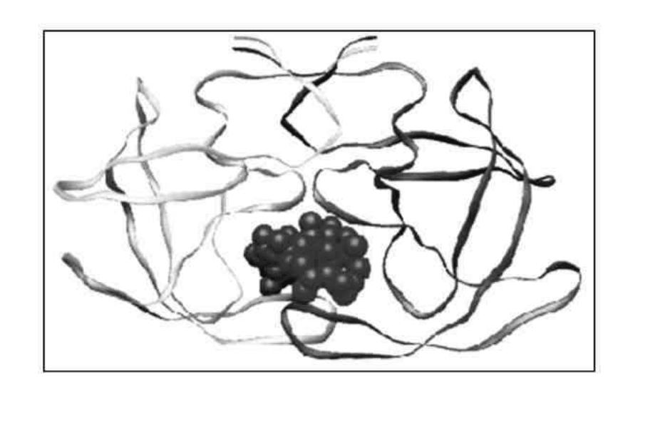
Protein folding
Proteins (polypeptides) are known to assume a variety of backbone conformations. In one conformation, the backbone is almost fully extended arrangement (as in a parallel or anti-parallel β-sheet), and in another conformation it is in a fully folded arrangement (as in an α-helix).
7.1 The end-to-end distance in a hexapeptide when it is in a fully extended conformation is approximately:
(a) \(10\ \text{Å}\) ☐
(b) \(15\ \text{Å}\) ☐
(c) \(20\ \text{Å}\) ☐
(d) \(25\ \text{Å}\) ☐
(Mark X in the correct box.)
7.2 Assuming that the following hexapeptide is a α-helix conformation, draw a connecting arrow between an oxygen atom and an amide \(\ce{NH}\) with which it is hydrogen bonded.
7.3 The following hexapeptides (A and B) display contrasting conformations in water at pH 7.0, especially when their serine hydroxyls are phosphorylated. A is moderately helical and it becomes a stronger helix on serine phosphorylation. B is weakly helical and it is completely disorded on serine phosphorylation. Draw connecting arrows to indicate the interactions between the residues that are responsible for this differing behaviour.
Consider the following process of folding/unfolding of a tripeptide segment in a large protein. The free energy change (\(\Delta G\)) will depend on the interaction of the unfolded tripeptide with the solvent (water) and with the rest of the protein in the folded state (see below). Assume that the tripeptide is made up of one non-polar (hydrophobic; shaded) and two polar (hydrophilic; unshaded) residues. Assume the following approximate changes in the interaction free energies :
(a) a non-polar residue and the solvent (water): \(\Delta G=+8\ \mathrm{kJ\,mol^{-1}}\)
(b) a non-polar residue and the rest of the protein: \(\Delta G=-4\ \mathrm{kJ\,mol^{-1}}\)
(c) a polar residue and the solvent (water): \(\Delta G=-16\ \mathrm{kJ\,mol^{-1}}\)
(d) a polar residue and the rest of the protein: \(\Delta G=-14\ \mathrm{kJ\,mol^{-1}}\)
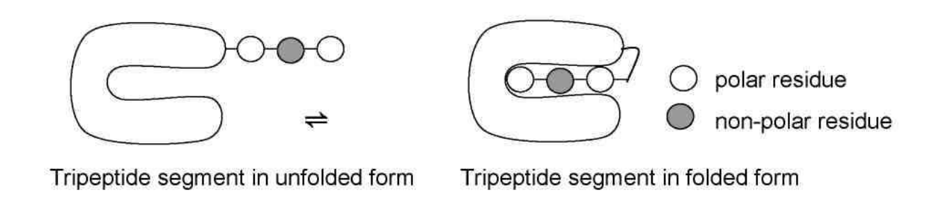
7.4 Calculate \(\Delta G\) for the folding of the tripeptide segment.
7.5 Calculate \(\Delta G\) for the folding of the tripeptide segment if all the three residues are polar.
β - lactamase and drug resistance
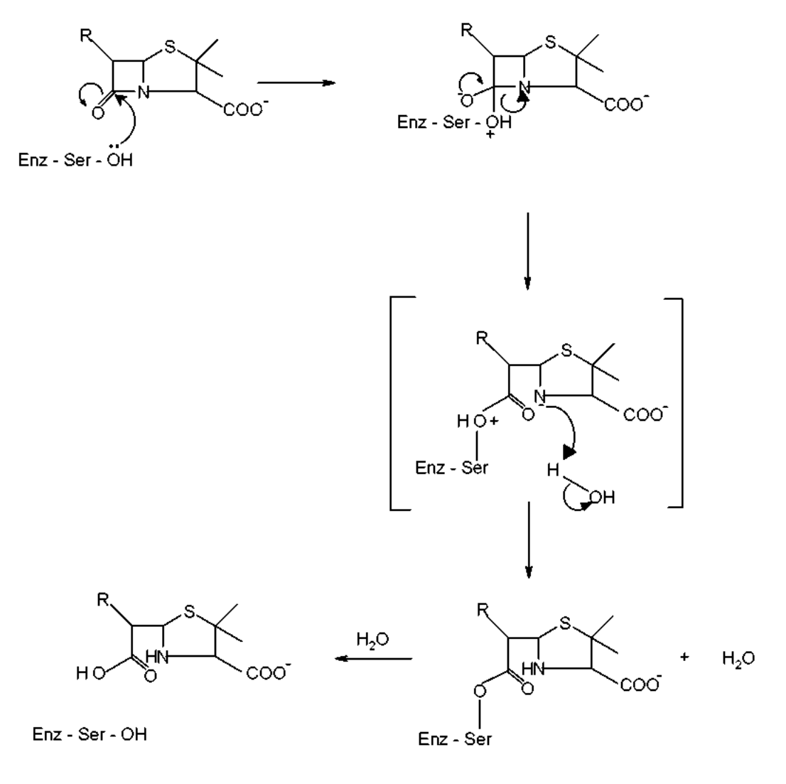
Penicillins are effective drugs in combating bacterial infections. An alarming development over the years has, however, been the emergence of drug resistant bacteria. Penicillin resistance arises due to the secretion of an enzyme called (3-lactamase (also known as penicillinase), which inactivates penicillin by opening its (3-lactam ring. The mechanism for this (3-lactam ring opening involves the nucleophilic attack by serine-\(\ce{OH}\) at the active site of the enzyme as shown below.
An attempt was made to characterize the b–lactamase from Staphylococcus aureus. When the pure enzyme was treated with a radiolabelled [\(\ce{^{32}P}\)] phosphorylating agent, only the active site serine got labelled. On analysis, this serine {molecular mass = 105 mass units (Da)} constituted 0.35% by weight of the b–lactamase.
7.6 Estimate the minimal molecular mass of this b–lactamase.
7.7 The approximate number of amino acid residues present in a protein of this size is :
(a) 100 ☐
(b) 150 ☐
(c) 275 ☐
(d) 375 ☐
[Mark X in the correct box.]
To map its active site, the b–lactamase was hydrolysed using trypsin, a specific enzyme. This resulted in a hexapeptide P1 containing the active site serine. Amino acid analysis revealed the following in equimolar proportion : Glu, Leu, Lys, Met, Phe and Ser. Treatment of P1 with Edman’s reagent (phenyl isothiocyanate) yielded phenyl thiohydantoin (PTH) derivative of phenyalanine and a peptide P2.
Treatment of P1 with cyanogen bromide (\(\ce{CNBr}\)) gave an acidic tetrapeptide P3 and a dipeptide P4.
Treatment of P2 with 1–fluoro–2,4–dinitrobenzene, followed by complete hydrolysis, yielded N–2,4–dinitrophenyl–Glu. P1, P2, and P3 contain the active site serine.
7.8 From the above information, deduce the amino acid sequence of P1, P2, P3 and P4.
7.9 Calculate the molecular mass of P3 in mass units (Da) from the information given in the attached Table.
The β–lactamase active site provides a unique microenvironment that makes the catalytic serine–\(\ce{OH}\) an unusually reactive nucleophile. The first order rate constant for β–lactamase catalysed reaction is \(350\ \mathrm{s^{-1}}\). For penicillin hydrolysis by free serine–\(\ce{OH}\) (at \(1\ \mathrm{M}\)) in solution, the pseudo first order rate constant is \(0.5\ \mathrm{s^{-1}}\).
7.10 From the information above, calculate the effective concentration of this nucleophile at the enzyme active site?
A molecule competing with penicillin for binding to the b–lactamase active site can inhibit the enzyme. Dissociation constants (\(K_D\)) for the inhibitor–lactamase complex for three different inhibitors are given below :
| Inhibitor | Dissociation constant (\(K_D\)) |
|---|---|
| A | \(2.0\times10^{-3}\) |
| B | \(1.0\times10^{-6}\) |
| C | \(5.0\times10^{-9}\) |
7.11 Indicate which of these inhibitors is most effective in protecting penicillin against β–lactamase. [Mark X in the correct box]
A ☐
B ☐
C ☐
A β–lactamase inhibitor was designed rationally. On binding to the enzyme active site, a nucleophilic attack by the \(\ce{OH}\) group of serine resulted in the opening of the β–lactam moiety of the inhibitor and elimination of \(\ce{Br^-}\). A reactive electrophile is generated as a result and it captures an active site residue X, inactivating the enzyme.
7.12 Based on the above information, identify the electrophile (A) generated and the final product (B) formed in the enzyme inactivation by the inhibitor shown.
Table
| Amino Acid | Structure | Molecular mass (Da) |
|---|---|---|
| Glu – Glutamic acid | S1 | 147 |
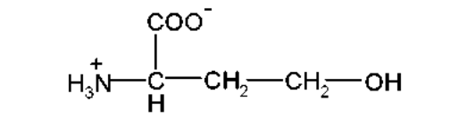
| Hms – Homoserine | S2 | 119 |
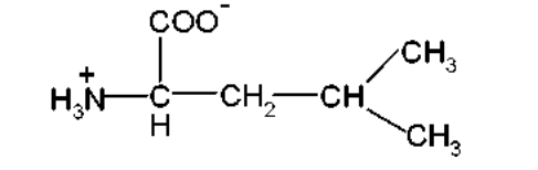
| Leu – Leucine | S3 | 131 |
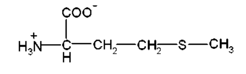
| Met – Methionine | S4 | 149 |
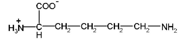
| Lys – Lysine | S5 | 146 |
| Phe – Phenylalanine | S6 | 165 |
S1:
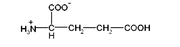
S2:
S3:
S4:
S5:
S6:
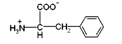
Table (continued)
| Amino Acid | Structure | Molecular mass (Da) |
|---|---|---|
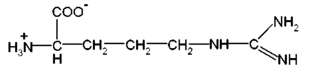
| Arg – Arginine | S7 | 174 |
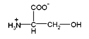
| Ser – Serine | S8 | 105 |
S7:
S8:
Official English text of the problem.
Answers
- 7.1: (c), approximately \(20\ \text{Å}\).
- 7.2: Connect the carbonyl oxygen of residue \(i\) to the amide hydrogen of residue \(i+4\). For the hexapeptide: \(1\rightarrow5\) and \(2\rightarrow6\).
- 7.3: Phosphorylation introduces negative charge. Favorable phosphate–cation interactions can strengthen A’s helix; unfavorable electrostatic interactions can destabilize B’s helix. The exact interacting residues cannot be identified without the missing sequences.
- 7.4: \(\boxed{\Delta G_{\mathrm{fold}}=-8\ \mathrm{kJ\,mol^{-1}}}\).
- 7.5: \(\boxed{\Delta G_{\mathrm{fold}}=+6\ \mathrm{kJ\,mol^{-1}}}\).
- 7.6: \(\boxed{M_{\min}=3.0\times10^{4}\ \mathrm{Da}=30\ \mathrm{kDa}}\).
- 7.7: (c), approximately 275 residues.
- 7.8: Sequences, written from N-terminus to C-terminus:
- P1: Phe–Glu–Ser–Met–Leu–Lys.
- P2: Glu–Ser–Met–Leu–Lys.
- P3: Phe–Glu–Ser–Hms, using the homoserine form.
- P4: Leu–Lys.
- 7.9: \(\boxed{M(\mathrm{P3})=482\ \mathrm{Da}}\) for the homoserine form indicated by the table.
- 7.10: \(\boxed{c_{\mathrm{eff}}=7\times10^{2}\ \mathrm{mol\,L^{-1}}}\).
- 7.11: C, which has the smallest \(K_D\).
- 7.12: A is the inhibitor-derived electrophilic intermediate generated after β-lactam opening and bromide elimination; B is its covalent capture product with residue X. Their specific structures cannot be determined without the missing inhibitor drawing.
Solution
7.1 — Length of the extended hexapeptide
An extended peptide backbone contributes approximately \(3.3\)–\(3.5\ \text{Å}\) along the chain per residue. Thus the approximate length of six residues is
\[ L\approx6(3.3\text{–}3.5)\ \text{Å} \approx20\text{–}21\ \text{Å}. \]
The appropriate choice is therefore (c), \(20\ \text{Å}\). This is an approximate chain length, rather than a distance between precisely specified terminal atoms.
7.2 — Hydrogen bonds in an α-helix
Number the residues from the N-terminus to the C-terminus. The characteristic α-helical hydrogen bond connects
\[ \text{carbonyl O of residue }i \quad\cdots\quad \text{H–N of residue }i+4. \]
For six residues, the two possible internal pairs are:
- Carbonyl oxygen of residue 1 with the amide hydrogen of residue 5.
- Carbonyl oxygen of residue 2 with the amide hydrogen of residue 6.
On the answer-sheet structure, draw the connecting arrows between those atoms. The carbonyl oxygen is the hydrogen-bond acceptor, and the amide group is the donor. These connections describe the helix’s spatial arrangement; ordinary SMILES does not encode that conformation or its hydrogen bonds.
7.3 — Effect of serine phosphorylation
At pH 7, an unmodified serine side chain is neutral. Its phosphate ester is negatively charged, predominantly as the dianionic phosphate group. Phosphorylation therefore changes both hydrogen bonding and electrostatic interactions.
The relevant amino-acid structures are given below as free amino-acid ions. In an actual peptide, their α-amino and α-carboxyl groups instead participate in the backbone.
| Name | SMILES |
|---|---|
| L-Serine, zwitterion | [NH3+][C@@H](CO)C(=O)[O-] |
| O-Phospho-L-serine, with a dianionic phosphate group | [NH3+][C@@H](COP(=O)([O-])[O-])C(=O)[O-] |
| L-Arginine, protonated guanidinium form | [NH3+][C@@H](CCCNC(=[NH2+])N)C(=O)[O-] |
| L-Lysine, protonated side-chain amino group | [NH3+][C@@H](CCCC[NH3+])C(=O)[O-] |
| L-Glutamate | [NH3+][C@@H](CCC(=O)[O-])C(=O)[O-] |
An electrostatic explanation consistent with the observations is:
- A: The phosphate group approaches a positively charged side chain, such as arginine’s guanidinium or lysine’s ammonium group. An attractive ion pair, reinforced by hydrogen bonding, stabilizes the helical arrangement.
- B: The phosphate group approaches another negatively charged group, such as a glutamate carboxylate. Repulsion destabilizes that arrangement.
Residues separated by three or four positions can approach one another on an α-helix, making their positions in the sequence important.
For such an arrangement, draw an attractive connection from a phosphate oxygen to the nearby cationic side chain in A, and indicate repulsion between the phosphate and nearby anionic group in B. Before phosphorylation, corresponding contacts involving serine’s hydroxyl would be hydrogen bonds rather than phosphate salt bridges.
These are conditional drawing instructions. The missing A and B sequences prevent assigning particular residue numbers or establishing that these particular side chains are present. Interactions with charged termini or the helix dipole could also contribute; the reported conformational changes alone do not uniquely identify the contacts.
7.4 — Folding with one non-polar and two polar residues
Use
\[ \Delta G_{\mathrm{fold}}=G_{\mathrm{folded}}-G_{\mathrm{unfolded}}. \]
For the unfolded segment, all three residues interact with water:
\[ G_{\mathrm{unfolded}} =8+2(-16) =-24\ \mathrm{kJ\,mol^{-1}}. \]
For the folded segment, they interact with the rest of the protein:
\[ G_{\mathrm{folded}} =-4+2(-14) =-32\ \mathrm{kJ\,mol^{-1}}. \]
Therefore,
\[ \boxed{\Delta G_{\mathrm{fold}} =-32-(-24) =-8\ \mathrm{kJ\,mol^{-1}}}. \]
Equivalently, burying the non-polar residue contributes \(-12\ \mathrm{kJ\,mol^{-1}}\), whereas burying each polar residue costs \(+2\ \mathrm{kJ\,mol^{-1}}\).
7.5 — Folding with three polar residues
Each polar residue changes its interaction free energy by
\[ -14-(-16)=+2\ \mathrm{kJ\,mol^{-1}}. \]
Consequently,
\[ \boxed{\Delta G_{\mathrm{fold}}=3(+2)=+6\ \mathrm{kJ\,mol^{-1}}}. \]
Folding is unfavorable under the simplified interaction-energy model supplied in the question.
7.6 — Minimal molecular mass of β-lactamase
The smallest possible enzyme molecule contains one labelled active-site serine. Using the stated serine mass of \(105\ \mathrm{Da}\),
\[ \frac{105\ \mathrm{Da}}{M_{\min}} =\frac{0.35}{100}=0.0035. \]
Thus
\[ \boxed{M_{\min} =\frac{105}{0.0035} =3.0\times10^{4}\ \mathrm{Da}}. \]
If there were \(n\) labelled serines per enzyme molecule, the inferred mass would be \(n\) times this value. Hence one labelled serine gives the minimum.
7.7 — Approximate number of residues
The standard approximate mean mass of an amino-acid residue in a protein is \(110\ \mathrm{Da}\). Therefore,
\[ N\approx\frac{30\,000\ \mathrm{Da}} {110\ \mathrm{Da\,residue^{-1}}} \approx273\ \text{residues}. \]
The nearest option is (c), 275.
7.8 — Sequences of P1–P4
The sequence follows from four cleavage and labelling observations.
-
Trypsin identifies the C-terminal residue.
Trypsin cleaves peptide bonds on the C-terminal side of lysine or arginine. P1 contains lysine and no arginine, so its C-terminal residue is Lys. -
Edman degradation identifies the first residue.
Release of the phenylalanine derivative establishes that P1 begins with Phe. P2 is the remaining pentapeptide. -
N-terminal labelling of P2 identifies the second residue of P1.
The dinitrophenyl-Glu product establishes that P2 begins with Glu. Thus P1 has the partial sequence \[ \mathrm{Phe-Glu-\ ?-\ ?-\ ?-Lys}. \] -
Cyanogen bromide locates methionine.
Cyanogen bromide,BrC#N, cleaves on the C-terminal side of methionine. A hexapeptide yielding fragments of lengths four and two would have Met at position 4 or position 2. Position 2 is already Glu, so Met must occupy position 4.
P3 is the four-residue N-terminal fragment and contains the active-site Ser. Its only remaining position is position 3. Leu consequently occupies position 5.
Hence
\[ \boxed{\mathrm{P1=Phe-Glu-Ser-Met-Leu-Lys}}. \]
Cyanogen-bromide cleavage changes the methionine at the end of the N-terminal fragment into a homoserine derivative. Taking the open-chain homoserine form indicated by the table gives
\[ \boxed{ \begin{aligned} \mathrm{P2}&=\mathrm{Glu-Ser-Met-Leu-Lys},\\ \mathrm{P3}&=\mathrm{Phe-Glu-Ser-Hms},\\ \mathrm{P4}&=\mathrm{Leu-Lys}. \end{aligned}} \]
The glutamate side chain accounts for P3’s acidic character.
The following SMILES show neutral peptide forms, with natural L stereochemistry and ordinary α-peptide linkages:
-
P1 — L-phenylalanyl-L-glutamyl-L-seryl-L-methionyl-L-leucyl-L-lysine:
N[C@@H](Cc1ccccc1)C(=O)N[C@@H](CCC(=O)O)C(=O)N[C@@H](CO)C(=O)N[C@@H](CCSC)C(=O)N[C@@H](CC(C)C)C(=O)N[C@@H](CCCCN)C(=O)O -
P2 — L-glutamyl-L-seryl-L-methionyl-L-leucyl-L-lysine:
N[C@@H](CCC(=O)O)C(=O)N[C@@H](CO)C(=O)N[C@@H](CCSC)C(=O)N[C@@H](CC(C)C)C(=O)N[C@@H](CCCCN)C(=O)O -
P3 — L-phenylalanyl-L-glutamyl-L-seryl-L-homoserine:
N[C@@H](Cc1ccccc1)C(=O)N[C@@H](CCC(=O)O)C(=O)N[C@@H](CO)C(=O)N[C@@H](CCO)C(=O)O -
P4 — L-leucyl-L-lysine:
N[C@@H](CC(C)C)C(=O)N[C@@H](CCCCN)C(=O)O
For conversion through to open-chain homoserine, the balanced overall cleavage, written using these neutral peptide forms, is
\[ \ce{P1 + BrCN + 2H2O -> P3 + P4 + CH3SCN + HBr}. \]
The additional species are water, O; methyl thiocyanate, CSC#N; and hydrogen bromide, [H]Br. The initially formed homoserine lactone accounts for the distinction discussed next.
7.9 — Molecular mass of P3
P3 contains Phe, Glu, Ser and Hms. L-Homoserine is N[C@@H](CCO)C(=O)O.
A linear tetrapeptide has three peptide bonds, so its mass equals the sum of the four free amino-acid masses minus three water molecules:
\[ \begin{aligned} M(\mathrm{P3}) &=165+147+105+119-3(18)\\ &=\boxed{482\ \mathrm{Da}}. \end{aligned} \]
Methionine’s mass must not be used for the terminal residue after cyanogen-bromide cleavage.
Chemically, cyanogen-bromide cleavage initially produces a terminal homoserine γ-lactone, which can hydrolyse to homoserine. The corresponding P3 lactone is L-phenylalanyl-L-glutamyl-L-seryl-L-homoserine γ-lactone:
N[C@@H](Cc1ccccc1)C(=O)N[C@@H](CCC(=O)O)C(=O)N[C@@H](CO)C(=O)N[C@@H](CCO1)C1=O
Its hydrolysis is
\[ \ce{P3(lactone) + H2O -> P3(homoserine)}. \]
Therefore the lactone would have mass \(482-18=464\ \mathrm{Da}\). The supplied homoserine table entry supports 482 Da as the intended answer.
7.10 — Effective concentration of the nucleophile
For the reaction with free serine, the pseudo-first-order rate constant is
\[ k_{\mathrm{obs}}=k_2[\text{serine}]. \]
At \(1\ \mathrm{mol\,L^{-1}}\),
\[ k_2=\frac{0.5\ \mathrm{s^{-1}}}{1\ \mathrm{mol\,L^{-1}}} =0.5\ \mathrm{L\,mol^{-1}\,s^{-1}}. \]
Define the effective concentration by equating the enzymatic rate constant to \(k_2c_{\mathrm{eff}}\):
\[ c_{\mathrm{eff}} =\frac{350\ \mathrm{s^{-1}}} {0.5\ \mathrm{L\,mol^{-1}\,s^{-1}}} =\boxed{7\times10^2\ \mathrm{mol\,L^{-1}}}. \]
This is a kinetic effective molarity: the solution concentration that would give the same rate under this comparison. It is not a literal bulk concentration inside the enzyme.
7.11 — Most effective inhibitor
For reversible binding,
\[ \ce{E + I <=> EI}, \qquad K_D=\frac{[\mathrm E][\mathrm I]}{[\mathrm{EI}]}. \]
A smaller \(K_D\) means stronger binding. At the same free inhibitor concentration, it also means a greater fraction of enzyme is inhibitor-bound:
\[ f_{\mathrm{bound}}=\frac{[\mathrm I]}{K_D+[\mathrm I]}. \]
Since
\[ 5.0\times10^{-9}<1.0\times10^{-6}<2.0\times10^{-3}, \]
inhibitor C is the most effective under comparable concentration conditions. Its molecular structure is not supplied, so no molecular name or SMILES can be inferred from its \(K_D\).
7.12 — Electrophile formation and enzyme inactivation
The missing inhibitor drawing is essential for assigning the structures of A and B. The information supplied establishes the following mechanistic framework:
-
Serine attacks the β-lactam carbonyl. Draw a curved arrow from the serine oxygen lone pair to the carbonyl carbon, and another from the carbonyl π bond to oxygen.
-
The β-lactam ring opens. Collapse of the tetrahedral intermediate breaks the lactam’s acyl carbon–nitrogen bond, with the necessary proton transfers. The initial product is a serine acyl-enzyme ester. Its ester linkage can be represented generically as
[*]C(=O)O[*], with the wildcards representing the inhibitor-derived and enzyme-derived portions. -
Bromide is eliminated. Bromide ion is
[Br-]. Its departure generates electrophile A. The omitted inhibitor connectivity determines which bond breaks, which new bond or π system forms, and where the electrophilic center lies. -
Residue X traps A. Draw an arrow from the nucleophilic lone pair on X to the electrophilic atom of A, with the corresponding electron redistribution. The resulting covalent adduct is B, which inactivates the enzyme.
The prose does not identify the bromine-bearing carbon, the inhibitor substituents, or the chemical identity of X. Consequently, a unique chemical name, complete SMILES, detailed product drawing, or atom-balanced overall equation for A and B cannot be supplied without inventing missing structural information.
The solution was checked against the official solution.
IChO 2001 · Theorieaufgabe 7
Peptide und Proteine
[In den Aminosäuretabellen beziehen sich S1–S8 auf die Strukturabbildungen, die unmittelbar unter der jeweiligen Tabelle wiedergegeben sind.]
AUFGABE 7
Peptide und Proteine
Proteinfaltung
Proteine (Polypeptide) können bekanntlich verschiedene Konformationen des Peptidrückgrats annehmen. In einer Konformation ist das Rückgrat nahezu vollständig gestreckt (wie in einem parallelen oder antiparallelen β-Faltblatt), in einer anderen liegt es in einer vollständig gefalteten Anordnung vor (wie in einer α-Helix).
7.1 Der Abstand zwischen den beiden Enden eines Hexapeptids in vollständig gestreckter Konformation beträgt ungefähr:
(a) \(10\ \text{Å}\) ☐
(b) \(15\ \text{Å}\) ☐
(c) \(20\ \text{Å}\) ☐
(d) \(25\ \text{Å}\) ☐
(Markieren Sie das richtige Kästchen mit X.)
7.2 Nehmen Sie an, dass das folgende Hexapeptid in einer α-Helix-Konformation vorliegt. Zeichnen Sie einen Verbindungspfeil zwischen einem Sauerstoffatom und einer Amid-\(\ce{NH}\)-Gruppe, mit der es eine Wasserstoffbrücke bildet.
7.3 Die folgenden Hexapeptide (A und B) zeigen in Wasser bei pH 7.0 unterschiedliche Konformationen, insbesondere wenn ihre Serin-Hydroxygruppen phosphoryliert sind. A ist mäßig helikal und bildet nach Phosphorylierung des Serins eine stärker ausgeprägte Helix. B ist schwach helikal und nach Phosphorylierung des Serins vollständig ungeordnet. Zeichnen Sie Verbindungspfeile ein, um die Wechselwirkungen zwischen den Resten anzugeben, die für dieses unterschiedliche Verhalten verantwortlich sind.
Betrachten Sie den folgenden Vorgang der Faltung/Entfaltung eines Tripeptidsegments in einem großen Protein. Die Änderung der freien Enthalpie (\(\Delta G\)) hängt von der Wechselwirkung des entfalteten Tripeptids mit dem Lösungsmittel (Wasser) und im gefalteten Zustand mit dem übrigen Protein ab (siehe unten). Nehmen Sie an, dass das Tripeptid aus einem unpolaren (hydrophoben; schattierten) und zwei polaren (hydrophilen; nicht schattierten) Resten besteht. Nehmen Sie die folgenden ungefähren Änderungen der freien Wechselwirkungsenthalpien an:
(a) ein unpolarer Rest und das Lösungsmittel (Wasser): \(\Delta G=+8\ \mathrm{kJ\,mol^{-1}}\)
(b) ein unpolarer Rest und das übrige Protein: \(\Delta G=-4\ \mathrm{kJ\,mol^{-1}}\)
(c) ein polarer Rest und das Lösungsmittel (Wasser): \(\Delta G=-16\ \mathrm{kJ\,mol^{-1}}\)
(d) ein polarer Rest und das übrige Protein: \(\Delta G=-14\ \mathrm{kJ\,mol^{-1}}\)
7.4 Berechnen Sie \(\Delta G\) für die Faltung des Tripeptidsegments.
7.5 Berechnen Sie \(\Delta G\) für die Faltung des Tripeptidsegments, wenn alle drei Reste polar sind.
β-Lactamase und Arzneimittelresistenz
Penicilline sind wirksame Arzneimittel zur Bekämpfung bakterieller Infektionen. Eine besorgniserregende Entwicklung im Laufe der Jahre ist jedoch das Auftreten arzneimittelresistenter Bakterien. Penicillinresistenz entsteht durch die Sekretion eines Enzyms namens β-Lactamase (auch als Penicillinase bekannt), das Penicillin durch Öffnung seines β-Lactamrings inaktiviert. Der Mechanismus dieser β-Lactamringöffnung umfasst den nukleophilen Angriff durch Serin-\(\ce{OH}\) im aktiven Zentrum des Enzyms, wie unten gezeigt.
Es wurde versucht, die β-Lactamase aus Staphylococcus aureus zu charakterisieren. Bei der Behandlung des reinen Enzyms mit einem radioaktiv markierten [\(\ce{^{32}P}\)] Phosphorylierungsreagenz wurde nur das Serin im aktiven Zentrum markiert. Die Analyse ergab, dass dieses Serin {Molekülmasse = 105 Masseneinheiten (Da)} einen Massenanteil von 0.35% an der β-Lactamase ausmachte.
7.6 Schätzen Sie die minimale Molekülmasse dieser β-Lactamase ab.
7.7 Die ungefähre Anzahl der Aminosäurereste in einem Protein dieser Größe beträgt:
(a) 100 ☐
(b) 150 ☐
(c) 275 ☐
(d) 375 ☐
[Markieren Sie das richtige Kästchen mit X.]
Zur Untersuchung ihres aktiven Zentrums wurde die β-Lactamase mit Trypsin, einem spezifischen Enzym, hydrolysiert. Dabei entstand ein Hexapeptid P1, das das Serin des aktiven Zentrums enthielt. Die Aminosäureanalyse ergab die folgenden Aminosäuren in äquimolaren Anteilen: Glu, Leu, Lys, Met, Phe und Ser. Die Behandlung von P1 mit dem Edman-Reagenz (Phenylisothiocyanat) lieferte das Phenylthiohydantoin-Derivat (PTH-Derivat) von Phenylalanin und ein Peptid P2.
Die Behandlung von P1 mit Bromcyan (\(\ce{CNBr}\)) ergab ein saures Tetrapeptid P3 und ein Dipeptid P4.
Die Behandlung von P2 mit 1-Fluor-2,4-dinitrobenzol mit anschließender vollständiger Hydrolyse ergab N-2,4-Dinitrophenyl-Glu. P1, P2 und P3 enthalten das Serin des aktiven Zentrums.
7.8 Leiten Sie aus den obigen Angaben die Aminosäuresequenzen von P1, P2, P3 und P4 ab.
7.9 Berechnen Sie anhand der Angaben in der beigefügten Tabelle die Molekülmasse von P3 in Masseneinheiten (Da).
Das aktive Zentrum der β-Lactamase bietet eine einzigartige Mikroumgebung, die das katalytische Serin-\(\ce{OH}\) zu einem ungewöhnlich reaktiven Nukleophil macht. Die Geschwindigkeitskonstante erster Ordnung für die durch β-Lactamase katalysierte Reaktion beträgt \(350\ \mathrm{s^{-1}}\). Für die Penicillinhydrolyse durch freies Serin-\(\ce{OH}\) (bei \(1\ \mathrm{M}\)) in Lösung beträgt die Geschwindigkeitskonstante pseudoerster Ordnung \(0.5\ \mathrm{s^{-1}}\).
7.10 Berechnen Sie anhand der obigen Angaben die effektive Konzentration dieses Nukleophils im aktiven Zentrum des Enzyms.
Ein Molekül, das mit Penicillin um die Bindung an das aktive Zentrum der β-Lactamase konkurriert, kann das Enzym hemmen. Die Dissoziationskonstanten (\(K_D\)) des Inhibitor-Lactamase-Komplexes für drei verschiedene Inhibitoren sind nachstehend angegeben:
| Inhibitor | Dissoziationskonstante (\(K_D\)) |
|---|---|
| A | \(2.0\times10^{-3}\) |
| B | \(1.0\times10^{-6}\) |
| C | \(5.0\times10^{-9}\) |
7.11 Geben Sie an, welcher dieser Inhibitoren Penicillin am wirksamsten vor β-Lactamase schützt. [Markieren Sie das richtige Kästchen mit X.]
A ☐
B ☐
C ☐
Ein β-Lactamase-Inhibitor wurde gezielt auf rationaler Grundlage entwickelt. Nach der Bindung an das aktive Zentrum des Enzyms führte ein nukleophiler Angriff der \(\ce{OH}\)-Gruppe des Serins zur Öffnung der β-Lactam-Einheit des Inhibitors und zur Abspaltung von \(\ce{Br^-}\). Dadurch entsteht ein reaktives Elektrophil, das einen Rest X im aktiven Zentrum bindet und so das Enzym inaktiviert.
7.12 Identifizieren Sie anhand der obigen Angaben das gebildete Elektrophil (A) und das Endprodukt (B), das bei der Inaktivierung des Enzyms durch den gezeigten Inhibitor entsteht.
Tabelle
| Aminosäure | Struktur | Molekülmasse (Da) |
|---|---|---|
| Glu – Glutaminsäure | S1 | 147 |
| Hms – Homoserin | S2 | 119 |
| Leu – Leucin | S3 | 131 |
| Met – Methionin | S4 | 149 |
| Lys – Lysin | S5 | 146 |
| Phe – Phenylalanin | S6 | 165 |
S1:
S2:
S3:
S4:
S5:
S6:
Tabelle (Fortsetzung)
| Aminosäure | Struktur | Molekülmasse (Da) |
|---|---|---|
| Arg – Arginin | S7 | 174 |
| Ser – Serin | S8 | 105 |
S7:
S8:
Aufgabentext aus dem offiziellen englischen Text übersetzt.
Antworten
- 7.1: (c), ungefähr \(20\ \text{Å}\).
- 7.2: Verbinden Sie den Carbonylsauerstoff des Restes \(i\) mit dem Amidwasserstoff des Restes \(i+4\). Für das Hexapeptid: \(1\rightarrow5\) und \(2\rightarrow6\).
- 7.3: Durch Phosphorylierung wird eine negative Ladung eingeführt. Günstige Phosphat-Kation-Wechselwirkungen können die Helix von A verstärken; ungünstige elektrostatische Wechselwirkungen können die Helix von B destabilisieren. Die genau miteinander wechselwirkenden Reste lassen sich ohne die fehlenden Sequenzen nicht bestimmen.
- 7.4: \(\boxed{\Delta G_{\mathrm{fold}}=-8\ \mathrm{kJ\,mol^{-1}}}\).
- 7.5: \(\boxed{\Delta G_{\mathrm{fold}}=+6\ \mathrm{kJ\,mol^{-1}}}\).
- 7.6: \(\boxed{M_{\min}=3.0\times10^{4}\ \mathrm{Da}=30\ \mathrm{kDa}}\).
- 7.7: (c), ungefähr 275 Reste.
- 7.8: Sequenzen, vom N-Terminus zum C-Terminus geschrieben:
- P1: Phe–Glu–Ser–Met–Leu–Lys.
- P2: Glu–Ser–Met–Leu–Lys.
- P3: Phe–Glu–Ser–Hms, unter Verwendung der Homoserinform.
- P4: Leu–Lys.
- 7.9: \(\boxed{M(\mathrm{P3})=482\ \mathrm{Da}}\) für die in der Tabelle angegebene Homoserinform.
- 7.10: \(\boxed{c_{\mathrm{eff}}=7\times10^{2}\ \mathrm{mol\,L^{-1}}}\).
- 7.11: C, das den kleinsten Wert von \(K_D\) aufweist.
- 7.12: A ist das vom Inhibitor abgeleitete elektrophile Zwischenprodukt, das nach Öffnung des β-Lactams und Abspaltung von Bromid entsteht; B ist das Produkt seiner kovalenten Bindung an den Rest X. Ihre konkreten Strukturen lassen sich ohne die fehlende Zeichnung des Inhibitors nicht bestimmen.
Lösung
7.1 — Länge des gestreckten Hexapeptids
Ein gestrecktes Peptidrückgrat trägt pro Rest ungefähr \(3.3\)–\(3.5\ \text{Å}\) zur Länge entlang der Kette bei. Die ungefähre Länge von sechs Resten beträgt somit
\[ L\approx6(3.3\text{–}3.5)\ \text{Å} \approx20\text{–}21\ \text{Å}. \]
Die passende Antwort ist daher (c), \(20\ \text{Å}\). Dies ist eine ungefähre Kettenlänge und kein Abstand zwischen genau festgelegten endständigen Atomen.
7.2 — Wasserstoffbrücken in einer α-Helix
Nummerieren Sie die Reste vom N-Terminus zum C-Terminus. Die für eine α-Helix charakteristische Wasserstoffbrücke verbindet
\[ \text{Carbonyl-O des Restes }i \quad\cdots\quad \text{H–N des Restes }i+4. \]
Bei sechs Resten sind die beiden möglichen internen Paare:
- Carbonylsauerstoff des Restes 1 mit dem Amidwasserstoff des Restes 5.
- Carbonylsauerstoff des Restes 2 mit dem Amidwasserstoff des Restes 6.
Zeichnen Sie in der Struktur auf dem Antwortblatt die Verbindungspfeile zwischen diesen Atomen ein. Der Carbonylsauerstoff ist der Wasserstoffbrückenakzeptor, die Amidgruppe der Donor. Diese Verbindungen beschreiben die räumliche Anordnung der Helix; gewöhnliche SMILES kodieren weder diese Konformation noch ihre Wasserstoffbrücken.
7.3 — Wirkung der Serinphosphorylierung
Bei pH 7 ist eine unmodifizierte Serinseitenkette neutral. Ihr Phosphatester ist negativ geladen und liegt überwiegend als dianionische Phosphatgruppe vor. Die Phosphorylierung verändert daher sowohl die Wasserstoffbrückenbildung als auch die elektrostatischen Wechselwirkungen.
Die relevanten Aminosäurestrukturen sind nachstehend als freie Aminosäureionen angegeben. In einem tatsächlichen Peptid sind ihre α-Amino- und α-Carboxylgruppen stattdessen am Aufbau des Rückgrats beteiligt.
| Name | SMILES |
|---|---|
| L-Serin, Zwitterion | [NH3+][C@@H](CO)C(=O)[O-] |
| O-Phospho-L-serin, mit einer dianionischen Phosphatgruppe | [NH3+][C@@H](COP(=O)([O-])[O-])C(=O)[O-] |
| L-Arginin, protonierte Guanidiniumform | [NH3+][C@@H](CCCNC(=[NH2+])N)C(=O)[O-] |
| L-Lysin, protonierte Aminogruppe der Seitenkette | [NH3+][C@@H](CCCC[NH3+])C(=O)[O-] |
| L-Glutamat | [NH3+][C@@H](CCC(=O)[O-])C(=O)[O-] |
Eine mit den Beobachtungen vereinbare elektrostatische Erklärung lautet:
- A: Die Phosphatgruppe nähert sich einer positiv geladenen Seitenkette, etwa der Guanidiniumgruppe von Arginin oder der Ammoniumgruppe von Lysin. Ein durch Wasserstoffbrücken verstärktes, anziehend wechselwirkendes Ionenpaar stabilisiert die helikale Anordnung.
- B: Die Phosphatgruppe nähert sich einer anderen negativ geladenen Gruppe, etwa der Carboxylatgruppe eines Glutamatrestes. Die Abstoßung destabilisiert diese Anordnung.
Reste, die in der Sequenz drei oder vier Positionen auseinanderliegen, können sich in einer α-Helix räumlich nahekommen; daher sind ihre Positionen in der Sequenz von Bedeutung.
Zeichnen Sie für eine solche Anordnung in A eine anziehende Verbindung von einem Phosphatsauerstoff zur benachbarten kationischen Seitenkette ein und kennzeichnen Sie in B die Abstoßung zwischen dem Phosphat und der benachbarten anionischen Gruppe. Vor der Phosphorylierung wären entsprechende Kontakte unter Beteiligung der Hydroxygruppe des Serins Wasserstoffbrücken anstelle von Phosphat-Salzbrücken.
Diese Zeichenanweisungen gelten unter den genannten Voraussetzungen. Da die Sequenzen von A und B fehlen, können weder bestimmte Restnummern zugeordnet noch das Vorhandensein dieser konkreten Seitenketten festgestellt werden. Auch Wechselwirkungen mit geladenen Termini oder dem Helixdipol könnten beitragen; allein aus den angegebenen Konformationsänderungen lassen sich die Kontakte nicht eindeutig bestimmen.
7.4 — Faltung mit einem unpolaren und zwei polaren Resten
Verwenden Sie
\[ \Delta G_{\mathrm{fold}}=G_{\mathrm{folded}}-G_{\mathrm{unfolded}}. \]
Im entfalteten Segment wechselwirken alle drei Reste mit Wasser:
\[ G_{\mathrm{unfolded}} =8+2(-16) =-24\ \mathrm{kJ\,mol^{-1}}. \]
Im gefalteten Segment wechselwirken sie mit dem übrigen Protein:
\[ G_{\mathrm{folded}} =-4+2(-14) =-32\ \mathrm{kJ\,mol^{-1}}. \]
Daher gilt
\[ \boxed{\Delta G_{\mathrm{fold}} =-32-(-24) =-8\ \mathrm{kJ\,mol^{-1}}}. \]
Gleichwertig dazu trägt die Einlagerung des unpolaren Restes in das Proteininnere \(-12\ \mathrm{kJ\,mol^{-1}}\) bei, während die Einlagerung jedes polaren Restes \(+2\ \mathrm{kJ\,mol^{-1}}\) kostet.
7.5 — Faltung mit drei polaren Resten
Bei jedem polaren Rest ändert sich die freie Wechselwirkungsenthalpie um
\[ -14-(-16)=+2\ \mathrm{kJ\,mol^{-1}}. \]
Folglich gilt
\[ \boxed{\Delta G_{\mathrm{fold}}=3(+2)=+6\ \mathrm{kJ\,mol^{-1}}}. \]
Im Rahmen des in der Aufgabe vorgegebenen vereinfachten Modells der Wechselwirkungsenergien ist die Faltung ungünstig.
7.6 — Minimale Molekülmasse der β-Lactamase
Das kleinstmögliche Enzymmolekül enthält ein markiertes Serin im aktiven Zentrum. Mit der angegebenen Serinmasse von \(105\ \mathrm{Da}\) gilt
\[ \frac{105\ \mathrm{Da}}{M_{\min}} =\frac{0.35}{100}=0.0035. \]
Somit ergibt sich
\[ \boxed{M_{\min} =\frac{105}{0.0035} =3.0\times10^{4}\ \mathrm{Da}}. \]
Gäbe es \(n\) markierte Serinreste pro Enzymmolekül, so wäre die daraus abgeleitete Masse das \(n\)-Fache dieses Wertes. Ein markiertes Serin liefert daher das Minimum.
7.7 — Ungefähre Anzahl der Reste
Die übliche Näherung für die mittlere Masse eines Aminosäurerestes in einem Protein beträgt \(110\ \mathrm{Da}\). Daher gilt
\[ N\approx\frac{30\,000\ \mathrm{Da}} {110\ \mathrm{Da\,residue^{-1}}} \approx273\ \text{Reste}. \]
Die nächstliegende Antwortmöglichkeit ist (c), 275.
7.8 — Sequenzen von P1–P4
Die Sequenz ergibt sich aus vier Beobachtungen zu Spaltung und Markierung.
-
Trypsin identifiziert den C-terminalen Rest.
Trypsin spaltet Peptidbindungen auf der C-terminalen Seite von Lysin oder Arginin. P1 enthält Lysin und kein Arginin, daher ist sein C-terminaler Rest Lys. -
Der Edman-Abbau identifiziert den ersten Rest.
Die Freisetzung des Phenylalaninderivats belegt, dass P1 mit Phe beginnt. P2 ist das verbleibende Pentapeptid. -
Die N-terminale Markierung von P2 identifiziert den zweiten Rest von P1.
Das Dinitrophenyl-Glu-Produkt belegt, dass P2 mit Glu beginnt. P1 hat somit die Teilsequenz \[ \mathrm{Phe-Glu-\ ?-\ ?-\ ?-Lys}. \] -
Bromcyan lokalisiert Methionin.
Bromcyan,BrC#N, spaltet auf der C-terminalen Seite von Methionin. Ein Hexapeptid, das Fragmente mit vier und zwei Resten liefert, müsste Met an Position 4 oder Position 2 enthalten. Position 2 ist bereits durch Glu besetzt, daher muss Met Position 4 einnehmen.
P3 ist das N-terminale Fragment aus vier Resten und enthält das Serin des aktiven Zentrums. Dafür bleibt nur Position 3 frei. Leu besetzt folglich Position 5.
Damit gilt
\[ \boxed{\mathrm{P1=Phe-Glu-Ser-Met-Leu-Lys}}. \]
Bei der Bromcyanspaltung wird das Methionin am Ende des N-terminalen Fragments in ein Homoserinderivat umgewandelt. Unter Verwendung der in der Tabelle angegebenen offenkettigen Homoserinform erhält man
\[ \boxed{ \begin{aligned} \mathrm{P2}&=\mathrm{Glu-Ser-Met-Leu-Lys},\\ \mathrm{P3}&=\mathrm{Phe-Glu-Ser-Hms},\\ \mathrm{P4}&=\mathrm{Leu-Lys}. \end{aligned}} \]
Die Glutamatseitenkette erklärt den sauren Charakter von P3.
Die folgenden SMILES zeigen neutrale Peptidformen mit natürlicher L-Stereochemie und gewöhnlichen α-Peptidbindungen:
-
P1 — L-Phenylalanyl-L-glutamyl-L-seryl-L-methionyl-L-leucyl-L-lysin:
N[C@@H](Cc1ccccc1)C(=O)N[C@@H](CCC(=O)O)C(=O)N[C@@H](CO)C(=O)N[C@@H](CCSC)C(=O)N[C@@H](CC(C)C)C(=O)N[C@@H](CCCCN)C(=O)O -
P2 — L-Glutamyl-L-seryl-L-methionyl-L-leucyl-L-lysin:
N[C@@H](CCC(=O)O)C(=O)N[C@@H](CO)C(=O)N[C@@H](CCSC)C(=O)N[C@@H](CC(C)C)C(=O)N[C@@H](CCCCN)C(=O)O -
P3 — L-Phenylalanyl-L-glutamyl-L-seryl-L-homoserin:
N[C@@H](Cc1ccccc1)C(=O)N[C@@H](CCC(=O)O)C(=O)N[C@@H](CO)C(=O)N[C@@H](CCO)C(=O)O -
P4 — L-Leucyl-L-lysin:
N[C@@H](CC(C)C)C(=O)N[C@@H](CCCCN)C(=O)O
Für die Umsetzung bis zum offenkettigen Homoserin lautet die ausgeglichene Gesamtgleichung der Spaltung unter Verwendung dieser neutralen Peptidformen
\[ \ce{P1 + BrCN + 2H2O -> P3 + P4 + CH3SCN + HBr}. \]
Die zusätzlichen Spezies sind Wasser, O; Methylthiocyanat, CSC#N; und Bromwasserstoff, [H]Br. Das zunächst gebildete Homoserinlacton erklärt die im Folgenden erörterte Unterscheidung.
7.9 — Molekülmasse von P3
P3 enthält Phe, Glu, Ser und Hms. L-Homoserin ist N[C@@H](CCO)C(=O)O.
Ein lineares Tetrapeptid besitzt drei Peptidbindungen. Seine Masse entspricht daher der Summe der Massen der vier freien Aminosäuren abzüglich dreier Wassermoleküle:
\[ \begin{aligned} M(\mathrm{P3}) &=165+147+105+119-3(18)\\ &=\boxed{482\ \mathrm{Da}}. \end{aligned} \]
Für den endständigen Rest nach der Bromcyanspaltung darf nicht die Masse von Methionin verwendet werden.
Chemisch entsteht bei der Bromcyanspaltung zunächst ein endständiges Homoserin-γ-lacton, das zu Homoserin hydrolysieren kann. Das entsprechende P3-Lacton ist L-Phenylalanyl-L-glutamyl-L-seryl-L-homoserin-γ-lacton:
N[C@@H](Cc1ccccc1)C(=O)N[C@@H](CCC(=O)O)C(=O)N[C@@H](CO)C(=O)N[C@@H](CCO1)C1=O
Seine Hydrolyse lautet
\[ \ce{P3(lactone) + H2O -> P3(homoserine)}. \]
Das Lacton hätte daher die Masse \(482-18=464\ \mathrm{Da}\). Der vorgegebene Tabelleneintrag für Homoserin spricht für 482 Da als beabsichtigte Antwort.
7.10 — Effektive Konzentration des Nukleophils
Für die Reaktion mit freiem Serin lautet die Geschwindigkeitskonstante pseudoerster Ordnung
\[ k_{\mathrm{obs}}=k_2[\text{Serin}]. \]
Bei \(1\ \mathrm{mol\,L^{-1}}\) gilt
\[ k_2=\frac{0.5\ \mathrm{s^{-1}}}{1\ \mathrm{mol\,L^{-1}}} =0.5\ \mathrm{L\,mol^{-1}\,s^{-1}}. \]
Definieren Sie die effektive Konzentration, indem Sie die enzymatische Geschwindigkeitskonstante mit \(k_2c_{\mathrm{eff}}\) gleichsetzen:
\[ c_{\mathrm{eff}} =\frac{350\ \mathrm{s^{-1}}} {0.5\ \mathrm{L\,mol^{-1}\,s^{-1}}} =\boxed{7\times10^2\ \mathrm{mol\,L^{-1}}}. \]
Dies ist eine kinetische effektive Molarität: die Konzentration in Lösung, die bei diesem Vergleich dieselbe Reaktionsgeschwindigkeit ergeben würde. Sie ist keine tatsächliche Volumenkonzentration innerhalb des Enzyms.
7.11 — Wirksamster Inhibitor
Für die reversible Bindung gilt
\[ \ce{E + I <=> EI}, \qquad K_D=\frac{[\mathrm E][\mathrm I]}{[\mathrm{EI}]}. \]
Ein kleinerer Wert von \(K_D\) bedeutet eine stärkere Bindung. Bei gleicher freier Inhibitorkonzentration bedeutet dies außerdem, dass ein größerer Anteil des Enzyms an den Inhibitor gebunden ist:
\[ f_{\mathrm{bound}}=\frac{[\mathrm I]}{K_D+[\mathrm I]}. \]
Da
\[ 5.0\times10^{-9}<1.0\times10^{-6}<2.0\times10^{-3}, \]
ist Inhibitor C unter vergleichbaren Konzentrationsbedingungen am wirksamsten. Seine Molekülstruktur ist nicht angegeben, daher lassen sich aus seinem \(K_D\) weder ein Molekülname noch SMILES ableiten.
7.12 — Elektrophilbildung und Enzyminaktivierung
Die fehlende Zeichnung des Inhibitors ist für die Zuordnung der Strukturen von A und B unverzichtbar. Die vorliegenden Angaben legen den folgenden mechanistischen Rahmen fest:
-
Serin greift die β-Lactam-Carbonylgruppe an. Zeichnen Sie einen gebogenen Pfeil vom freien Elektronenpaar des Serinsauerstoffs zum Carbonylkohlenstoff und einen weiteren von der Carbonyl-π-Bindung zum Sauerstoff.
-
Der β-Lactamring öffnet sich. Beim Zerfall des tetraedrischen Zwischenprodukts wird die Bindung zwischen dem Acylkohlenstoff und dem Stickstoff des Lactams unter den erforderlichen Protonenübertragungen gespalten. Das erste Produkt ist ein Serin-Acyl-Enzym-Ester. Seine Esterbindung lässt sich allgemein als
[*]C(=O)O[*]darstellen, wobei die Platzhalter für die vom Inhibitor beziehungsweise vom Enzym stammenden Anteile stehen. -
Bromid wird abgespalten. Das Bromidion ist
[Br-]. Durch seinen Austritt entsteht das Elektrophil A. Die nicht angegebene Verknüpfung der Atome im Inhibitor bestimmt, welche Bindung gespalten wird, welche neue Bindung oder welches π-System entsteht und wo das elektrophile Zentrum liegt. -
Rest X fängt A ab. Zeichnen Sie einen Pfeil vom nukleophilen freien Elektronenpaar an X zum elektrophilen Atom von A sowie die entsprechende Elektronenverschiebung. Das entstehende kovalente Addukt ist B, das das Enzym inaktiviert.
Der Text benennt weder das bromtragende Kohlenstoffatom noch die Substituenten des Inhibitors oder die chemische Identität von X. Folglich lassen sich ein eindeutiger chemischer Name, vollständige SMILES, eine detaillierte Produktzeichnung oder eine atomar ausgeglichene Gesamtgleichung für A und B nicht angeben, ohne fehlende Strukturinformationen zu erfinden.
Die Lösung wurde mit der offiziellen Lösung abgeglichen.
IChO 2001 · Teori sorusu 7
Peptitler ve Proteinler
[Amino asit tablolarında S1–S8, her tablonun hemen altında verilen yapı şekillerini ifade eder.]
SORU 7
Peptitler ve Proteinler
Protein katlanması
Proteinlerin (polipeptitlerin) çeşitli omurga konformasyonları alabildiği bilinmektedir. Bir konformasyonda omurga neredeyse tamamen uzamış bir düzen alırken (paralel veya antiparalel β-tabakada olduğu gibi), başka bir konformasyonda tamamen katlanmış bir düzen alır (α-sarmalda olduğu gibi).
7.1 Tamamen uzamış konformasyondaki bir hekzapeptidin iki ucu arasındaki uzaklık yaklaşık olarak ne kadardır?
(a) \(10\ \text{Å}\) ☐
(b) \(15\ \text{Å}\) ☐
(c) \(20\ \text{Å}\) ☐
(d) \(25\ \text{Å}\) ☐
(Doğru kutuyu X ile işaretleyiniz.)
7.2 Aşağıdaki hekzapeptidin α-sarmal konformasyonunda olduğunu varsayarak, bir oksijen atomu ile hidrojen bağı yaptığı amit \(\ce{NH}\) grubu arasına bir bağlantı oku çiziniz.
7.3 Aşağıdaki hekzapeptitler (A ve B), pH 7.0'de suda, özellikle serin hidroksilleri fosforillendiğinde, birbirine zıt konformasyon davranışları sergiler. A orta derecede sarmal yapıdadır ve serin fosforillendiğinde sarmal yapısı güçlenir. B zayıf derecede sarmal yapıdadır ve serin fosforillendiğinde tamamen düzensiz hâle gelir. Bu farklı davranıştan sorumlu kalıntılar arasındaki etkileşimleri göstermek üzere bağlantı okları çiziniz.
Büyük bir proteindeki bir tripeptit parçasının aşağıdaki katlanma/açılma sürecini ele alınız. Serbest enerji değişimi (\(\Delta G\)), açılmış tripeptidin çözücüyle (su) etkileşimine ve katlanmış durumdayken proteinin geri kalanıyla etkileşimine bağlı olacaktır (aşağıya bakınız). Tripeptidin bir apolar (hidrofobik; gölgeli) ve iki polar (hidrofilik; gölgesiz) kalıntıdan oluştuğunu varsayınız. Etkileşim serbest enerjilerindeki yaklaşık değişimlerin aşağıdaki gibi olduğunu kabul ediniz:
(a) apolar bir kalıntı ile çözücü (su): \(\Delta G=+8\ \mathrm{kJ\,mol^{-1}}\)
(b) apolar bir kalıntı ile proteinin geri kalanı: \(\Delta G=-4\ \mathrm{kJ\,mol^{-1}}\)
(c) polar bir kalıntı ile çözücü (su): \(\Delta G=-16\ \mathrm{kJ\,mol^{-1}}\)
(d) polar bir kalıntı ile proteinin geri kalanı: \(\Delta G=-14\ \mathrm{kJ\,mol^{-1}}\)
7.4 Tripeptit parçasının katlanması için \(\Delta G\) değerini hesaplayınız.
7.5 Üç kalıntının da polar olması durumunda tripeptit parçasının katlanması için \(\Delta G\) değerini hesaplayınız.
β-laktamaz ve ilaç direnci
Penisilinler, bakteriyel enfeksiyonlarla mücadelede etkili ilaçlardır. Ancak yıllar içinde ilaca dirençli bakterilerin ortaya çıkması kaygı verici bir gelişme olmuştur. Penisilin direnci, penisilinin β-laktam halkasını açarak onu etkisiz hâle getiren β-laktamaz (penisilinaz olarak da bilinir) adlı bir enzimin salgılanmasından kaynaklanır. Bu β-laktam halkasının açılma mekanizması, aşağıda gösterildiği gibi enzimin aktif bölgesindeki serin-\(\ce{OH}\) grubunun nükleofilik saldırısını içerir.
Staphylococcus aureus bakterisinden elde edilen β-laktamazı karakterize etmek için bir çalışma yapılmıştır. Saf enzim, radyoaktif olarak işaretlenmiş [\(\ce{^{32}P}\)] bir fosforilleme reaktifiyle muamele edildiğinde yalnızca aktif bölgedeki serin işaretlenmiştir. Analiz sonucunda bu serinin {molekül kütlesi = 105 kütle birimi (Da)}, β-laktamazın kütlece %0.35'ini oluşturduğu belirlenmiştir.
7.6 Bu β-laktamazın en küçük molekül kütlesini tahmin ediniz.
7.7 Bu büyüklükte bir proteinde bulunan amino asit kalıntılarının yaklaşık sayısı şudur:
(a) 100 ☐
(b) 150 ☐
(c) 275 ☐
(d) 375 ☐
[Doğru kutuyu X ile işaretleyiniz.]
Aktif bölgesini haritalamak için β-laktamaz, özgül bir enzim olan tripsin kullanılarak hidroliz edilmiştir. Bunun sonucunda aktif bölge serinini içeren P1 hekzapeptidi elde edilmiştir. Amino asit analizi, eşmolar oranlarda şu amino asitlerin bulunduğunu göstermiştir: Glu, Leu, Lys, Met, Phe ve Ser. P1'in Edman reaktifiyle (fenil izotiyosiyanat) muamelesi sonucunda fenilalaninin fenil tiyohidantoin (PTH) türevi ve P2 peptidi elde edilmiştir.
P1'in siyanojen bromür (\(\ce{CNBr}\)) ile muamelesi, asidik bir tetrapeptit olan P3'ü ve bir dipeptit olan P4'ü vermiştir.
P2'nin 1–floro–2,4–dinitrobenzen ile muamelesinin ardından tam hidroliz uygulanmasıyla N–2,4–dinitrofenil–Glu elde edilmiştir. P1, P2 ve P3, aktif bölge serinini içerir.
7.8 Yukarıdaki bilgilerden hareketle P1, P2, P3 ve P4'ün amino asit dizilerini belirleyiniz.
7.9 Ekteki tabloda verilen bilgilerden yararlanarak P3'ün molekül kütlesini kütle birimi (Da) cinsinden hesaplayınız.
β-laktamazın aktif bölgesi, katalitik serin–\(\ce{OH}\) grubunu alışılmadık derecede reaktif bir nükleofil hâline getiren benzersiz bir mikroçevre sağlar. β-laktamazın katalizlediği tepkimenin birinci dereceden hız sabiti \(350\ \mathrm{s^{-1}}\) değerindedir. Çözeltide serbest serin–\(\ce{OH}\) (\(1\ \mathrm{M}\) derişimde) ile penisilin hidrolizi için yalancı birinci dereceden hız sabiti \(0.5\ \mathrm{s^{-1}}\) değerindedir.
7.10 Yukarıdaki bilgilerden yararlanarak bu nükleofilin enzimin aktif bölgesindeki etkin derişimini hesaplayınız.
β-laktamazın aktif bölgesine bağlanmak için penisilinle yarışan bir molekül enzimi inhibe edebilir. Üç farklı inhibitör için inhibitör–laktamaz kompleksinin ayrışma sabitleri (\(K_D\)) aşağıda verilmiştir:
| İnhibitör | Ayrışma sabiti (\(K_D\)) |
|---|---|
| A | \(2.0\times10^{-3}\) |
| B | \(1.0\times10^{-6}\) |
| C | \(5.0\times10^{-9}\) |
7.11 Bu inhibitörlerden hangisinin penisilini β-laktamaza karşı korumada en etkili olduğunu belirtiniz. [Doğru kutuyu X ile işaretleyiniz]
A ☐
B ☐
C ☐
Akılcı tasarım yoluyla bir β-laktamaz inhibitörü geliştirilmiştir. İnhibitör enzimin aktif bölgesine bağlandığında, serinin \(\ce{OH}\) grubunun nükleofilik saldırısı inhibitörün β-laktam kısmının açılmasına ve \(\ce{Br^-}\) eliminasyonuna yol açmıştır. Bunun sonucunda reaktif bir elektrofil oluşur ve bu elektrofil aktif bölgedeki X kalıntısını yakalayarak enzimi etkisiz hâle getirir.
7.12 Yukarıdaki bilgilere dayanarak, gösterilen inhibitörün enzimi etkisiz hâle getirmesi sırasında oluşan elektrofili (A) ve son ürünü (B) belirleyiniz.
Tablo
| Amino asit | Yapı | Molekül kütlesi (Da) |
|---|---|---|
| Glu – Glutamik asit | S1 | 147 |
| Hms – Homoserin | S2 | 119 |
| Leu – Lösin | S3 | 131 |
| Met – Metiyonin | S4 | 149 |
| Lys – Lizin | S5 | 146 |
| Phe – Fenilalanin | S6 | 165 |
S1:
S2:
S3:
S4:
S5:
S6:
Tablo (devamı)
| Amino asit | Yapı | Molekül kütlesi (Da) |
|---|---|---|
| Arg – Arjinin | S7 | 174 |
| Ser – Serin | S8 | 105 |
S7:
S8:
Soru metni resmi İngilizce metinden çevrilmiştir.
Cevaplar
- 7.1: (c), yaklaşık \(20\ \text{Å}\).
- 7.2: \(i\) kalıntısının karbonil oksijenini \(i+4\) kalıntısının amit hidrojenine bağlayınız. Hekzapeptit için: \(1\rightarrow5\) ve \(2\rightarrow6\).
- 7.3: Fosforillenme negatif yük kazandırır. Elverişli fosfat–katyon etkileşimleri A'nın sarmalını güçlendirebilir; elverişsiz elektrostatik etkileşimler ise B'nin sarmalını kararsızlaştırabilir. Eksik diziler olmadan etkileşen kalıntılar kesin olarak belirlenemez.
- 7.4: \(\boxed{\Delta G_{\mathrm{fold}}=-8\ \mathrm{kJ\,mol^{-1}}}\).
- 7.5: \(\boxed{\Delta G_{\mathrm{fold}}=+6\ \mathrm{kJ\,mol^{-1}}}\).
- 7.6: \(\boxed{M_{\min}=3.0\times10^{4}\ \mathrm{Da}=30\ \mathrm{kDa}}\).
- 7.7: (c), yaklaşık 275 kalıntı.
- 7.8: N-ucundan C-ucuna doğru yazılmış diziler:
- P1: Phe–Glu–Ser–Met–Leu–Lys.
- P2: Glu–Ser–Met–Leu–Lys.
- P3: Homoserin biçimi kullanıldığında Phe–Glu–Ser–Hms.
- P4: Leu–Lys.
- 7.9: Tabloda belirtilen homoserin biçimi için \(\boxed{M(\mathrm{P3})=482\ \mathrm{Da}}\).
- 7.10: \(\boxed{c_{\mathrm{eff}}=7\times10^{2}\ \mathrm{mol\,L^{-1}}}\).
- 7.11: En küçük \(K_D\) değerine sahip olan C.
- 7.12: A, β-laktam halkasının açılması ve bromür eliminasyonunun ardından oluşan, inhibitörden türemiş elektrofilik ara üründür; B, bu ara ürünün X kalıntısı tarafından kovalent olarak yakalanmasıyla oluşan üründür. Eksik inhibitör çizimi olmadan bunların özgül yapıları belirlenemez.
Çözüm
7.1 — Uzamış hekzapeptidin uzunluğu
Uzamış bir peptit omurgasında her kalıntı, zincir boyunca uzunluğa yaklaşık \(3.3\)–\(3.5\ \text{Å}\) katkıda bulunur. Dolayısıyla altı kalıntının yaklaşık uzunluğu şöyledir:
\[ L\approx6(3.3\text{–}3.5)\ \text{Å} \approx20\text{–}21\ \text{Å}. \]
Bu nedenle uygun seçenek (c), \(20\ \text{Å}\) olur. Bu değer, kesin olarak belirtilmiş uç atomlar arasındaki uzaklıktan ziyade yaklaşık bir zincir uzunluğudur.
7.2 — α-sarmaldaki hidrojen bağları
Kalıntıları N-ucundan C-ucuna doğru numaralandırınız. α-sarmala özgü hidrojen bağı şu grupları birbirine bağlar:
\[ \text{karbonil O, kalıntı }i \quad\cdots\quad \text{H–N, kalıntı }i+4. \]
Altı kalıntı için mümkün olan iki iç bağ çifti şunlardır:
- 1 numaralı kalıntının karbonil oksijeni ile 5 numaralı kalıntının amit hidrojeni.
- 2 numaralı kalıntının karbonil oksijeni ile 6 numaralı kalıntının amit hidrojeni.
Cevap kâğıdındaki yapı üzerinde bu atomların arasına bağlantı oklarını çiziniz. Karbonil oksijeni hidrojen bağı alıcısı, amit grubu ise vericisidir. Bu bağlantılar sarmalın uzaysal düzenini tanımlar; sıradan SMILES gösterimi bu konformasyonu veya hidrojen bağlarını kodlamaz.
7.3 — Serin fosforillenmesinin etkisi
pH 7'de, modifiye edilmemiş bir serin yan zinciri nötrdür. Fosfat esteri ise ağırlıklı olarak dianyonik fosfat grubu biçiminde bulunur ve negatif yüklüdür. Dolayısıyla fosforillenme hem hidrojen bağlarını hem de elektrostatik etkileşimleri değiştirir.
İlgili amino asit yapıları aşağıda serbest amino asit iyonları olarak verilmiştir. Gerçek bir peptitte ise bunların α-amino ve α-karboksil grupları omurga yapısına katılır.
| Ad | SMILES |
|---|---|
| L-Serin, zwitteriyon | [NH3+][C@@H](CO)C(=O)[O-] |
| O-Fosfo-L-serin, dianyonik fosfat grubu içeren | [NH3+][C@@H](COP(=O)([O-])[O-])C(=O)[O-] |
| L-Arjinin, protonlanmış guanidinyum biçimi | [NH3+][C@@H](CCCNC(=[NH2+])N)C(=O)[O-] |
| L-Lizin, yan zincir amino grubu protonlanmış | [NH3+][C@@H](CCCC[NH3+])C(=O)[O-] |
| L-Glutamat | [NH3+][C@@H](CCC(=O)[O-])C(=O)[O-] |
Gözlemlerle tutarlı bir elektrostatik açıklama şöyledir:
- A: Fosfat grubu, arjininin guanidinyum veya lizinin amonyum grubu gibi pozitif yüklü bir yan zincire yaklaşır. Hidrojen bağlarıyla güçlenen çekici bir iyon çifti, sarmal düzeni kararlı hâle getirir.
- B: Fosfat grubu, glutamatın karboksilat grubu gibi başka bir negatif yüklü gruba yaklaşır. İtme, bu düzeni kararsızlaştırır.
Dizide üç veya dört konum arayla bulunan kalıntılar bir α-sarmal üzerinde birbirine yaklaşabilir; bu nedenle dizideki konumları önemlidir.
Böyle bir düzen için A'da bir fosfat oksijeninden yakındaki katyonik yan zincire çekici bir bağlantı çiziniz; B'de ise fosfat ile yakındaki anyonik grup arasındaki itmeyi gösteriniz. Fosforillenmeden önce, serinin hidroksilini içeren karşılık gelen temaslar fosfat tuz köprüleri değil, hidrojen bağları olurdu.
Bu çizim yönergeleri koşulludur. A ve B dizilerinin eksik olması, belirli kalıntı numaralarının atanmasını veya bu belirli yan zincirlerin mevcut olduğunun ortaya konmasını engeller. Yüklü uçlarla veya sarmal dipolüyle etkileşimler de katkıda bulunabilir; bildirilen konformasyon değişiklikleri tek başına temasları kesin olarak belirlemez.
7.4 — Bir apolar ve iki polar kalıntıyla katlanma
Şu bağıntıyı kullanınız:
\[ \Delta G_{\mathrm{fold}}=G_{\mathrm{folded}}-G_{\mathrm{unfolded}}. \]
Açılmış parçada üç kalıntının tümü suyla etkileşir:
\[ G_{\mathrm{unfolded}} =8+2(-16) =-24\ \mathrm{kJ\,mol^{-1}}. \]
Katlanmış parçada ise proteinin geri kalanıyla etkileşirler:
\[ G_{\mathrm{folded}} =-4+2(-14) =-32\ \mathrm{kJ\,mol^{-1}}. \]
Dolayısıyla,
\[ \boxed{\Delta G_{\mathrm{fold}} =-32-(-24) =-8\ \mathrm{kJ\,mol^{-1}}}. \]
Eşdeğer olarak, apolar kalıntının gömülmesi \(-12\ \mathrm{kJ\,mol^{-1}}\) katkı sağlarken, her bir polar kalıntının gömülmesinin enerji maliyeti \(+2\ \mathrm{kJ\,mol^{-1}}\) olur.
7.5 — Üç polar kalıntıyla katlanma
Her bir polar kalıntının etkileşim serbest enerjisindeki değişim şöyledir:
\[ -14-(-16)=+2\ \mathrm{kJ\,mol^{-1}}. \]
Buna göre,
\[ \boxed{\Delta G_{\mathrm{fold}}=3(+2)=+6\ \mathrm{kJ\,mol^{-1}}}. \]
Soruda verilen basitleştirilmiş etkileşim enerjisi modeline göre katlanma elverişsizdir.
7.6 — β-laktamazın en küçük molekül kütlesi
Mümkün olan en küçük enzim molekülü, aktif bölgede bir adet işaretli serin içerir. Verilen \(105\ \mathrm{Da}\) serin kütlesi kullanıldığında,
\[ \frac{105\ \mathrm{Da}}{M_{\min}} =\frac{0.35}{100}=0.0035. \]
Böylece
\[ \boxed{M_{\min} =\frac{105}{0.0035} =3.0\times10^{4}\ \mathrm{Da}}. \]
Enzim molekülü başına \(n\) işaretli serin olsaydı, çıkarılan kütle bu değerin \(n\) katı olurdu. Bu nedenle bir işaretli serin en küçük değeri verir.
7.7 — Yaklaşık kalıntı sayısı
Bir proteindeki amino asit kalıntısının standart yaklaşık ortalama kütlesi \(110\ \mathrm{Da}\) değerindedir. Dolayısıyla,
\[ N\approx\frac{30\,000\ \mathrm{Da}} {110\ \mathrm{Da\,residue^{-1}}} \approx273\ \text{kalıntı}. \]
En yakın seçenek (c), 275 olur.
7.8 — P1–P4 dizileri
Dizi, dört parçalama ve işaretleme gözleminden çıkarılır.
-
Tripsin, C-ucundaki kalıntıyı belirler.
Tripsin, lizin veya arjininin C-ucu tarafındaki peptit bağlarını keser. P1 lizin içerir ve arjinin içermez; dolayısıyla C-ucundaki kalıntı Lys'tir. -
Edman parçalanması ilk kalıntıyı belirler.
Fenilalanin türevinin açığa çıkması, P1'in Phe ile başladığını gösterir. P2, geriye kalan pentapeptittir. -
P2'nin N-ucunun işaretlenmesi, P1'in ikinci kalıntısını belirler.
Dinitrofenil-Glu ürünü, P2'nin Glu ile başladığını gösterir. Böylece P1'in kısmi dizisi şöyledir: \[ \mathrm{Phe-Glu-\ ?-\ ?-\ ?-Lys}. \] -
Siyanojen bromür metiyoninin yerini belirler.
Siyanojen bromür,BrC#N, metiyoninin C-ucu tarafından keser. Uzunlukları dört ve iki olan parçalar veren bir hekzapeptitte Met, 4. veya 2. konumda bulunmalıdır. 2. konumda zaten Glu bulunduğundan Met, 4. konumda olmalıdır.
P3, N-ucunu içeren dört kalıntılı parçadır ve aktif bölgedeki Ser'i içerir. Bunun için geriye kalan tek konum 3. konumdur. Sonuç olarak Leu, 5. konumda yer alır.
Böylece
\[ \boxed{\mathrm{P1=Phe-Glu-Ser-Met-Leu-Lys}}. \]
Siyanojen bromürle parçalama, N-ucunu içeren parçanın sonundaki metiyonini bir homoserin türevine dönüştürür. Tabloda belirtilen açık zincirli homoserin biçimi esas alındığında şu diziler elde edilir:
\[ \boxed{ \begin{aligned} \mathrm{P2}&=\mathrm{Glu-Ser-Met-Leu-Lys},\\ \mathrm{P3}&=\mathrm{Phe-Glu-Ser-Hms},\\ \mathrm{P4}&=\mathrm{Leu-Lys}. \end{aligned}} \]
P3'ün asidik karakteri glutamat yan zincirinden kaynaklanır.
Aşağıdaki SMILES gösterimleri, doğal L stereokimyasına ve olağan α-peptit bağlarına sahip nötr peptit biçimlerini gösterir:
-
P1 — L-fenilalanil-L-glutamil-L-seril-L-metiyonil-L-lösil-L-lizin:
N[C@@H](Cc1ccccc1)C(=O)N[C@@H](CCC(=O)O)C(=O)N[C@@H](CO)C(=O)N[C@@H](CCSC)C(=O)N[C@@H](CC(C)C)C(=O)N[C@@H](CCCCN)C(=O)O -
P2 — L-glutamil-L-seril-L-metiyonil-L-lösil-L-lizin:
N[C@@H](CCC(=O)O)C(=O)N[C@@H](CO)C(=O)N[C@@H](CCSC)C(=O)N[C@@H](CC(C)C)C(=O)N[C@@H](CCCCN)C(=O)O -
P3 — L-fenilalanil-L-glutamil-L-seril-L-homoserin:
N[C@@H](Cc1ccccc1)C(=O)N[C@@H](CCC(=O)O)C(=O)N[C@@H](CO)C(=O)N[C@@H](CCO)C(=O)O -
P4 — L-lösil-L-lizin:
N[C@@H](CC(C)C)C(=O)N[C@@H](CCCCN)C(=O)O
Açık zincirli homoserine kadar ilerleyen dönüşüm için, bu nötr peptit biçimleri kullanılarak yazılan denkleştirilmiş toplam parçalanma tepkimesi şöyledir:
\[ \ce{P1 + BrCN + 2H2O -> P3 + P4 + CH3SCN + HBr}. \]
Diğer türler su, O; metil tiyosiyanat, CSC#N; ve hidrojen bromürdür, [H]Br. İlk oluşan homoserin lakton, aşağıda ele alınan ayrımın nedenidir.
7.9 — P3'ün molekül kütlesi
P3; Phe, Glu, Ser ve Hms içerir. L-Homoserinin gösterimi N[C@@H](CCO)C(=O)O şeklindedir.
Doğrusal bir tetrapeptit üç peptit bağı içerir; dolayısıyla kütlesi, dört serbest amino asidin kütleleri toplamından üç su molekülünün kütlesinin çıkarılmasıyla bulunur:
\[ \begin{aligned} M(\mathrm{P3}) &=165+147+105+119-3(18)\\ &=\boxed{482\ \mathrm{Da}}. \end{aligned} \]
Siyanojen bromürle parçalamadan sonra uçtaki kalıntı için metiyoninin kütlesi kullanılmamalıdır.
Kimyasal olarak, siyanojen bromürle parçalama başlangıçta uçta bir homoserin γ-laktonu oluşturur; bu yapı hidroliz olarak homoserine dönüşebilir. Karşılık gelen P3 laktonu, L-fenilalanil-L-glutamil-L-seril-L-homoserin γ-laktonudur:
N[C@@H](Cc1ccccc1)C(=O)N[C@@H](CCC(=O)O)C(=O)N[C@@H](CO)C(=O)N[C@@H](CCO1)C1=O
Hidrolizi şöyledir:
\[ \ce{P3(lactone) + H2O -> P3(homoserine)}. \]
Dolayısıyla laktonun kütlesi \(482-18=464\ \mathrm{Da}\) olurdu. Tabloda homoserinin verilmesi, amaçlanan cevabın 482 Da olduğunu destekler.
7.10 — Nükleofilin etkin derişimi
Serbest serinle tepkime için yalancı birinci dereceden hız sabiti şöyledir:
\[ k_{\mathrm{obs}}=k_2[\text{serin}]. \]
\(1\ \mathrm{mol\,L^{-1}}\) derişimde,
\[ k_2=\frac{0.5\ \mathrm{s^{-1}}}{1\ \mathrm{mol\,L^{-1}}} =0.5\ \mathrm{L\,mol^{-1}\,s^{-1}}. \]
Enzimatik hız sabitini \(k_2c_{\mathrm{eff}}\) ifadesine eşitleyerek etkin derişimi tanımlayınız:
\[ c_{\mathrm{eff}} =\frac{350\ \mathrm{s^{-1}}} {0.5\ \mathrm{L\,mol^{-1}\,s^{-1}}} =\boxed{7\times10^2\ \mathrm{mol\,L^{-1}}}. \]
Bu değer kinetik etkin molaritedir: bu karşılaştırma koşullarında aynı hızı verecek çözelti derişimidir. Enzimin içindeki gerçek bir hacimsel derişim değildir.
7.11 — En etkili inhibitör
Tersinir bağlanma için,
\[ \ce{E + I <=> EI}, \qquad K_D=\frac{[\mathrm E][\mathrm I]}{[\mathrm{EI}]}. \]
Daha küçük bir \(K_D\), daha güçlü bağlanma anlamına gelir. Aynı serbest inhibitör derişiminde, enzimin daha büyük bir kesrinin inhibitöre bağlı olması anlamına da gelir:
\[ f_{\mathrm{bound}}=\frac{[\mathrm I]}{K_D+[\mathrm I]}. \]
Şu eşitsizlik geçerli olduğundan,
\[ 5.0\times10^{-9}<1.0\times10^{-6}<2.0\times10^{-3}, \]
karşılaştırılabilir derişim koşullarında en etkili olan C inhibitörüdür. Molekül yapısı verilmediğinden, \(K_D\) değerinden bir molekül adı veya SMILES gösterimi çıkarılamaz.
7.12 — Elektrofil oluşumu ve enzimin etkisiz hâle gelmesi
A ve B'nin yapılarını belirlemek için eksik inhibitör çizimi gereklidir. Verilen bilgiler aşağıdaki mekanizma çerçevesini ortaya koyar:
-
Serin, β-laktam karboniline saldırır. Serin oksijeninin ortaklanmamış elektron çiftinden karbonil karbonuna bir kıvrımlı ok ve karbonil π bağından oksijene başka bir kıvrımlı ok çiziniz.
-
β-laktam halkası açılır. Tetrahedral ara ürünün çökmesi, gerekli proton aktarımlarıyla birlikte laktamın açil karbonu–azot bağını koparır. İlk ürün, serin üzerinden oluşmuş bir açil-enzim esteridir. Ester bağı genel olarak
[*]C(=O)O[*]biçiminde gösterilebilir; burada joker simgeler inhibitörden ve enzimden türeyen kısımları temsil eder. -
Bromür elimine olur. Bromür iyonu
[Br-]şeklindedir. Ayrılması, A elektrofilini oluşturur. Verilmeyen inhibitörün bağlanma düzeni, hangi bağın koptuğunu, hangi yeni bağın veya π sisteminin oluştuğunu ve elektrofilik merkezin nerede bulunduğunu belirler. -
X kalıntısı A'yı yakalar. X üzerindeki nükleofilik ortaklanmamış elektron çiftinden A'nın elektrofilik atomuna bir ok çiziniz ve buna karşılık gelen elektron yeniden dağılımını gösteriniz. Oluşan kovalent katılma ürünü B'dir ve enzimi etkisiz hâle getirir.
Metin, brom taşıyan karbonu, inhibitörün sübstitüentlerini veya X'in kimyasal kimliğini belirtmemektedir. Sonuç olarak, eksik yapısal bilgiler uydurulmadan A ve B için tek ve kesin bir kimyasal ad, tam SMILES gösterimi, ayrıntılı ürün çizimi veya atom sayıları bakımından denkleştirilmiş toplam tepkime denklemi verilemez.
Çözüm, resmi çözümle karşılaştırılarak kontrol edildi.
IChO 2001 · Problema teórico 7
Péptidos y proteínas
[En las tablas de aminoácidos, S1–S8 se refieren a las figuras de las estructuras reproducidas inmediatamente debajo de cada tabla.]
PROBLEMA 7
Péptidos y proteínas
Plegamiento de proteínas
Se sabe que las proteínas (polipéptidos) adoptan diversas conformaciones de la cadena principal. En una de ellas, la cadena principal presenta una disposición casi completamente extendida (como en una lámina β paralela o antiparalela), y en otra presenta una disposición completamente plegada (como en una hélice α).
7.1 La distancia entre los extremos de un hexapéptido cuando se encuentra en una conformación completamente extendida es aproximadamente:
(a) \(10\ \text{Å}\) ☐
(b) \(15\ \text{Å}\) ☐
(c) \(20\ \text{Å}\) ☐
(d) \(25\ \text{Å}\) ☐
(Marque con una X la casilla correcta.)
7.2 Suponiendo que el siguiente hexapéptido adopta una conformación de hélice α, dibuje una flecha que conecte un átomo de oxígeno con un grupo amida \(\ce{NH}\) con el que forme un enlace de hidrógeno.
7.3 Los siguientes hexapéptidos (A y B) presentan conformaciones contrastantes en agua a pH 7.0, especialmente cuando se fosforilan los grupos hidroxilo de sus serinas. A es moderadamente helicoidal y su hélice se refuerza al fosforilarse la serina. B es débilmente helicoidal y queda completamente desordenado al fosforilarse la serina. Dibuje flechas de conexión para indicar las interacciones entre los residuos responsables de este comportamiento diferente.
Considere el siguiente proceso de plegamiento/desplegamiento de un segmento tripeptídico en una proteína grande. La variación de energía libre (\(\Delta G\)) dependerá de la interacción del tripéptido desplegado con el disolvente (agua) y con el resto de la proteína en el estado plegado (véase a continuación). Suponga que el tripéptido está formado por un residuo apolar (hidrófobo; sombreado) y dos residuos polares (hidrófilos; sin sombrear). Suponga las siguientes variaciones aproximadas de las energías libres de interacción:
(a) un residuo apolar y el disolvente (agua): \(\Delta G=+8\ \mathrm{kJ\,mol^{-1}}\)
(b) un residuo apolar y el resto de la proteína: \(\Delta G=-4\ \mathrm{kJ\,mol^{-1}}\)
(c) un residuo polar y el disolvente (agua): \(\Delta G=-16\ \mathrm{kJ\,mol^{-1}}\)
(d) un residuo polar y el resto de la proteína: \(\Delta G=-14\ \mathrm{kJ\,mol^{-1}}\)
7.4 Calcule \(\Delta G\) para el plegamiento del segmento tripeptídico.
7.5 Calcule \(\Delta G\) para el plegamiento del segmento tripeptídico si los tres residuos son polares.
β-lactamasa y resistencia a los fármacos
Las penicilinas son fármacos eficaces para combatir las infecciones bacterianas. Sin embargo, a lo largo de los años ha surgido un fenómeno alarmante: la aparición de bacterias resistentes a los fármacos. La resistencia a la penicilina se debe a la secreción de una enzima denominada β-lactamasa (también conocida como penicilinasa), que inactiva la penicilina al abrir su anillo β-lactámico. El mecanismo de apertura de este anillo β-lactámico implica el ataque nucleófilo del grupo \(\ce{OH}\) de la serina en el centro activo de la enzima, como se muestra a continuación.
Se intentó caracterizar la β-lactamasa de Staphylococcus aureus. Al tratar la enzima pura con un agente fosforilante marcado radiactivamente [\(\ce{^{32}P}\)], solo quedó marcada la serina del centro activo. El análisis reveló que esta serina {masa molecular = 105 unidades de masa (Da)} constituía el 0.35% en masa de la β-lactamasa.
7.6 Estime la masa molecular mínima de esta β-lactamasa.
7.7 El número aproximado de residuos de aminoácidos presentes en una proteína de este tamaño es:
(a) 100 ☐
(b) 150 ☐
(c) 275 ☐
(d) 375 ☐
[Marque con una X la casilla correcta.]
Para localizar su centro activo, se hidrolizó la β-lactamasa utilizando tripsina, una enzima específica. Así se obtuvo un hexapéptido P1 que contenía la serina del centro activo. El análisis de aminoácidos reveló los siguientes en proporciones equimolares: Glu, Leu, Lys, Met, Phe y Ser. El tratamiento de P1 con el reactivo de Edman (isotiocianato de fenilo) produjo el derivado de feniltiohidantoína (PTH) de la fenilalanina y un péptido P2.
El tratamiento de P1 con bromuro de cianógeno (\(\ce{CNBr}\)) produjo un tetrapéptido ácido P3 y un dipéptido P4.
El tratamiento de P2 con 1–fluoro–2,4–dinitrobenceno, seguido de una hidrólisis completa, produjo N–2,4–dinitrofenil–Glu. P1, P2 y P3 contienen la serina del centro activo.
7.8 A partir de la información anterior, deduzca la secuencia de aminoácidos de P1, P2, P3 y P4.
7.9 Calcule la masa molecular de P3 en unidades de masa (Da) a partir de la información proporcionada en la tabla adjunta.
El centro activo de la β-lactamasa proporciona un microentorno singular que convierte el grupo \(\ce{OH}\) de la serina catalítica en un nucleófilo extraordinariamente reactivo. La constante de velocidad de primer orden de la reacción catalizada por la β-lactamasa es \(350\ \mathrm{s^{-1}}\). Para la hidrólisis de la penicilina por el grupo \(\ce{OH}\) de la serina libre (a \(1\ \mathrm{M}\)) en disolución, la constante de velocidad de pseudo primer orden es \(0.5\ \mathrm{s^{-1}}\).
7.10 A partir de la información anterior, ¿cuál es la concentración efectiva de este nucleófilo en el centro activo de la enzima?
Una molécula que compita con la penicilina por la unión al centro activo de la β-lactamasa puede inhibir la enzima. A continuación se indican las constantes de disociación (\(K_D\)) del complejo inhibidor–lactamasa para tres inhibidores distintos:
| Inhibidor | Constante de disociación (\(K_D\)) |
|---|---|
| A | \(2.0\times10^{-3}\) |
| B | \(1.0\times10^{-6}\) |
| C | \(5.0\times10^{-9}\) |
7.11 Indique cuál de estos inhibidores es el más eficaz para proteger la penicilina frente a la β-lactamasa. [Marque con una X la casilla correcta]
A ☐
B ☐
C ☐
Se diseñó racionalmente un inhibidor de la β-lactamasa. Tras su unión al centro activo de la enzima, el ataque nucleófilo del grupo \(\ce{OH}\) de la serina provocó la apertura de la porción β-lactámica del inhibidor y la eliminación de \(\ce{Br^-}\). Como resultado se genera un electrófilo reactivo que captura un residuo X del centro activo e inactiva la enzima.
7.12 A partir de la información anterior, identifique el electrófilo (A) generado y el producto final (B) formado durante la inactivación de la enzima por el inhibidor mostrado.
Tabla
| Aminoácido | Estructura | Masa molecular (Da) |
|---|---|---|
| Glu – Ácido glutámico | S1 | 147 |
| Hms – Homoserina | S2 | 119 |
| Leu – Leucina | S3 | 131 |
| Met – Metionina | S4 | 149 |
| Lys – Lisina | S5 | 146 |
| Phe – Fenilalanina | S6 | 165 |
S1:
S2:
S3:
S4:
S5:
S6:
Tabla (continuación)
| Aminoácido | Estructura | Masa molecular (Da) |
|---|---|---|
| Arg – Arginina | S7 | 174 |
| Ser – Serina | S8 | 105 |
S7:
S8:
Enunciado traducido del texto oficial en inglés.
Respuestas
- 7.1: (c), aproximadamente \(20\ \text{Å}\).
- 7.2: Conecte el oxígeno carbonílico del residuo \(i\) con el hidrógeno amídico del residuo \(i+4\). Para el hexapéptido: \(1\rightarrow5\) y \(2\rightarrow6\).
- 7.3: La fosforilación introduce carga negativa. Las interacciones favorables entre el fosfato y los cationes pueden reforzar la hélice de A; las interacciones electrostáticas desfavorables pueden desestabilizar la hélice de B. No es posible identificar los residuos concretos que interaccionan sin las secuencias que faltan.
- 7.4: \(\boxed{\Delta G_{\mathrm{fold}}=-8\ \mathrm{kJ\,mol^{-1}}}\).
- 7.5: \(\boxed{\Delta G_{\mathrm{fold}}=+6\ \mathrm{kJ\,mol^{-1}}}\).
- 7.6: \(\boxed{M_{\min}=3.0\times10^{4}\ \mathrm{Da}=30\ \mathrm{kDa}}\).
- 7.7: (c), aproximadamente 275 residuos.
- 7.8: Secuencias, escritas del extremo N al extremo C:
- P1: Phe–Glu–Ser–Met–Leu–Lys.
- P2: Glu–Ser–Met–Leu–Lys.
- P3: Phe–Glu–Ser–Hms, utilizando la forma de homoserina.
- P4: Leu–Lys.
- 7.9: \(\boxed{M(\mathrm{P3})=482\ \mathrm{Da}}\) para la forma de homoserina indicada en la tabla.
- 7.10: \(\boxed{c_{\mathrm{eff}}=7\times10^{2}\ \mathrm{mol\,L^{-1}}}\).
- 7.11: C, que tiene el menor \(K_D\).
- 7.12: A es el intermedio electrófilo derivado del inhibidor que se genera tras la apertura del anillo β-lactámico y la eliminación de bromuro; B es el producto de su captura covalente por el residuo X. Sus estructuras específicas no pueden determinarse sin el dibujo del inhibidor que falta.
Solución
7.1 — Longitud del hexapéptido extendido
Una cadena principal peptídica extendida aporta aproximadamente \(3.3\)–\(3.5\ \text{Å}\) a lo largo de la cadena por residuo. Por tanto, la longitud aproximada de seis residuos es
\[ L\approx6(3.3\text{–}3.5)\ \text{Å} \approx20\text{–}21\ \text{Å}. \]
La opción adecuada es, por tanto, (c), \(20\ \text{Å}\). Se trata de una longitud aproximada de la cadena, no de una distancia entre átomos terminales especificados con precisión.
7.2 — Enlaces de hidrógeno en una hélice α
Numere los residuos desde el extremo N hasta el extremo C. El enlace de hidrógeno característico de una hélice α conecta
\[ \text{O carbonílico del residuo }i \quad\cdots\quad \text{H–N del residuo }i+4. \]
Para seis residuos, los dos pares internos posibles son:
- El oxígeno carbonílico del residuo 1 con el hidrógeno amídico del residuo 5.
- El oxígeno carbonílico del residuo 2 con el hidrógeno amídico del residuo 6.
En la estructura de la hoja de respuestas, dibuje las flechas de conexión entre esos átomos. El oxígeno carbonílico es el aceptor del enlace de hidrógeno y el grupo amida es el donador. Estas conexiones describen la disposición espacial de la hélice; la notación SMILES ordinaria no codifica esa conformación ni sus enlaces de hidrógeno.
7.3 — Efecto de la fosforilación de la serina
A pH 7, una cadena lateral de serina sin modificar es neutra. Su éster fosfato tiene carga negativa, predominantemente en forma de grupo fosfato dianiónico. Por tanto, la fosforilación modifica tanto los enlaces de hidrógeno como las interacciones electrostáticas.
Las estructuras de los aminoácidos pertinentes se muestran a continuación como iones de aminoácidos libres. En un péptido real, sus grupos α-amino y α-carboxilo forman parte de la cadena principal.
| Nombre | SMILES |
|---|---|
| L-Serina, zwitterión | [NH3+][C@@H](CO)C(=O)[O-] |
| O-Fosfo-L-serina, con un grupo fosfato dianiónico | [NH3+][C@@H](COP(=O)([O-])[O-])C(=O)[O-] |
| L-Arginina, forma de guanidinio protonado | [NH3+][C@@H](CCCNC(=[NH2+])N)C(=O)[O-] |
| L-Lisina, grupo amino de la cadena lateral protonado | [NH3+][C@@H](CCCC[NH3+])C(=O)[O-] |
| L-Glutamato | [NH3+][C@@H](CCC(=O)[O-])C(=O)[O-] |
Una explicación electrostática compatible con las observaciones es:
- A: El grupo fosfato se aproxima a una cadena lateral con carga positiva, como el grupo guanidinio de la arginina o el grupo amonio de la lisina. Un par iónico atractivo, reforzado por enlaces de hidrógeno, estabiliza la disposición helicoidal.
- B: El grupo fosfato se aproxima a otro grupo con carga negativa, como un carboxilato del glutamato. La repulsión desestabiliza esa disposición.
Los residuos separados por tres o cuatro posiciones pueden aproximarse entre sí en una hélice α, por lo que sus posiciones en la secuencia son importantes.
Para una disposición de este tipo, dibuje una conexión atractiva desde un oxígeno del fosfato hasta la cadena lateral catiónica próxima en A, e indique la repulsión entre el fosfato y el grupo aniónico próximo en B. Antes de la fosforilación, los contactos correspondientes en los que interviene el hidroxilo de la serina serían enlaces de hidrógeno en lugar de puentes salinos del fosfato.
Estas instrucciones de dibujo son condicionales. La ausencia de las secuencias de A y B impide asignar números de residuo concretos o establecer que estas cadenas laterales específicas estén presentes. Las interacciones con los extremos cargados o con el dipolo de la hélice también podrían contribuir; los cambios conformacionales descritos, por sí solos, no identifican de forma unívoca los contactos.
7.4 — Plegamiento con un residuo apolar y dos polares
Utilice
\[ \Delta G_{\mathrm{fold}}=G_{\mathrm{folded}}-G_{\mathrm{unfolded}}. \]
En el segmento desplegado, los tres residuos interaccionan con el agua:
\[ G_{\mathrm{unfolded}} =8+2(-16) =-24\ \mathrm{kJ\,mol^{-1}}. \]
En el segmento plegado, interaccionan con el resto de la proteína:
\[ G_{\mathrm{folded}} =-4+2(-14) =-32\ \mathrm{kJ\,mol^{-1}}. \]
Por tanto,
\[ \boxed{\Delta G_{\mathrm{fold}} =-32-(-24) =-8\ \mathrm{kJ\,mol^{-1}}}. \]
De forma equivalente, enterrar el residuo apolar aporta \(-12\ \mathrm{kJ\,mol^{-1}}\), mientras que enterrar cada residuo polar supone un coste de \(+2\ \mathrm{kJ\,mol^{-1}}\).
7.5 — Plegamiento con tres residuos polares
La energía libre de interacción de cada residuo polar cambia en
\[ -14-(-16)=+2\ \mathrm{kJ\,mol^{-1}}. \]
En consecuencia,
\[ \boxed{\Delta G_{\mathrm{fold}}=3(+2)=+6\ \mathrm{kJ\,mol^{-1}}}. \]
El plegamiento es desfavorable según el modelo simplificado de energía de interacción proporcionado en el enunciado.
7.6 — Masa molecular mínima de la β-lactamasa
La molécula de enzima más pequeña posible contiene una serina marcada en el centro activo. Utilizando la masa de serina indicada, \(105\ \mathrm{Da}\),
\[ \frac{105\ \mathrm{Da}}{M_{\min}} =\frac{0.35}{100}=0.0035. \]
Así,
\[ \boxed{M_{\min} =\frac{105}{0.0035} =3.0\times10^{4}\ \mathrm{Da}}. \]
Si hubiera \(n\) serinas marcadas por molécula de enzima, la masa deducida sería \(n\) veces este valor. Por tanto, una serina marcada da el mínimo.
7.7 — Número aproximado de residuos
La masa media aproximada habitual de un residuo de aminoácido en una proteína es \(110\ \mathrm{Da}\). Por tanto,
\[ N\approx\frac{30\,000\ \mathrm{Da}} {110\ \mathrm{Da\,residue^{-1}}} \approx273\ \text{residuos}. \]
La opción más cercana es (c), 275.
7.8 — Secuencias de P1–P4
La secuencia se deduce de cuatro observaciones de escisión y marcaje.
-
La tripsina identifica el residuo C-terminal.
La tripsina escinde los enlaces peptídicos por el lado C-terminal de la lisina o la arginina. P1 contiene lisina y no contiene arginina, por lo que su residuo C-terminal es Lys. -
La degradación de Edman identifica el primer residuo.
La liberación del derivado de fenilalanina establece que P1 comienza por Phe. P2 es el pentapéptido restante. -
El marcaje N-terminal de P2 identifica el segundo residuo de P1.
El producto dinitrofenil-Glu establece que P2 comienza por Glu. Por tanto, P1 tiene la secuencia parcial \[ \mathrm{Phe-Glu-\ ?-\ ?-\ ?-Lys}. \] -
El bromuro de cianógeno localiza la metionina.
El bromuro de cianógeno,BrC#N, escinde por el lado C-terminal de la metionina. Un hexapéptido que produzca fragmentos de cuatro y dos residuos tendría Met en la posición 4 o en la posición 2. La posición 2 ya es Glu, de modo que Met debe ocupar la posición 4.
P3 es el fragmento N-terminal de cuatro residuos y contiene la Ser del centro activo. La única posición que queda disponible para ella es la posición 3. En consecuencia, Leu ocupa la posición 5.
Por tanto,
\[ \boxed{\mathrm{P1=Phe-Glu-Ser-Met-Leu-Lys}}. \]
La escisión con bromuro de cianógeno transforma la metionina del extremo del fragmento N-terminal en un derivado de homoserina. Si se toma la forma de homoserina de cadena abierta indicada en la tabla, se obtiene
\[ \boxed{ \begin{aligned} \mathrm{P2}&=\mathrm{Glu-Ser-Met-Leu-Lys},\\ \mathrm{P3}&=\mathrm{Phe-Glu-Ser-Hms},\\ \mathrm{P4}&=\mathrm{Leu-Lys}. \end{aligned}} \]
La cadena lateral del glutamato explica el carácter ácido de P3.
Los siguientes SMILES muestran formas peptídicas neutras, con la estereoquímica L natural y enlaces α-peptídicos ordinarios:
-
P1 — L-fenilalanil-L-glutamil-L-seril-L-metionil-L-leucil-L-lisina:
N[C@@H](Cc1ccccc1)C(=O)N[C@@H](CCC(=O)O)C(=O)N[C@@H](CO)C(=O)N[C@@H](CCSC)C(=O)N[C@@H](CC(C)C)C(=O)N[C@@H](CCCCN)C(=O)O -
P2 — L-glutamil-L-seril-L-metionil-L-leucil-L-lisina:
N[C@@H](CCC(=O)O)C(=O)N[C@@H](CO)C(=O)N[C@@H](CCSC)C(=O)N[C@@H](CC(C)C)C(=O)N[C@@H](CCCCN)C(=O)O -
P3 — L-fenilalanil-L-glutamil-L-seril-L-homoserina:
N[C@@H](Cc1ccccc1)C(=O)N[C@@H](CCC(=O)O)C(=O)N[C@@H](CO)C(=O)N[C@@H](CCO)C(=O)O -
P4 — L-leucil-L-lisina:
N[C@@H](CC(C)C)C(=O)N[C@@H](CCCCN)C(=O)O
Para la conversión hasta homoserina de cadena abierta, la reacción global de escisión ajustada, escrita utilizando estas formas peptídicas neutras, es
\[ \ce{P1 + BrCN + 2H2O -> P3 + P4 + CH3SCN + HBr}. \]
Las especies adicionales son agua, O; tiocianato de metilo, CSC#N; y bromuro de hidrógeno, [H]Br. La lactona de homoserina formada inicialmente explica la distinción que se analiza a continuación.
7.9 — Masa molecular de P3
P3 contiene Phe, Glu, Ser y Hms. La L-homoserina es N[C@@H](CCO)C(=O)O.
Un tetrapéptido lineal tiene tres enlaces peptídicos, por lo que su masa es igual a la suma de las masas de los cuatro aminoácidos libres menos tres moléculas de agua:
\[ \begin{aligned} M(\mathrm{P3}) &=165+147+105+119-3(18)\\ &=\boxed{482\ \mathrm{Da}}. \end{aligned} \]
No debe utilizarse la masa de la metionina para el residuo terminal después de la escisión con bromuro de cianógeno.
Químicamente, la escisión con bromuro de cianógeno produce inicialmente una γ-lactona de homoserina terminal, que puede hidrolizarse a homoserina. La lactona de P3 correspondiente es la γ-lactona de L-fenilalanil-L-glutamil-L-seril-L-homoserina:
N[C@@H](Cc1ccccc1)C(=O)N[C@@H](CCC(=O)O)C(=O)N[C@@H](CO)C(=O)N[C@@H](CCO1)C1=O
Su hidrólisis es
\[ \ce{P3(lactone) + H2O -> P3(homoserine)}. \]
Por tanto, la lactona tendría una masa de \(482-18=464\ \mathrm{Da}\). La entrada de homoserina en la tabla proporcionada respalda 482 Da como respuesta prevista.
7.10 — Concentración efectiva del nucleófilo
Para la reacción con serina libre, la constante de velocidad de pseudo primer orden es
\[ k_{\mathrm{obs}}=k_2[\text{serina}]. \]
A \(1\ \mathrm{mol\,L^{-1}}\),
\[ k_2=\frac{0.5\ \mathrm{s^{-1}}}{1\ \mathrm{mol\,L^{-1}}} =0.5\ \mathrm{L\,mol^{-1}\,s^{-1}}. \]
Defina la concentración efectiva igualando la constante de velocidad enzimática a \(k_2c_{\mathrm{eff}}\):
\[ c_{\mathrm{eff}} =\frac{350\ \mathrm{s^{-1}}} {0.5\ \mathrm{L\,mol^{-1}\,s^{-1}}} =\boxed{7\times10^2\ \mathrm{mol\,L^{-1}}}. \]
Esta es una molaridad efectiva cinética: la concentración en disolución que daría la misma velocidad en esta comparación. No es una concentración real en el interior de la enzima.
7.11 — Inhibidor más eficaz
Para una unión reversible,
\[ \ce{E + I <=> EI}, \qquad K_D=\frac{[\mathrm E][\mathrm I]}{[\mathrm{EI}]}. \]
Un \(K_D\) menor significa una unión más fuerte. A la misma concentración de inhibidor libre, también significa que una fracción mayor de la enzima está unida al inhibidor:
\[ f_{\mathrm{bound}}=\frac{[\mathrm I]}{K_D+[\mathrm I]}. \]
Puesto que
\[ 5.0\times10^{-9}<1.0\times10^{-6}<2.0\times10^{-3}, \]
el inhibidor C es el más eficaz en condiciones de concentración comparables. No se proporciona su estructura molecular, por lo que no puede deducirse ningún nombre molecular ni SMILES a partir de su \(K_D\).
7.12 — Formación del electrófilo e inactivación de la enzima
El dibujo del inhibidor que falta es esencial para asignar las estructuras de A y B. La información proporcionada establece el siguiente esquema mecanístico:
-
La serina ataca el carbonilo β-lactámico. Dibuje una flecha curva desde el par de electrones no compartidos del oxígeno de la serina hasta el carbono carbonílico, y otra desde el enlace π del carbonilo hasta el oxígeno.
-
Se abre el anillo β-lactámico. El colapso del intermedio tetraédrico rompe el enlace entre el carbono acílico y el nitrógeno de la lactama, con las transferencias de protones necesarias. El producto inicial es un éster de acil-enzima formado con la serina. Su enlace éster puede representarse de forma genérica como
[*]C(=O)O[*], donde los comodines representan las porciones derivadas del inhibidor y de la enzima. -
Se elimina bromuro. El ion bromuro es
[Br-]. Su salida genera el electrófilo A. La conectividad del inhibidor omitida determina qué enlace se rompe, qué nuevo enlace o sistema π se forma y dónde se encuentra el centro electrófilo. -
El residuo X captura A. Dibuje una flecha desde el par de electrones no compartidos nucleófilo de X hasta el átomo electrófilo de A, con la correspondiente redistribución electrónica. El aducto covalente resultante es B, que inactiva la enzima.
El texto no identifica el carbono que porta el bromo, los sustituyentes del inhibidor ni la identidad química de X. En consecuencia, no es posible proporcionar un nombre químico único, un SMILES completo, un dibujo detallado de los productos ni una ecuación global con balance atómico para A y B sin inventar la información estructural que falta.
La solución se comprobó con la solución oficial.
IChO 2001 · 理论题 7
肽与蛋白质
[在氨基酸表中,S1–S8 指紧接各表下方列出的结构图。]
题目 7
肽与蛋白质
蛋白质折叠
已知蛋白质(多肽)的主链可以采取多种构象。在一种构象中,主链几乎完全伸展(如平行或反平行 β-折叠片);在另一种构象中,主链则处于完全折叠的状态(如 α-螺旋)。
7.1 六肽处于完全伸展构象时,其两端之间的距离约为:
(a) \(10\ \text{Å}\) ☐
(b) \(15\ \text{Å}\) ☐
(c) \(20\ \text{Å}\) ☐
(d) \(25\ \text{Å}\) ☐
(在正确的方框内标记 X。)
7.2 假设下列六肽采取 α-螺旋构象,请在一个氧原子与同它形成氢键的酰胺 \(\ce{NH}\) 之间画出连接箭头。
7.3 下列六肽(A 和 B)在 pH 7.0 的水溶液中表现出截然不同的构象,尤其是在其丝氨酸羟基被磷酸化之后。A 具有中等程度的螺旋结构,丝氨酸磷酸化后,其螺旋结构增强。B 的螺旋结构较弱,丝氨酸磷酸化后则完全无序。请画出连接箭头,标示导致这种不同表现的残基间相互作用。
考虑大分子蛋白质中一个三肽片段的如下折叠/解折叠过程。自由能变化(\(\Delta G\))取决于未折叠三肽与溶剂(水)的相互作用,以及折叠状态下三肽与蛋白质其余部分的相互作用(见下图)。假设该三肽由一个非极性(疏水性;阴影表示)残基和两个极性(亲水性;无阴影)残基组成。假设相互作用自由能的近似变化如下:
(a) 非极性残基与溶剂(水):\(\Delta G=+8\ \mathrm{kJ\,mol^{-1}}\)
(b) 非极性残基与蛋白质其余部分:\(\Delta G=-4\ \mathrm{kJ\,mol^{-1}}\)
(c) 极性残基与溶剂(水):\(\Delta G=-16\ \mathrm{kJ\,mol^{-1}}\)
(d) 极性残基与蛋白质其余部分:\(\Delta G=-14\ \mathrm{kJ\,mol^{-1}}\)
7.4 计算该三肽片段折叠的 \(\Delta G\)。
7.5 如果三个残基均为极性残基,计算该三肽片段折叠的 \(\Delta G\)。
β-内酰胺酶与耐药性
青霉素类是治疗细菌感染的有效药物。然而,多年来耐药细菌的出现成为令人担忧的问题。对青霉素的耐药性源于细菌分泌一种称为 β-内酰胺酶(也称青霉素酶)的酶,该酶通过打开青霉素的 β-内酰胺环而使其失活。这一 β-内酰胺环开环机制涉及酶活性位点的丝氨酸–\(\ce{OH}\) 的亲核进攻,如下图所示。
研究人员尝试对来自金黄色葡萄球菌(Staphylococcus aureus)的 β-内酰胺酶进行表征。用放射性标记的 [\(\ce{^{32}P}\)] 磷酸化试剂处理纯酶时,只有活性位点的丝氨酸被标记。分析表明,该丝氨酸{分子质量 = 105 质量单位(Da)}占 β-内酰胺酶质量的 0.35%。
7.6 估算该 β-内酰胺酶的最小分子质量。
7.7 这种大小的蛋白质中所含氨基酸残基的数目约为:
(a) 100 ☐
(b) 150 ☐
(c) 275 ☐
(d) 375 ☐
[在正确的方框内标记 X。]
为确定其活性位点的结构,使用具有专一性的胰蛋白酶水解该 β-内酰胺酶,得到含有活性位点丝氨酸的六肽 P1。氨基酸分析表明,下列氨基酸以等摩尔比例存在:Glu、Leu、Lys、Met、Phe 和 Ser。用 Edman 试剂(异硫氰酸苯酯)处理 P1,得到苯丙氨酸的苯基硫乙内酰脲(PTH)衍生物和肽 P2。
用溴化氰(\(\ce{CNBr}\))处理 P1,得到酸性四肽 P3 和二肽 P4。
用 1–氟–2,4–二硝基苯处理 P2,随后完全水解,得到 N–2,4–二硝基苯基–Glu。P1、P2 和 P3 均含有活性位点丝氨酸。
7.8 根据以上信息,推导 P1、P2、P3 和 P4 的氨基酸序列。
7.9 根据附表所给信息,计算 P3 的分子质量,以质量单位(Da)表示。
β-内酰胺酶的活性位点提供了一种独特的微环境,使参与催化的丝氨酸–\(\ce{OH}\) 成为反应性异常强的亲核试剂。β-内酰胺酶催化反应的一级速率常数为 \(350\ \mathrm{s^{-1}}\)。溶液中游离丝氨酸–\(\ce{OH}\)(浓度为 \(1\ \mathrm{M}\))促使青霉素水解的准一级速率常数为 \(0.5\ \mathrm{s^{-1}}\)。
7.10 根据以上信息,计算该亲核试剂在酶活性位点的有效浓度?
能够与青霉素竞争结合 β-内酰胺酶活性位点的分子可以抑制该酶。三种不同抑制剂形成的抑制剂–内酰胺酶复合物的解离常数(\(K_D\))如下:
| 抑制剂 | 解离常数(\(K_D\)) |
|---|---|
| A | \(2.0\times10^{-3}\) |
| B | \(1.0\times10^{-6}\) |
| C | \(5.0\times10^{-9}\) |
7.11 指出这些抑制剂中哪一种能最有效地保护青霉素免遭 β-内酰胺酶作用。[在正确的方框内标记 X]
A ☐
B ☐
C ☐
通过理性设计得到了一种 β-内酰胺酶抑制剂。该抑制剂与酶活性位点结合后,丝氨酸的 \(\ce{OH}\) 基团发起亲核进攻,使抑制剂的 β-内酰胺部分开环,并消去 \(\ce{Br^-}\)。由此生成一种活泼的亲电试剂,它捕获活性位点的残基 X,使酶失活。
7.12 根据以上信息,确定图示抑制剂使酶失活过程中生成的亲电试剂(A)以及形成的最终产物(B)。
表
| 氨基酸 | 结构 | 分子质量(Da) |
|---|---|---|
| Glu – 谷氨酸 | S1 | 147 |
| Hms – 高丝氨酸 | S2 | 119 |
| Leu – 亮氨酸 | S3 | 131 |
| Met – 甲硫氨酸 | S4 | 149 |
| Lys – 赖氨酸 | S5 | 146 |
| Phe – 苯丙氨酸 | S6 | 165 |
S1:
S2:
S3:
S4:
S5:
S6:
表(续)
| 氨基酸 | 结构 | 分子质量(Da) |
|---|---|---|
| Arg – 精氨酸 | S7 | 174 |
| Ser – 丝氨酸 | S8 | 105 |
S7:
S8:
题目译自官方英文原文。
答案
- 7.1: (c),约 \(20\ \text{Å}\)。
- 7.2: 将残基 \(i\) 的羰基氧与残基 \(i+4\) 的酰胺氢相连。对于该六肽:\(1\rightarrow5\) 和 \(2\rightarrow6\)。
- 7.3: 磷酸化引入负电荷。有利的磷酸基团–阳离子相互作用可增强 A 的螺旋结构;不利的静电相互作用可降低 B 的螺旋结构的稳定性。缺少序列,无法确定具体发生相互作用的残基。
- 7.4: \(\boxed{\Delta G_{\mathrm{fold}}=-8\ \mathrm{kJ\,mol^{-1}}}\)。
- 7.5: \(\boxed{\Delta G_{\mathrm{fold}}=+6\ \mathrm{kJ\,mol^{-1}}}\)。
- 7.6: \(\boxed{M_{\min}=3.0\times10^{4}\ \mathrm{Da}=30\ \mathrm{kDa}}\)。
- 7.7: (c),约 275 个残基。
- 7.8: 从 N 端至 C 端书写的序列:
- P1: Phe–Glu–Ser–Met–Leu–Lys。
- P2: Glu–Ser–Met–Leu–Lys。
- P3: Phe–Glu–Ser–Hms,采用高丝氨酸形式。
- P4: Leu–Lys。
- 7.9: 对于表中所指的高丝氨酸形式,\(\boxed{M(\mathrm{P3})=482\ \mathrm{Da}}\)。
- 7.10: \(\boxed{c_{\mathrm{eff}}=7\times10^{2}\ \mathrm{mol\,L^{-1}}}\)。
- 7.11: C,其 \(K_D\) 最小。
- 7.12: A 是 β-内酰胺开环并消去溴离子后,由抑制剂衍生而来的亲电中间体;B 是它被残基 X 共价捕获后形成的产物。缺少抑制剂结构图,无法确定它们的具体结构。
解答
7.1 — 伸展六肽的长度
在伸展的肽主链中,每个残基沿肽链方向贡献约 \(3.3\)–\(3.5\ \text{Å}\) 的长度。因此,六个残基的近似长度为
\[ L\approx6(3.3\text{–}3.5)\ \text{Å} \approx20\text{–}21\ \text{Å}. \]
因此,合适的选项是 (c),\(20\ \text{Å}\)。这是肽链的近似长度,而不是两个精确指定的末端原子之间的距离。
7.2 — α-螺旋中的氢键
从 N 端至 C 端对残基编号。α-螺旋的特征氢键连接
\[ \text{羰基 O,来自残基 }i \quad\cdots\quad \text{H–N,来自残基 }i+4. \]
对于六个残基,可能形成的两对内部氢键为:
- 残基 1 的羰基氧与残基 5 的酰胺氢。
- 残基 2 的羰基氧与残基 6 的酰胺氢。
在答题纸的结构图上,在这些原子之间画出连接箭头。羰基氧是氢键受体,酰胺基是氢键供体。这些连接描述了螺旋的空间排列;普通 SMILES 不编码该构象及其氢键。
7.3 — 丝氨酸磷酸化的影响
在 pH 7 时,未修饰的丝氨酸侧链呈电中性。其磷酸酯带负电荷,主要以带两个负电荷的磷酸基团形式存在。因此,磷酸化同时改变氢键和静电相互作用。
下表以游离氨基酸离子的形式给出了相关氨基酸的结构。在实际肽链中,它们的 α-氨基和 α-羧基则参与构成主链。
| 名称 | SMILES |
|---|---|
| L-丝氨酸,两性离子 | [NH3+][C@@H](CO)C(=O)[O-] |
| O-磷酸-L-丝氨酸,含带两个负电荷的磷酸基团 | [NH3+][C@@H](COP(=O)([O-])[O-])C(=O)[O-] |
| L-精氨酸,质子化胍基形式 | [NH3+][C@@H](CCCNC(=[NH2+])N)C(=O)[O-] |
| L-赖氨酸,侧链氨基质子化 | [NH3+][C@@H](CCCC[NH3+])C(=O)[O-] |
| L-谷氨酸根 | [NH3+][C@@H](CCC(=O)[O-])C(=O)[O-] |
与观察结果一致的一种静电解释如下:
- A: 磷酸基团靠近带正电荷的侧链,例如精氨酸的胍基正离子或赖氨酸的铵基。相互吸引的离子对在氢键的增强作用下,使螺旋排列稳定。
- B: 磷酸基团靠近另一个带负电荷的基团,例如谷氨酸的羧酸根。排斥作用使这种排列失稳。
在 α-螺旋中,序列位置相差三个或四个的残基可以相互靠近,因此它们在序列中的位置很重要。
对于这样的排列,在 A 中画出从磷酸基团的氧到附近阳离子侧链的吸引连接,并在 B 中标出磷酸基团与附近阴离子基团之间的排斥作用。磷酸化之前,涉及丝氨酸羟基的相应接触是氢键,而不是磷酸基团形成的盐桥。
这些绘图说明是有条件的。 由于缺少 A 和 B 的序列,无法指定具体残基编号,也无法确定这些特定侧链是否存在。与带电末端或螺旋偶极的相互作用也可能有所贡献;仅凭所述构象变化,不能唯一确定这些接触。
7.4 — 含一个非极性残基和两个极性残基时的折叠
使用
\[ \Delta G_{\mathrm{fold}}=G_{\mathrm{folded}}-G_{\mathrm{unfolded}}. \]
对于未折叠片段,三个残基均与水相互作用:
\[ G_{\mathrm{unfolded}} =8+2(-16) =-24\ \mathrm{kJ\,mol^{-1}}. \]
对于折叠片段,它们与蛋白质其余部分相互作用:
\[ G_{\mathrm{folded}} =-4+2(-14) =-32\ \mathrm{kJ\,mol^{-1}}. \]
因此,
\[ \boxed{\Delta G_{\mathrm{fold}} =-32-(-24) =-8\ \mathrm{kJ\,mol^{-1}}}. \]
等价地说,将非极性残基埋入蛋白质内部贡献 \(-12\ \mathrm{kJ\,mol^{-1}}\),而将每个极性残基埋入内部则需要付出 \(+2\ \mathrm{kJ\,mol^{-1}}\) 的自由能代价。
7.5 — 含三个极性残基时的折叠
每个极性残基的相互作用自由能变化为
\[ -14-(-16)=+2\ \mathrm{kJ\,mol^{-1}}. \]
因此,
\[ \boxed{\Delta G_{\mathrm{fold}}=3(+2)=+6\ \mathrm{kJ\,mol^{-1}}}. \]
在题目给出的简化相互作用能模型下,折叠是不利的。
7.6 — β-内酰胺酶的最小分子质量
最小的可能酶分子含有一个被标记的活性位点丝氨酸。使用题目给出的丝氨酸质量 \(105\ \mathrm{Da}\),
\[ \frac{105\ \mathrm{Da}}{M_{\min}} =\frac{0.35}{100}=0.0035. \]
于是
\[ \boxed{M_{\min} =\frac{105}{0.0035} =3.0\times10^{4}\ \mathrm{Da}}. \]
如果每个酶分子中有 \(n\) 个被标记的丝氨酸,则推算出的质量为此值的 \(n\) 倍。因此,一个被标记的丝氨酸给出最小值。
7.7 — 残基数目的近似值
蛋白质中氨基酸残基的常用近似平均质量为 \(110\ \mathrm{Da}\)。因此,
\[ N\approx\frac{30\,000\ \mathrm{Da}} {110\ \mathrm{Da\,residue^{-1}}} \approx273\ \text{个残基}. \]
最接近的选项为 (c),275。
7.8 — P1–P4 的序列
根据四项裂解和标记观察结果,可以确定序列。
-
胰蛋白酶确定 C 端残基。
胰蛋白酶切断赖氨酸或精氨酸 C 端一侧的肽键。P1 含有赖氨酸而不含精氨酸,因此其 C 端残基为 Lys。 -
Edman 降解确定第一个残基。
释放出苯丙氨酸衍生物,说明 P1 以 Phe 开头。P2 是剩余的五肽。 -
P2 的 N 端标记确定 P1 的第二个残基。
二硝基苯基–Glu 产物说明 P2 以 Glu 开头。因此,P1 的部分序列为 \[ \mathrm{Phe-Glu-\ ?-\ ?-\ ?-Lys}. \] -
溴化氰确定甲硫氨酸的位置。
溴化氰BrC#N在甲硫氨酸的 C 端一侧切断肽键。六肽若产生长度分别为四和二的片段,则 Met 应位于第 4 位或第 2 位。第 2 位已确定为 Glu,因此 Met 必须位于第 4 位。
P3 是含四个残基的 N 端片段,并含有活性位点 Ser。唯一剩余的位置是第 3 位。因此,Leu 位于第 5 位。
所以
\[ \boxed{\mathrm{P1=Phe-Glu-Ser-Met-Leu-Lys}}. \]
溴化氰裂解将 N 端片段末端的甲硫氨酸转变为高丝氨酸衍生物。采用表中所指的开链高丝氨酸形式,得到
\[ \boxed{ \begin{aligned} \mathrm{P2}&=\mathrm{Glu-Ser-Met-Leu-Lys},\\ \mathrm{P3}&=\mathrm{Phe-Glu-Ser-Hms},\\ \mathrm{P4}&=\mathrm{Leu-Lys}. \end{aligned}} \]
谷氨酸侧链使 P3 呈酸性。
以下 SMILES 表示中性肽形式,具有天然 L 构型和普通 α-肽键:
-
P1 — L-苯丙氨酰-L-谷氨酰-L-丝氨酰-L-甲硫氨酰-L-亮氨酰-L-赖氨酸:
N[C@@H](Cc1ccccc1)C(=O)N[C@@H](CCC(=O)O)C(=O)N[C@@H](CO)C(=O)N[C@@H](CCSC)C(=O)N[C@@H](CC(C)C)C(=O)N[C@@H](CCCCN)C(=O)O -
P2 — L-谷氨酰-L-丝氨酰-L-甲硫氨酰-L-亮氨酰-L-赖氨酸:
N[C@@H](CCC(=O)O)C(=O)N[C@@H](CO)C(=O)N[C@@H](CCSC)C(=O)N[C@@H](CC(C)C)C(=O)N[C@@H](CCCCN)C(=O)O -
P3 — L-苯丙氨酰-L-谷氨酰-L-丝氨酰-L-高丝氨酸:
N[C@@H](Cc1ccccc1)C(=O)N[C@@H](CCC(=O)O)C(=O)N[C@@H](CO)C(=O)N[C@@H](CCO)C(=O)O -
P4 — L-亮氨酰-L-赖氨酸:
N[C@@H](CC(C)C)C(=O)N[C@@H](CCCCN)C(=O)O
对于最终转化至开链高丝氨酸的过程,用这些中性肽形式书写的配平总裂解反应为
\[ \ce{P1 + BrCN + 2H2O -> P3 + P4 + CH3SCN + HBr}. \]
其他物种为水 O、硫氰酸甲酯 CSC#N 和溴化氢 [H]Br。最初形成的高丝氨酸内酯是下文所讨论差别的原因。
7.9 — P3 的分子质量
P3 含有 Phe、Glu、Ser 和 Hms。L-高丝氨酸为 N[C@@H](CCO)C(=O)O。
线性四肽含有三个肽键,因此其质量等于四个游离氨基酸的质量之和减去三个水分子的质量:
\[ \begin{aligned} M(\mathrm{P3}) &=165+147+105+119-3(18)\\ &=\boxed{482\ \mathrm{Da}}. \end{aligned} \]
对于溴化氰裂解后的末端残基,不能使用甲硫氨酸的质量。
从化学过程来看,溴化氰裂解最初在末端生成高丝氨酸 γ-内酯,它可以水解为高丝氨酸。相应的 P3 内酯为 L-苯丙氨酰-L-谷氨酰-L-丝氨酰-L-高丝氨酸 γ-内酯:
N[C@@H](Cc1ccccc1)C(=O)N[C@@H](CCC(=O)O)C(=O)N[C@@H](CO)C(=O)N[C@@H](CCO1)C1=O
其水解反应为
\[ \ce{P3(lactone) + H2O -> P3(homoserine)}. \]
因此,该内酯的质量为 \(482-18=464\ \mathrm{Da}\)。所给表格中的高丝氨酸条目支持以 482 Da 作为预期答案。
7.10 — 亲核试剂的有效浓度
对于游离丝氨酸参与的反应,准一级速率常数为
\[ k_{\mathrm{obs}}=k_2[\text{丝氨酸}]. \]
在 \(1\ \mathrm{mol\,L^{-1}}\) 时,
\[ k_2=\frac{0.5\ \mathrm{s^{-1}}}{1\ \mathrm{mol\,L^{-1}}} =0.5\ \mathrm{L\,mol^{-1}\,s^{-1}}. \]
令酶促反应的速率常数等于 \(k_2c_{\mathrm{eff}}\),以此定义有效浓度:
\[ c_{\mathrm{eff}} =\frac{350\ \mathrm{s^{-1}}} {0.5\ \mathrm{L\,mol^{-1}\,s^{-1}}} =\boxed{7\times10^2\ \mathrm{mol\,L^{-1}}}. \]
这是一个动力学有效摩尔浓度:在这种比较条件下,使溶液反应达到相同速率所需的浓度。它并不是酶内部实际的体相浓度。
7.11 — 最有效的抑制剂
对于可逆结合,
\[ \ce{E + I <=> EI}, \qquad K_D=\frac{[\mathrm E][\mathrm I]}{[\mathrm{EI}]}. \]
较小的 \(K_D\) 意味着较强的结合。在相同的游离抑制剂浓度下,它也意味着与抑制剂结合的酶所占比例更大:
\[ f_{\mathrm{bound}}=\frac{[\mathrm I]}{K_D+[\mathrm I]}. \]
由于
\[ 5.0\times10^{-9}<1.0\times10^{-6}<2.0\times10^{-3}, \]
在可比的浓度条件下,抑制剂 C 最有效。题目未给出其分子结构,因此不能由其 \(K_D\) 推断分子名称或 SMILES。
7.12 — 亲电试剂的形成与酶的失活
要确定 A 和 B 的结构,缺失的抑制剂结构图是必不可少的。所给信息确定了以下机理框架:
-
丝氨酸进攻 β-内酰胺羰基。 从丝氨酸氧原子的孤电子对画一条弯箭头指向羰基碳,再从羰基 π 键画另一条弯箭头指向氧原子。
-
β-内酰胺环打开。 四面体中间体的解离伴随着必要的质子转移,断开内酰胺的酰基碳–氮键。初始产物是丝氨酸酰化酶酯。其酯键可用通式
[*]C(=O)O[*]表示,其中通配符分别代表来源于抑制剂和酶的部分。 -
溴离子被消去。 溴离子为
[Br-]。它的离去生成亲电试剂 A。未给出的抑制剂原子连接方式决定了哪一根键断裂、形成哪一根新键或哪一种 π 体系,以及亲电中心位于何处。 -
残基 X 捕获 A。 从 X 上具有亲核性的孤电子对画一条箭头指向 A 的亲电原子,并画出相应的电子重新分布。生成的共价加合物即为 B,它使酶失活。
文字说明并未指明与溴相连的碳原子、抑制剂的取代基或 X 的化学身份。因此,在不凭空编造缺失结构信息的前提下,无法给出 A 和 B 的唯一化学名称、完整 SMILES、详细产物结构图或原子守恒的总反应方程式。
解答已与官方解答核对。
IChO 2001 · Теоретическая задача 7
Пептиды и белки
[В таблицах аминокислот обозначения S1–S8 относятся к рисункам структур, приведённым непосредственно под каждой таблицей.]
ЗАДАЧА 7
Пептиды и белки
Сворачивание белка
Известно, что белки (полипептиды) могут принимать различные конформации основной цепи. В одной конформации основная цепь почти полностью вытянута (как в параллельном или антипараллельном β-слое), а в другой — полностью свёрнута (как в α-спирали).
7.1 Расстояние между концами гексапептида в полностью вытянутой конформации приблизительно равно:
(a) \(10\ \text{Å}\) ☐
(b) \(15\ \text{Å}\) ☐
(c) \(20\ \text{Å}\) ☐
(d) \(25\ \text{Å}\) ☐
(Отметьте знаком X правильный вариант.)
7.2 Предполагая, что приведённый ниже гексапептид находится в конформации α-спирали, нарисуйте стрелку, соединяющую атом кислорода с амидной группой \(\ce{NH}\), с которой он образует водородную связь.
7.3 Приведённые ниже гексапептиды (A и B) имеют существенно различающиеся конформации в воде при pH 7.0, особенно после фосфорилирования гидроксильных групп серина. A обладает умеренно выраженной спиральной структурой, которая усиливается при фосфорилировании серина. B обладает слабо выраженной спиральной структурой и полностью разупорядочивается при фосфорилировании серина. Нарисуйте соединяющие стрелки, указывающие взаимодействия между остатками, которые обусловливают это различие в поведении.
Рассмотрим следующий процесс сворачивания/разворачивания трипептидного участка в большом белке. Изменение свободной энергии (\(\Delta G\)) будет зависеть от взаимодействия развёрнутого трипептида с растворителем (водой) и от его взаимодействия с остальной частью белка в свёрнутом состоянии (см. ниже). Предположим, что трипептид состоит из одного неполярного (гидрофобного; заштрихованного) и двух полярных (гидрофильных; незаштрихованных) остатков. Примем следующие приближённые значения изменений свободной энергии взаимодействия:
(a) неполярного остатка с растворителем (водой): \(\Delta G=+8\ \mathrm{kJ\,mol^{-1}}\)
(b) неполярного остатка с остальной частью белка: \(\Delta G=-4\ \mathrm{kJ\,mol^{-1}}\)
(c) полярного остатка с растворителем (водой): \(\Delta G=-16\ \mathrm{kJ\,mol^{-1}}\)
(d) полярного остатка с остальной частью белка: \(\Delta G=-14\ \mathrm{kJ\,mol^{-1}}\)
7.4 Рассчитайте \(\Delta G\) сворачивания трипептидного участка.
7.5 Рассчитайте \(\Delta G\) сворачивания трипептидного участка, если все три остатка полярны.
β-лактамаза и лекарственная устойчивость
Пенициллины — эффективные препараты для борьбы с бактериальными инфекциями. Однако с годами тревожной тенденцией стало появление бактерий, устойчивых к лекарственным препаратам. Устойчивость к пенициллину возникает вследствие секреции фермента β-лактамазы (также известной как пенициллиназа), который инактивирует пенициллин, раскрывая его β-лактамное кольцо. Механизм раскрытия этого β-лактамного кольца включает нуклеофильную атаку гидроксильной группы серина, серин–\(\ce{OH}\), в активном центре фермента, как показано ниже.
Была предпринята попытка охарактеризовать β-лактамазу из Staphylococcus aureus. При обработке чистого фермента фосфорилирующим реагентом с радиоактивной меткой [\(\ce{^{32}P}\)] метка включалась только в серин активного центра. Анализ показал, что этот серин {молекулярная масса = 105 единиц массы (Da)} составлял 0.35% массы β-лактамазы.
7.6 Оцените минимальную молекулярную массу этой β-лактамазы.
7.7 Приблизительное число аминокислотных остатков в белке такого размера составляет:
(a) 100 ☐
(b) 150 ☐
(c) 275 ☐
(d) 375 ☐
[Отметьте знаком X правильный вариант.]
Для установления структуры активного центра β-лактамазу гидролизовали с помощью специфического фермента трипсина. В результате был получен гексапептид P1, содержащий серин активного центра. Аминокислотный анализ выявил следующие аминокислоты в эквимолярном соотношении: Glu, Leu, Lys, Met, Phe и Ser. Обработка P1 реагентом Эдмана (фенилизотиоцианатом) дала фенилтиогидантоиновое (PTH) производное фенилаланина и пептид P2.
Обработка P1 бромцианом (\(\ce{CNBr}\)) дала кислый тетрапептид P3 и дипептид P4.
Обработка P2 1-фтор-2,4-динитробензолом с последующим полным гидролизом дала N–2,4-динитрофенил–Glu. P1, P2 и P3 содержат серин активного центра.
7.8 На основании приведённых сведений определите аминокислотные последовательности P1, P2, P3 и P4.
7.9 Рассчитайте молекулярную массу P3 в единицах массы (Da), используя данные прилагаемой таблицы.
Активный центр β-лактамазы создаёт уникальное микроокружение, благодаря которому каталитическая группа серин–\(\ce{OH}\) становится необычайно реакционноспособным нуклеофилом. Константа скорости первого порядка для реакции, катализируемой β-лактамазой, равна \(350\ \mathrm{s^{-1}}\). Для гидролиза пенициллина свободной группой серин–\(\ce{OH}\) (при концентрации \(1\ \mathrm{M}\)) в растворе константа скорости псевдопервого порядка равна \(0.5\ \mathrm{s^{-1}}\).
7.10 На основании приведённых сведений рассчитайте эффективную концентрацию этого нуклеофила в активном центре фермента.
Молекула, конкурирующая с пенициллином за связывание с активным центром β-лактамазы, может ингибировать фермент. Ниже приведены константы диссоциации (\(K_D\)) комплекса ингибитор–лактамаза для трёх различных ингибиторов:
| Ингибитор | Константа диссоциации (\(K_D\)) |
|---|---|
| A | \(2.0\times10^{-3}\) |
| B | \(1.0\times10^{-6}\) |
| C | \(5.0\times10^{-9}\) |
7.11 Укажите, какой из этих ингибиторов наиболее эффективно защищает пенициллин от β-лактамазы. [Отметьте знаком X правильный вариант]
A ☐
B ☐
C ☐
Методом рационального конструирования был разработан ингибитор β-лактамазы. После его связывания с активным центром фермента нуклеофильная атака группы \(\ce{OH}\) серина приводила к раскрытию β-лактамного фрагмента ингибитора и отщеплению \(\ce{Br^-}\). В результате образуется реакционноспособный электрофил, который связывает остаток X активного центра, инактивируя фермент.
7.12 На основании приведённых сведений определите электрофил (A) и конечный продукт (B), образующиеся при инактивации фермента показанным ингибитором.
Таблица
| Аминокислота | Структура | Молекулярная масса (Da) |
|---|---|---|
| Glu – Глутаминовая кислота | S1 | 147 |
| Hms – Гомосерин | S2 | 119 |
| Leu – Лейцин | S3 | 131 |
| Met – Метионин | S4 | 149 |
| Lys – Лизин | S5 | 146 |
| Phe – Фенилаланин | S6 | 165 |
S1:
S2:
S3:
S4:
S5:
S6:
Таблица (продолжение)
| Аминокислота | Структура | Молекулярная масса (Da) |
|---|---|---|
| Arg – Аргинин | S7 | 174 |
| Ser – Серин | S8 | 105 |
S7:
S8:
Условие переведено с официального английского текста.
Ответы
- 7.1: (c), приблизительно \(20\ \text{Å}\).
- 7.2: Соедините карбонильный атом кислорода остатка \(i\) с амидным атомом водорода остатка \(i+4\). Для гексапептида: \(1\rightarrow5\) и \(2\rightarrow6\).
- 7.3: Фосфорилирование вносит отрицательный заряд. Благоприятные взаимодействия фосфат–катион могут усиливать спиральную структуру A; неблагоприятные электростатические взаимодействия могут дестабилизировать спиральную структуру B. Без отсутствующих последовательностей точно определить взаимодействующие остатки невозможно.
- 7.4: \(\boxed{\Delta G_{\mathrm{fold}}=-8\ \mathrm{kJ\,mol^{-1}}}\).
- 7.5: \(\boxed{\Delta G_{\mathrm{fold}}=+6\ \mathrm{kJ\,mol^{-1}}}\).
- 7.6: \(\boxed{M_{\min}=3.0\times10^{4}\ \mathrm{Da}=30\ \mathrm{kDa}}\).
- 7.7: (c), приблизительно 275 остатков.
- 7.8: Последовательности, записанные от N-конца к C-концу:
- P1: Phe–Glu–Ser–Met–Leu–Lys.
- P2: Glu–Ser–Met–Leu–Lys.
- P3: Phe–Glu–Ser–Hms, для гомосериновой формы.
- P4: Leu–Lys.
- 7.9: \(\boxed{M(\mathrm{P3})=482\ \mathrm{Da}}\) для гомосериновой формы, указанной в таблице.
- 7.10: \(\boxed{c_{\mathrm{eff}}=7\times10^{2}\ \mathrm{mol\,L^{-1}}}\).
- 7.11: C, имеющий наименьшее значение \(K_D\).
- 7.12: A — электрофильный интермедиат, образующийся из ингибитора после раскрытия β-лактамного кольца и отщепления бромида; B — продукт его ковалентного связывания с остатком X. Их конкретные структуры невозможно определить без отсутствующего рисунка ингибитора.
Решение
7.1 — Длина вытянутого гексапептида
В вытянутой основной цепи пептида на каждый остаток приходится приблизительно \(3.3\)–\(3.5\ \text{Å}\) длины вдоль цепи. Следовательно, приблизительная длина цепи из шести остатков составляет
\[ L\approx6(3.3\text{–}3.5)\ \text{Å} \approx20\text{–}21\ \text{Å}. \]
Таким образом, правильный вариант — (c), \(20\ \text{Å}\). Это приблизительная длина цепи, а не расстояние между точно заданными концевыми атомами.
7.2 — Водородные связи в α-спирали
Пронумеруем остатки от N-конца к C-концу. Характерная для α-спирали водородная связь соединяет
\[ \text{карбонильный O остатка }i \quad\cdots\quad \text{H–N остатка }i+4. \]
Для шести остатков возможны две внутренние пары:
- Карбонильный атом кислорода остатка 1 и амидный атом водорода остатка 5.
- Карбонильный атом кислорода остатка 2 и амидный атом водорода остатка 6.
На структуре в бланке ответов нарисуйте стрелки, соединяющие эти атомы. Карбонильный атом кислорода является акцептором водородной связи, а амидная группа — донором. Эти связи описывают пространственное строение спирали; обычная запись SMILES не кодирует эту конформацию или её водородные связи.
7.3 — Влияние фосфорилирования серина
При pH 7 немодифицированная боковая цепь серина нейтральна. Его фосфатный эфир заряжен отрицательно, причём фосфатная группа преимущественно находится в дианионной форме. Поэтому фосфорилирование изменяет как образование водородных связей, так и электростатические взаимодействия.
Структуры соответствующих аминокислот приведены ниже в виде ионов свободных аминокислот. В реальном пептиде их α-аминогруппы и α-карбоксильные группы вместо этого участвуют в образовании основной цепи.
| Название | SMILES |
|---|---|
| L-Серин, цвиттер-ион | [NH3+][C@@H](CO)C(=O)[O-] |
| O-Фосфо-L-серин с дианионной фосфатной группой | [NH3+][C@@H](COP(=O)([O-])[O-])C(=O)[O-] |
| L-Аргинин, протонированная гуанидиниевая форма | [NH3+][C@@H](CCCNC(=[NH2+])N)C(=O)[O-] |
| L-Лизин с протонированной аминогруппой боковой цепи | [NH3+][C@@H](CCCC[NH3+])C(=O)[O-] |
| L-Глутамат | [NH3+][C@@H](CCC(=O)[O-])C(=O)[O-] |
Электростатическое объяснение, согласующееся с наблюдениями, состоит в следующем:
- A: Фосфатная группа сближается с положительно заряженной боковой цепью, например с гуанидиниевой группой аргинина или аммонийной группой лизина. Притяжение в образующейся ионной паре, усиленное водородными связями, стабилизирует спиральную структуру.
- B: Фосфатная группа сближается с другой отрицательно заряженной группой, например с карбоксилатной группой глутамата. Отталкивание дестабилизирует эту структуру.
Остатки, положения которых в последовательности различаются на три или четыре позиции, могут сближаться в α-спирали, поэтому их расположение в последовательности имеет значение.
Для такого расположения нарисуйте связь, обозначающую притяжение, от атома кислорода фосфатной группы к соседней катионной боковой цепи в A и укажите отталкивание между фосфатной и соседней анионной группами в B. До фосфорилирования соответствующие контакты с участием гидроксильной группы серина представляли бы собой водородные связи, а не солевые мостики с участием фосфата.
Эти указания по построению рисунка носят условный характер. Отсутствие последовательностей A и B не позволяет указать конкретные номера остатков или установить, что именно эти боковые цепи присутствуют. Вклад также могут вносить взаимодействия с заряженными концами цепи или диполем спирали; одни лишь описанные конформационные изменения не позволяют однозначно определить контакты.
7.4 — Сворачивание участка с одним неполярным и двумя полярными остатками
Используем выражение
\[ \Delta G_{\mathrm{fold}}=G_{\mathrm{folded}}-G_{\mathrm{unfolded}}. \]
В развёрнутом участке все три остатка взаимодействуют с водой:
\[ G_{\mathrm{unfolded}} =8+2(-16) =-24\ \mathrm{kJ\,mol^{-1}}. \]
В свёрнутом участке они взаимодействуют с остальной частью белка:
\[ G_{\mathrm{folded}} =-4+2(-14) =-32\ \mathrm{kJ\,mol^{-1}}. \]
Следовательно,
\[ \boxed{\Delta G_{\mathrm{fold}} =-32-(-24) =-8\ \mathrm{kJ\,mol^{-1}}}. \]
То же самое можно выразить иначе: погружение неполярного остатка внутрь белка даёт вклад \(-12\ \mathrm{kJ\,mol^{-1}}\), тогда как погружение каждого полярного остатка требует \(+2\ \mathrm{kJ\,mol^{-1}}\).
7.5 — Сворачивание участка с тремя полярными остатками
Для каждого полярного остатка свободная энергия взаимодействия изменяется на
\[ -14-(-16)=+2\ \mathrm{kJ\,mol^{-1}}. \]
Следовательно,
\[ \boxed{\Delta G_{\mathrm{fold}}=3(+2)=+6\ \mathrm{kJ\,mol^{-1}}}. \]
В рамках упрощённой модели энергии взаимодействий, приведённой в условии, сворачивание энергетически невыгодно.
7.6 — Минимальная молекулярная масса β-лактамазы
Наименьшая возможная молекула фермента содержит один меченый остаток серина в активном центре. Используя указанную массу серина \(105\ \mathrm{Da}\), получаем
\[ \frac{105\ \mathrm{Da}}{M_{\min}} =\frac{0.35}{100}=0.0035. \]
Отсюда
\[ \boxed{M_{\min} =\frac{105}{0.0035} =3.0\times10^{4}\ \mathrm{Da}}. \]
Если бы на одну молекулу фермента приходилось \(n\) меченых остатков серина, рассчитанная масса была бы в \(n\) раз больше этого значения. Поэтому один меченый остаток серина соответствует минимуму.
7.7 — Приблизительное число остатков
Стандартное приближённое значение средней массы аминокислотного остатка в белке равно \(110\ \mathrm{Da}\). Поэтому
\[ N\approx\frac{30\,000\ \mathrm{Da}} {110\ \mathrm{Da\,residue^{-1}}} \approx273\ \text{остатка}. \]
Ближайший вариант — (c), 275.
7.8 — Последовательности P1–P4
Последовательность устанавливается на основании четырёх наблюдений при расщеплении и введении меток.
-
Трипсин позволяет определить C-концевой остаток.
Трипсин расщепляет пептидные связи с C-концевой стороны лизина или аргинина. P1 содержит лизин и не содержит аргинина, поэтому его C-концевой остаток — Lys. -
Деградация по Эдману позволяет определить первый остаток.
Выделение производного фенилаланина показывает, что P1 начинается с Phe. P2 — оставшийся пентапептид. -
Мечение N-конца P2 позволяет определить второй остаток P1.
Образование динитрофенил-Glu показывает, что P2 начинается с Glu. Таким образом, частичная последовательность P1 имеет вид \[ \mathrm{Phe-Glu-\ ?-\ ?-\ ?-Lys}. \] -
Бромциан позволяет установить положение метионина.
Бромциан,BrC#N, расщепляет цепь с C-концевой стороны метионина. В гексапептиде, дающем фрагменты длиной четыре и два остатка, Met должен находиться в положении 4 или в положении 2. Положение 2 уже занято Glu, поэтому Met должен занимать положение 4.
P3 — N-концевой фрагмент из четырёх остатков, содержащий Ser активного центра. Единственное оставшееся для него место — положение 3. Следовательно, Leu занимает положение 5.
Отсюда
\[ \boxed{\mathrm{P1=Phe-Glu-Ser-Met-Leu-Lys}}. \]
При расщеплении бромцианом метионин на конце N-концевого фрагмента превращается в производное гомосерина. Если принять указанную в таблице форму гомосерина с открытой цепью, получаем
\[ \boxed{ \begin{aligned} \mathrm{P2}&=\mathrm{Glu-Ser-Met-Leu-Lys},\\ \mathrm{P3}&=\mathrm{Phe-Glu-Ser-Hms},\\ \mathrm{P4}&=\mathrm{Leu-Lys}. \end{aligned}} \]
Боковая цепь глутамата обусловливает кислый характер P3.
Следующие записи SMILES представляют нейтральные формы пептидов с природной L-стереохимией и обычными α-пептидными связями:
-
P1 — L-фенилаланил-L-глутамил-L-серил-L-метионил-L-лейцил-L-лизин:
N[C@@H](Cc1ccccc1)C(=O)N[C@@H](CCC(=O)O)C(=O)N[C@@H](CO)C(=O)N[C@@H](CCSC)C(=O)N[C@@H](CC(C)C)C(=O)N[C@@H](CCCCN)C(=O)O -
P2 — L-глутамил-L-серил-L-метионил-L-лейцил-L-лизин:
N[C@@H](CCC(=O)O)C(=O)N[C@@H](CO)C(=O)N[C@@H](CCSC)C(=O)N[C@@H](CC(C)C)C(=O)N[C@@H](CCCCN)C(=O)O -
P3 — L-фенилаланил-L-глутамил-L-серил-L-гомосерин:
N[C@@H](Cc1ccccc1)C(=O)N[C@@H](CCC(=O)O)C(=O)N[C@@H](CO)C(=O)N[C@@H](CCO)C(=O)O -
P4 — L-лейцил-L-лизин:
N[C@@H](CC(C)C)C(=O)N[C@@H](CCCCN)C(=O)O
Для превращения, доведённого до образования гомосерина с открытой цепью, суммарное уравнение расщепления с расставленными коэффициентами, записанное с использованием этих нейтральных форм пептидов, имеет вид
\[ \ce{P1 + BrCN + 2H2O -> P3 + P4 + CH3SCN + HBr}. \]
Дополнительные вещества — вода, O; метилтиоцианат, CSC#N; и бромоводород, [H]Br. Первоначально образующийся лактон гомосерина обусловливает различие, обсуждаемое далее.
7.9 — Молекулярная масса P3
P3 содержит Phe, Glu, Ser и Hms. L-Гомосерин имеет запись N[C@@H](CCO)C(=O)O.
Линейный тетрапептид имеет три пептидные связи, поэтому его масса равна сумме масс четырёх свободных аминокислот за вычетом масс трёх молекул воды:
\[ \begin{aligned} M(\mathrm{P3}) &=165+147+105+119-3(18)\\ &=\boxed{482\ \mathrm{Da}}. \end{aligned} \]
После расщепления бромцианом для концевого остатка нельзя использовать массу метионина.
С химической точки зрения при расщеплении бромцианом первоначально образуется концевой γ-лактон гомосерина, который может гидролизоваться до гомосерина. Соответствующий лактон P3 — γ-лактон L-фенилаланил-L-глутамил-L-серил-L-гомосерина:
N[C@@H](Cc1ccccc1)C(=O)N[C@@H](CCC(=O)O)C(=O)N[C@@H](CO)C(=O)N[C@@H](CCO1)C1=O
Его гидролиз описывается уравнением
\[ \ce{P3(lactone) + H2O -> P3(homoserine)}. \]
Следовательно, масса лактона составляла бы \(482-18=464\ \mathrm{Da}\). Приведённые в таблице данные для гомосерина указывают на 482 Da как на предполагаемый ответ.
7.10 — Эффективная концентрация нуклеофила
Для реакции со свободным серином константа скорости псевдопервого порядка равна
\[ k_{\mathrm{obs}}=k_2[\text{серин}]. \]
При \(1\ \mathrm{mol\,L^{-1}}\)
\[ k_2=\frac{0.5\ \mathrm{s^{-1}}}{1\ \mathrm{mol\,L^{-1}}} =0.5\ \mathrm{L\,mol^{-1}\,s^{-1}}. \]
Определим эффективную концентрацию, приравняв константу скорости ферментативной реакции к \(k_2c_{\mathrm{eff}}\):
\[ c_{\mathrm{eff}} =\frac{350\ \mathrm{s^{-1}}} {0.5\ \mathrm{L\,mol^{-1}\,s^{-1}}} =\boxed{7\times10^2\ \mathrm{mol\,L^{-1}}}. \]
Это кинетическая эффективная молярная концентрация: концентрация в растворе, которая при таком сравнении обеспечивала бы ту же скорость. Она не является буквальной объёмной концентрацией внутри фермента.
7.11 — Наиболее эффективный ингибитор
Для обратимого связывания
\[ \ce{E + I <=> EI}, \qquad K_D=\frac{[\mathrm E][\mathrm I]}{[\mathrm{EI}]}. \]
Меньшее значение \(K_D\) означает более прочное связывание. При одинаковой концентрации свободного ингибитора оно также означает большую долю фермента, связанного с ингибитором:
\[ f_{\mathrm{bound}}=\frac{[\mathrm I]}{K_D+[\mathrm I]}. \]
Поскольку
\[ 5.0\times10^{-9}<1.0\times10^{-6}<2.0\times10^{-3}, \]
ингибитор C наиболее эффективен при сопоставимых концентрационных условиях. Его молекулярная структура не приведена, поэтому по его \(K_D\) нельзя определить ни название молекулы, ни её SMILES.
7.12 — Образование электрофила и инактивация фермента
Отсутствующий рисунок ингибитора необходим для определения структур A и B. Приведённые сведения позволяют установить следующую общую схему механизма:
-
Серин атакует карбонильную группу β-лактама. Нарисуйте изогнутую стрелку от неподелённой электронной пары атома кислорода серина к карбонильному атому углерода и ещё одну — от карбонильной π-связи к атому кислорода.
-
β-Лактамное кольцо раскрывается. Распад тетраэдрического интермедиата приводит к разрыву связи между ацильным атомом углерода и атомом азота лактама и сопровождается необходимыми переносами протонов. Первоначальный продукт — ацилфермент со сложноэфирной связью по серину. Его сложноэфирную связь можно в общем виде представить как
[*]C(=O)O[*], где символы подстановки обозначают фрагменты, происходящие из ингибитора и фермента. -
Отщепляется бромид. Бромид-ион имеет запись
[Br-]. Его отщепление приводит к образованию электрофила A. Отсутствующая схема связей в ингибиторе определяет, какая связь разрывается, какая новая связь или π-система образуется и где расположен электрофильный центр. -
Остаток X связывает A. Нарисуйте стрелку от нуклеофильной неподелённой электронной пары X к электрофильному атому A с соответствующим перераспределением электронов. Образующийся ковалентный аддукт — B, который инактивирует фермент.
Текст не указывает атом углерода, несущий бром, заместители ингибитора или химическую природу X. Поэтому невозможно привести однозначное химическое название, полную запись SMILES, подробный рисунок продуктов или суммарное уравнение с атомным балансом для A и B, не выдумывая отсутствующие структурные сведения.
Решение проверено по официальному решению.
IChO 2001 · مسألة نظرية 7
الببتيدات والبروتينات
[في جداول الأحماض الأمينية، تشير S1–S8 إلى أشكال البنى المعروضة مباشرةً أسفل كل جدول.]
المسألة 7
الببتيدات والبروتينات
طيّ البروتين
من المعروف أن البروتينات (عديدات الببتيد) تتخذ تشكيلات فراغية متنوعة للسلسلة الرئيسية. ففي أحد هذه التشكيلات، تكون السلسلة الرئيسية ممتدة امتدادًا شبه كامل (كما في صفيحة β المتوازية أو المتعاكسة التوازي)، وفي تشكيل آخر تكون مطوية تمامًا (كما في حلزون α).
7.1 تبلغ المسافة التقريبية بين طرفَي ببتيد سداسي، عندما يكون في تشكيل ممتد تمامًا:
(a) \(10\ \text{Å}\) ☐
(b) \(15\ \text{Å}\) ☐
(c) \(20\ \text{Å}\) ☐
(d) \(25\ \text{Å}\) ☐
(ضع علامة X في المربع الصحيح.)
7.2 بافتراض أن الببتيد السداسي الآتي يتخذ تشكيل حلزون α، ارسم سهمًا يصل بين ذرة أكسجين ومجموعة أميد \(\ce{NH}\) ترتبط بها برابطة هيدروجينية.
7.3 يُظهر الببتيدان السداسيان الآتيان (A وB) تشكيلات فراغية متباينة في الماء عند pH 7.0، ولا سيما عند فسفرة مجموعات هيدروكسيل السيرين فيهما. يتخذ A تشكيلًا حلزونيًا بدرجة متوسطة، ويزداد طابعه الحلزوني عند فسفرة السيرين. أما B فطابعه الحلزوني ضعيف، ويصبح غير منتظم تمامًا عند فسفرة السيرين. ارسم أسهمًا تصل بين البقايا لتوضيح التآثرات المسؤولة عن هذا الاختلاف في السلوك.
تأمل العملية الآتية لطيّ/انبساط مقطع ثلاثي الببتيد في بروتين كبير. يعتمد تغير الطاقة الحرة (\(\Delta G\)) على تآثر ثلاثي الببتيد غير المطوي مع المذيب (الماء)، وعلى تآثره مع بقية البروتين في الحالة المطوية (انظر أدناه). افترض أن ثلاثي الببتيد يتكون من بقية واحدة غير قطبية (كارهة للماء؛ مظللة) وبقيتين قطبيتين (محبتين للماء؛ غير مظللتين). وافترض التغيرات التقريبية الآتية في الطاقات الحرة للتآثر:
(a) بقية غير قطبية والمذيب (الماء): \(\Delta G=+8\ \mathrm{kJ\,mol^{-1}}\)
(b) بقية غير قطبية وبقية البروتين: \(\Delta G=-4\ \mathrm{kJ\,mol^{-1}}\)
(c) بقية قطبية والمذيب (الماء): \(\Delta G=-16\ \mathrm{kJ\,mol^{-1}}\)
(d) بقية قطبية وبقية البروتين: \(\Delta G=-14\ \mathrm{kJ\,mol^{-1}}\)
7.4 احسب \(\Delta G\) لطيّ المقطع الثلاثي الببتيد.
7.5 احسب \(\Delta G\) لطيّ المقطع الثلاثي الببتيد إذا كانت البقايا الثلاث جميعها قطبية.
β-لاكتاماز ومقاومة الأدوية
البنسلينات أدوية فعالة في مكافحة العدوى البكتيرية. غير أن ظهور بكتيريا مقاومة للأدوية على مر السنين كان تطورًا مثيرًا للقلق. تنشأ مقاومة البنسلين نتيجة إفراز إنزيم يُسمى β-لاكتاماز (ويُعرف أيضًا بالبنسليناز)، يعطّل البنسلين بفتح حلقة β-لاكتام فيه. وتنطوي آلية فتح حلقة β-لاكتام هذه على هجوم محب للنواة تقوم به مجموعة \(\ce{OH}\) للسيرين في الموقع الفعال للإنزيم، كما هو موضح أدناه.
جرت محاولة لتوصيف إنزيم β-لاكتاماز من المكورات العنقودية الذهبية. وعند معالجة الإنزيم النقي بعامل فسفرة موسوم إشعاعيًا [\(\ce{^{32}P}\)]، لم يُوسم إلا سيرين الموقع الفعال. وأظهر التحليل أن هذا السيرين {الكتلة الجزيئية = 105 وحدات كتلة (Da)} يشكّل 0.35% من كتلة β-لاكتاماز.
7.6 قدّر الحد الأدنى للكتلة الجزيئية لإنزيم β-لاكتاماز هذا.
7.7 العدد التقريبي لبقايا الأحماض الأمينية الموجودة في بروتين بهذا الحجم هو:
(a) 100 ☐
(b) 150 ☐
(c) 275 ☐
(d) 375 ☐
[ضع علامة X في المربع الصحيح.]
لتحديد موضع موقعه الفعال، حُلّل β-لاكتاماز مائيًا باستخدام التربسين، وهو إنزيم نوعي. وأنتج ذلك ببتيدًا سداسيًا P1 يحتوي على سيرين الموقع الفعال. وكشف تحليل الأحماض الأمينية عن وجود الأحماض الآتية بنسب مولية متساوية: Glu وLeu وLys وMet وPhe وSer. وأدت معالجة P1 بكاشف إدمان (إيزوثيوسيانات الفينيل) إلى إنتاج مشتق فينيل ثيوهيدانتوين (PTH) للفينيل ألانين وببتيد P2.
أعطت معالجة P1 ببروميد السيانوجين (\(\ce{CNBr}\)) ببتيدًا رباعيًا حمضيًا P3 وببتيدًا ثنائيًا P4.
أعطت معالجة P2 بـ1–فلورو–2,4–ثنائي نيتروبنزين، ثم إجراء تحلل مائي كامل، N–2,4–ثنائي نيتروفينيل–Glu. وتحتوي P1 وP2 وP3 على سيرين الموقع الفعال.
7.8 استنتج من المعلومات السابقة تسلسل الأحماض الأمينية في P1 وP2 وP3 وP4.
7.9 احسب الكتلة الجزيئية لـP3 بوحدات الكتلة (Da) من المعلومات الواردة في الجدول المرفق.
يوفّر الموقع الفعال لإنزيم β-لاكتاماز بيئة مجهرية فريدة تجعل مجموعة \(\ce{OH}\) للسيرين الحفّاز محبًا للنواة ذا تفاعلية عالية على نحو غير معتاد. ويبلغ ثابت سرعة التفاعل من الرتبة الأولى المحفَّز بإنزيم β-لاكتاماز \(350\ \mathrm{s^{-1}}\). أما التحلل المائي للبنسلين بواسطة مجموعة \(\ce{OH}\) للسيرين الحر (عند \(1\ \mathrm{M}\)) في المحلول، فيبلغ ثابت سرعته من الرتبة الأولى الظاهرية \(0.5\ \mathrm{s^{-1}}\).
7.10 احسب، من المعلومات السابقة، التركيز الفعال لهذا المحب للنواة في الموقع الفعال للإنزيم؟
يمكن لجزيء ينافس البنسلين على الارتباط بالموقع الفعال لإنزيم β-لاكتاماز أن يثبّط الإنزيم. وترد أدناه ثوابت التفكك (\(K_D\)) لمعقّد المثبّط–لاكتاماز لثلاثة مثبّطات مختلفة:
| المثبّط | ثابت التفكك (\(K_D\)) |
|---|---|
| A | \(2.0\times10^{-3}\) |
| B | \(1.0\times10^{-6}\) |
| C | \(5.0\times10^{-9}\) |
7.11 حدّد أي هذه المثبّطات هو الأكثر فعالية في حماية البنسلين من β-لاكتاماز. [ضع علامة X في المربع الصحيح]
A ☐
B ☐
C ☐
صُمّم مثبّط لإنزيم β-لاكتاماز تصميمًا عقلانيًا. وعند ارتباطه بالموقع الفعال للإنزيم، أدى هجوم محب للنواة قامت به مجموعة \(\ce{OH}\) للسيرين إلى فتح جزء β-لاكتام في المثبّط وحذف \(\ce{Br^-}\). وينشأ نتيجة لذلك محب للإلكترونات شديد التفاعل، يلتقط بقية X في الموقع الفعال، فيعطّل الإنزيم.
7.12 استنادًا إلى المعلومات السابقة، حدّد المحب للإلكترونات (A) المتولّد والناتج النهائي (B) المتكوّن أثناء تعطيل الإنزيم بالمثبّط الموضّح.
جدول
| الحمض الأميني | البنية | الكتلة الجزيئية (Da) |
|---|---|---|
| Glu – حمض الغلوتاميك | S1 | 147 |
| Hms – هوموسيرين | S2 | 119 |
| Leu – ليوسين | S3 | 131 |
| Met – ميثيونين | S4 | 149 |
| Lys – ليسين | S5 | 146 |
| Phe – فينيل ألانين | S6 | 165 |
S1:
S2:
S3:
S4:
S5:
S6:
جدول (تابع)
| الحمض الأميني | البنية | الكتلة الجزيئية (Da) |
|---|---|---|
| Arg – أرجينين | S7 | 174 |
| Ser – سيرين | S8 | 105 |
S7:
S8:
نص المسألة مترجم عن النص الإنجليزي الرسمي.
الإجابات
- 7.1: (c)، نحو \(20\ \text{Å}\).
- 7.2: صِل أكسجين الكربونيل في البقية \(i\) بهيدروجين الأميد في البقية \(i+4\). وللببتيد السداسي: \(1\rightarrow5\) و\(2\rightarrow6\).
- 7.3: تُدخل الفسفرة شحنة سالبة. ويمكن لتآثرات الفوسفات–الكاتيون المواتية أن تعزّز حلزون A؛ أما التآثرات الكهروستاتيكية غير المواتية فيمكن أن تزعزع استقرار حلزون B. لا يمكن تحديد البقايا المتآثرة بدقة دون التسلسلات المفقودة.
- 7.4: \(\boxed{\Delta G_{\mathrm{fold}}=-8\ \mathrm{kJ\,mol^{-1}}}\).
- 7.5: \(\boxed{\Delta G_{\mathrm{fold}}=+6\ \mathrm{kJ\,mol^{-1}}}\).
- 7.6: \(\boxed{M_{\min}=3.0\times10^{4}\ \mathrm{Da}=30\ \mathrm{kDa}}\).
- 7.7: (c)، نحو 275 بقية.
- 7.8: التسلسلات، مكتوبة من النهاية N إلى النهاية C:
- P1: Phe–Glu–Ser–Met–Leu–Lys.
- P2: Glu–Ser–Met–Leu–Lys.
- P3: Phe–Glu–Ser–Hms، باستخدام صيغة الهوموسيرين.
- P4: Leu–Lys.
- 7.9: \(\boxed{M(\mathrm{P3})=482\ \mathrm{Da}}\) لصيغة الهوموسيرين التي يشير إليها الجدول.
- 7.10: \(\boxed{c_{\mathrm{eff}}=7\times10^{2}\ \mathrm{mol\,L^{-1}}}\).
- 7.11: C، إذ له أصغر قيمة لـ\(K_D\).
- 7.12: A هو الوسيط المحب للإلكترونات المشتق من المثبّط والمتولّد بعد فتح β-لاكتام وحذف البروميد؛ وB هو ناتج التقاطه تساهميًا بواسطة البقية X. لا يمكن تحديد بنيتيهما المحددتين دون الرسم المفقود للمثبّط.
الحل
7.1 — طول الببتيد السداسي الممتد
تسهم السلسلة الرئيسية للببتيد الممتد بنحو \(3.3\)–\(3.5\ \text{Å}\) على امتداد السلسلة لكل بقية. ومن ثم يكون الطول التقريبي لست بقايا:
\[ L\approx6(3.3\text{–}3.5)\ \text{Å} \approx20\text{–}21\ \text{Å}. \]
إذن فالاختيار المناسب هو (c)، \(20\ \text{Å}\). وهذا طول تقريبي للسلسلة، وليس مسافة بين ذرات طرفية محددة بدقة.
7.2 — الروابط الهيدروجينية في حلزون α
رقّم البقايا من النهاية N إلى النهاية C. تصل الرابطة الهيدروجينية المميزة لحلزون α بين:
\[ \text{O الكربونيل في البقية }i \quad\cdots\quad \text{H–N في البقية }i+4. \]
بالنسبة إلى ست بقايا، يكون الزوجان الداخليان الممكنان:
- أكسجين الكربونيل في البقية 1 مع هيدروجين الأميد في البقية 5.
- أكسجين الكربونيل في البقية 2 مع هيدروجين الأميد في البقية 6.
ارسم الأسهم الواصلة بين هذه الذرات على البنية الموجودة في ورقة الإجابة. أكسجين الكربونيل هو مستقبِل الرابطة الهيدروجينية، ومجموعة الأميد هي المانحة. وتصف هذه الوصلات الترتيب الفراغي للحلزون؛ ولا يرمّز تمثيل SMILES المعتاد هذا التشكيل الفراغي أو روابطه الهيدروجينية.
7.3 — أثر فسفرة السيرين
عند pH 7، تكون السلسلة الجانبية للسيرين غير المعدّل متعادلة. أما إستر الفوسفات الخاص به فيحمل شحنة سالبة، ويكون في الغالب على هيئة مجموعة فوسفات ثنائية الشحنة السالبة. لذلك تغيّر الفسفرة كلاً من الترابط الهيدروجيني والتآثرات الكهروستاتيكية.
ترد أدناه بنى الأحماض الأمينية ذات الصلة على هيئة أيونات أحماض أمينية حرة. أما في الببتيد الفعلي، فتشارك مجموعتا α-أمينو وα-كربوكسيل فيها في السلسلة الرئيسية بدلًا من ذلك.
| الاسم | SMILES |
|---|---|
| L-سيرين، أيون مزدوج الشحنة | [NH3+][C@@H](CO)C(=O)[O-] |
| O-فوسفو-L-سيرين، مع مجموعة فوسفات ثنائية الشحنة السالبة | [NH3+][C@@H](COP(=O)([O-])[O-])C(=O)[O-] |
| L-أرجينين، بصيغة الغوانيدينيوم المبرتنة | [NH3+][C@@H](CCCNC(=[NH2+])N)C(=O)[O-] |
| L-ليسين، مع مجموعة أمينو مبرتنة في السلسلة الجانبية | [NH3+][C@@H](CCCC[NH3+])C(=O)[O-] |
| L-غلوتامات | [NH3+][C@@H](CCC(=O)[O-])C(=O)[O-] |
يمكن تقديم تفسير كهروستاتيكي يتفق مع الملاحظات كما يأتي:
- A: تقترب مجموعة الفوسفات من سلسلة جانبية موجبة الشحنة، مثل مجموعة الغوانيدينيوم في الأرجينين أو مجموعة الأمونيوم في الليسين. ويثبّت زوج أيوني متجاذب، يعزّزه الترابط الهيدروجيني، الترتيب الحلزوني.
- B: تقترب مجموعة الفوسفات من مجموعة أخرى سالبة الشحنة، مثل كربوكسيلات الغلوتامات. ويزعزع التنافر استقرار ذلك الترتيب.
يمكن للبقايا التي تفصل بينها ثلاثة أو أربعة مواضع أن تقترب بعضها من بعض على حلزون α، مما يجعل مواضعها في التسلسل مهمة.
لمثل هذا الترتيب، ارسم وصلة تجاذب من أكسجين الفوسفات إلى السلسلة الجانبية الكاتيونية القريبة في A، وبيّن التنافر بين الفوسفات والمجموعة الأنيونية القريبة في B. وقبل الفسفرة، تكون التلامسات المناظرة التي تشمل هيدروكسيل السيرين روابط هيدروجينية بدلًا من جسور ملحية للفوسفات.
هذه تعليمات رسم مشروطة. فغياب تسلسلَي A وB يمنع إسناد أرقام محددة للبقايا أو إثبات وجود هذه السلاسل الجانبية بعينها. وقد تسهم أيضًا التآثرات مع النهايات المشحونة أو ثنائي قطب الحلزون؛ ولا تكفي التغيرات المبلغ عنها في التشكيل الفراغي وحدها لتحديد التلامسات تحديدًا وحيدًا.
7.4 — الطيّ مع بقية واحدة غير قطبية وبقيتين قطبيتين
نستخدم:
\[ \Delta G_{\mathrm{fold}}=G_{\mathrm{folded}}-G_{\mathrm{unfolded}}. \]
في المقطع غير المطوي، تتآثر البقايا الثلاث جميعها مع الماء:
\[ G_{\mathrm{unfolded}} =8+2(-16) =-24\ \mathrm{kJ\,mol^{-1}}. \]
أما في المقطع المطوي، فتتآثر مع بقية البروتين:
\[ G_{\mathrm{folded}} =-4+2(-14) =-32\ \mathrm{kJ\,mol^{-1}}. \]
وعليه،
\[ \boxed{\Delta G_{\mathrm{fold}} =-32-(-24) =-8\ \mathrm{kJ\,mol^{-1}}}. \]
وبصورة مكافئة، يسهم دفن البقية غير القطبية بمقدار \(-12\ \mathrm{kJ\,mol^{-1}}\)، في حين يتطلب دفن كل بقية قطبية طاقة مقدارها \(+2\ \mathrm{kJ\,mol^{-1}}\).
7.5 — الطيّ مع ثلاث بقايا قطبية
تتغير الطاقة الحرة لتآثر كل بقية قطبية بمقدار:
\[ -14-(-16)=+2\ \mathrm{kJ\,mol^{-1}}. \]
وبالتالي،
\[ \boxed{\Delta G_{\mathrm{fold}}=3(+2)=+6\ \mathrm{kJ\,mol^{-1}}}. \]
الطيّ غير مواتٍ وفق نموذج طاقة التآثر المبسّط المعطى في السؤال.
7.6 — الحد الأدنى للكتلة الجزيئية لإنزيم β-لاكتاماز
يحتوي أصغر جزيء إنزيم ممكن على بقية سيرين موسومة واحدة في الموقع الفعال. وباستخدام كتلة السيرين المعطاة، وهي \(105\ \mathrm{Da}\)،
\[ \frac{105\ \mathrm{Da}}{M_{\min}} =\frac{0.35}{100}=0.0035. \]
إذن،
\[ \boxed{M_{\min} =\frac{105}{0.0035} =3.0\times10^{4}\ \mathrm{Da}}. \]
لو وُجدت \(n\) بقايا سيرين موسومة في كل جزيء إنزيم، لكانت الكتلة المستنتجة تساوي \(n\) أمثال هذه القيمة. ولذلك تعطي بقية سيرين موسومة واحدة الحد الأدنى.
7.7 — العدد التقريبي للبقايا
المتوسط التقريبي المعتمد لكتلة بقية حمض أميني في بروتين هو \(110\ \mathrm{Da}\). ومن ثم،
\[ N\approx\frac{30\,000\ \mathrm{Da}} {110\ \mathrm{Da\,residue^{-1}}} \approx273\ \text{بقية}. \]
أقرب خيار هو (c)، 275.
7.8 — تسلسلات P1–P4
يُستنتج التسلسل من أربع ملاحظات تتعلق بالشطر والوسم.
-
يحدّد التربسين البقية عند النهاية C.
يشطر التربسين الروابط الببتيدية من جهة النهاية C لليسين أو الأرجينين. ويحتوي P1 على الليسين ولا يحتوي على الأرجينين، لذا فإن بقيته عند النهاية C هي Lys. -
يحدّد تحلّل إدمان البقية الأولى.
يثبت تحرر مشتق الفينيل ألانين أن P1 يبدأ بـPhe. وP2 هو الببتيد الخماسي المتبقي. -
يحدّد وسم النهاية N في P2 البقية الثانية في P1.
يثبت ناتج ثنائي نيتروفينيل-Glu أن P2 يبدأ بـGlu. ومن ثم يكون لـP1 التسلسل الجزئي: \[ \mathrm{Phe-Glu-\ ?-\ ?-\ ?-Lys}. \] -
يحدّد بروميد السيانوجين موضع الميثيونين.
يشطر بروميد السيانوجين،BrC#N، من جهة النهاية C للميثيونين. وإذا أعطى ببتيد سداسي قطعتين طولهما أربع بقايا وبقيتان، فيكون Met في الموضع 4 أو الموضع 2. ولأن الموضع 2 مشغول بالفعل بـGlu، فلا بد أن يشغل Met الموضع 4.
P3 هو القطعة المكوّنة من أربع بقايا عند النهاية N، ويحتوي على Ser الخاص بالموقع الفعال. والموضع الوحيد المتبقي له هو الموضع 3. وبناءً على ذلك، يشغل Leu الموضع 5.
ومن ثم،
\[ \boxed{\mathrm{P1=Phe-Glu-Ser-Met-Leu-Lys}}. \]
يحوّل الشطر ببروميد السيانوجين الميثيونين عند نهاية القطعة ذات النهاية N إلى مشتق هوموسيرين. وباعتماد صيغة الهوموسيرين مفتوحة السلسلة التي يشير إليها الجدول، نحصل على:
\[ \boxed{ \begin{aligned} \mathrm{P2}&=\mathrm{Glu-Ser-Met-Leu-Lys},\\ \mathrm{P3}&=\mathrm{Phe-Glu-Ser-Hms},\\ \mathrm{P4}&=\mathrm{Leu-Lys}. \end{aligned}} \]
تفسّر السلسلة الجانبية للغلوتامات الطابع الحمضي لـP3.
تُظهر تمثيلات SMILES الآتية صيغ الببتيدات المتعادلة، ذات الكيمياء الفراغية الطبيعية L والروابط الببتيدية α المعتادة:
-
P1 — L-فينيل ألانيل-L-غلوتاميل-L-سيريل-L-ميثيونيل-L-ليوسيل-L-ليسين:
N[C@@H](Cc1ccccc1)C(=O)N[C@@H](CCC(=O)O)C(=O)N[C@@H](CO)C(=O)N[C@@H](CCSC)C(=O)N[C@@H](CC(C)C)C(=O)N[C@@H](CCCCN)C(=O)O -
P2 — L-غلوتاميل-L-سيريل-L-ميثيونيل-L-ليوسيل-L-ليسين:
N[C@@H](CCC(=O)O)C(=O)N[C@@H](CO)C(=O)N[C@@H](CCSC)C(=O)N[C@@H](CC(C)C)C(=O)N[C@@H](CCCCN)C(=O)O -
P3 — L-فينيل ألانيل-L-غلوتاميل-L-سيريل-L-هوموسيرين:
N[C@@H](Cc1ccccc1)C(=O)N[C@@H](CCC(=O)O)C(=O)N[C@@H](CO)C(=O)N[C@@H](CCO)C(=O)O -
P4 — L-ليوسيل-L-ليسين:
N[C@@H](CC(C)C)C(=O)N[C@@H](CCCCN)C(=O)O
للتحول وصولًا إلى الهوموسيرين مفتوح السلسلة، تكون معادلة الشطر الكلية الموزونة، المكتوبة باستخدام صيغ الببتيدات المتعادلة هذه:
\[ \ce{P1 + BrCN + 2H2O -> P3 + P4 + CH3SCN + HBr}. \]
الأنواع الإضافية هي الماء، O؛ وثيوسيانات الميثيل، CSC#N؛ وبروميد الهيدروجين، [H]Br. ويُفسّر لاكتون الهوموسيرين المتكوّن أولًا الفرق الذي ستجري مناقشته فيما يأتي.
7.9 — الكتلة الجزيئية لـP3
يحتوي P3 على Phe وGlu وSer وHms. وتمثيل L-هوموسيرين هو N[C@@H](CCO)C(=O)O.
يحتوي الببتيد الرباعي الخطي على ثلاث روابط ببتيدية، لذا تساوي كتلته مجموع كتل الأحماض الأمينية الحرة الأربعة مطروحًا منه كتلة ثلاثة جزيئات ماء:
\[ \begin{aligned} M(\mathrm{P3}) &=165+147+105+119-3(18)\\ &=\boxed{482\ \mathrm{Da}}. \end{aligned} \]
يجب عدم استخدام كتلة الميثيونين للبقية الطرفية بعد الشطر ببروميد السيانوجين.
كيميائيًا، ينتج الشطر ببروميد السيانوجين أولًا γ-لاكتون الهوموسيرين عند النهاية، ويمكن أن يتحلل مائيًا إلى هوموسيرين. ولاكتون P3 المناظر هو γ-لاكتون L-فينيل ألانيل-L-غلوتاميل-L-سيريل-L-هوموسيرين:
N[C@@H](Cc1ccccc1)C(=O)N[C@@H](CCC(=O)O)C(=O)N[C@@H](CO)C(=O)N[C@@H](CCO1)C1=O
ويكون تحلله المائي:
\[ \ce{P3(lactone) + H2O -> P3(homoserine)}. \]
لذلك تكون كتلة اللاكتون \(482-18=464\ \mathrm{Da}\). وتؤيد بيانات الهوموسيرين الواردة في الجدول أن 482 Da هي الإجابة المقصودة.
7.10 — التركيز الفعال للمحب للنواة
بالنسبة إلى التفاعل مع السيرين الحر، يكون ثابت السرعة من الرتبة الأولى الظاهرية:
\[ k_{\mathrm{obs}}=k_2[\text{السيرين}]. \]
عند \(1\ \mathrm{mol\,L^{-1}}\)،
\[ k_2=\frac{0.5\ \mathrm{s^{-1}}}{1\ \mathrm{mol\,L^{-1}}} =0.5\ \mathrm{L\,mol^{-1}\,s^{-1}}. \]
نعرّف التركيز الفعال بمساواة ثابت سرعة التفاعل الإنزيمي بـ\(k_2c_{\mathrm{eff}}\):
\[ c_{\mathrm{eff}} =\frac{350\ \mathrm{s^{-1}}} {0.5\ \mathrm{L\,mol^{-1}\,s^{-1}}} =\boxed{7\times10^2\ \mathrm{mol\,L^{-1}}}. \]
هذه مولارية فعالة حركيًا: أي التركيز في المحلول الذي يعطي السرعة نفسها وفق هذه المقارنة. وهي ليست تركيزًا حجميًا فعليًا داخل الإنزيم.
7.11 — المثبّط الأكثر فعالية
للارتباط العكوس،
\[ \ce{E + I <=> EI}, \qquad K_D=\frac{[\mathrm E][\mathrm I]}{[\mathrm{EI}]}. \]
تعني القيمة الأصغر لـ\(K_D\) ارتباطًا أقوى. وعند تساوي تركيز المثبّط الحر، تعني أيضًا أن نسبة أكبر من الإنزيم تكون مرتبطة بالمثبّط:
\[ f_{\mathrm{bound}}=\frac{[\mathrm I]}{K_D+[\mathrm I]}. \]
وبما أن:
\[ 5.0\times10^{-9}<1.0\times10^{-6}<2.0\times10^{-3}, \]
فإن المثبّط C هو الأكثر فعالية في ظروف تركيز قابلة للمقارنة. ولم تُعطَ بنيته الجزيئية، لذا لا يمكن استنتاج اسم جزيئي أو تمثيل SMILES من قيمة \(K_D\) الخاصة به.
7.12 — تكوّن المحب للإلكترونات وتعطيل الإنزيم
الرسم المفقود للمثبّط ضروري لتعيين بنيتَي A وB. وتثبت المعلومات المعطاة الإطار الآلي الآتي:
-
يهاجم السيرين كربونيل β-لاكتام. ارسم سهمًا منحنيًا من زوج الإلكترونات غير الرابط على أكسجين السيرين إلى كربون الكربونيل، وسهمًا آخر من رابطة π للكربونيل إلى الأكسجين.
-
تنفتح حلقة β-لاكتام. يؤدي انهيار الوسيط رباعي السطوح إلى كسر رابطة كربون الأسيل–النيتروجين في اللاكتام، مع انتقالات البروتون اللازمة. والناتج الأولي هو إستر أسيل–إنزيم مرتبط بالسيرين. ويمكن تمثيل رابطته الإسترية بصورة عامة بالصيغة
[*]C(=O)O[*]، حيث تمثّل الرموز البديلة الجزأين المشتقين من المثبّط ومن الإنزيم. -
يُحذف البروميد. أيون البروميد هو
[Br-]. ويولّد خروجه المحب للإلكترونات A. ويحدّد اتصال ذرات المثبّط المحذوف أي رابطة تنكسر، وأي رابطة جديدة أو نظام π يتكوّن، وأين يقع المركز المحب للإلكترونات. -
تلتقط البقية X المركّب A. ارسم سهمًا من زوج الإلكترونات غير الرابط المحب للنواة على X إلى الذرة المحبة للإلكترونات في A، مع إعادة توزيع الإلكترونات الموافقة. وناتج الإضافة التساهمي المتكوّن هو B، الذي يعطّل الإنزيم.
لا يحدّد النص ذرة الكربون الحاملة للبروم، ولا مستبدلات المثبّط، ولا الهوية الكيميائية لـX. ولذلك، لا يمكن تقديم اسم كيميائي وحيد، أو تمثيل SMILES كامل، أو رسم تفصيلي للناتج، أو معادلة كلية موزونة ذريًا لـA وB دون اختلاق المعلومات البنيوية المفقودة.
تمت مراجعة الحل بمقارنته بالحل الرسمي.