International Chemistry Olympiad
IChO 2009
Cambridge, United Kingdom · 6 theory problems with complete solutions
Problem 1: Estimating the Avogadro constant
Many different methods have been used to determine the Avogadro constant. Three different methods are given below.
Method A – from X-ray diffraction data (modern)
The unit cell is the smallest repeating unit in a crystal structure. The unit cell of a gold crystal is found by X-ray diffraction to have the face-centred cubic unit structure (i.e. where the centre of an atom is located at each corner of a cube and in the middle of each face). The side of the unit cell is found to be 0.408 nm.
1.1 Sketch the unit cell and calculate how many \(\ce{Au}\) atoms the cell contains.
1.2 The density of \(\ce{Au}\) is \(1.93\cdot10^4\ \mathrm{kg\,m^{-3}}\). Calculate the volume and mass of the cubic unit cell.
1.3 Hence calculate the mass of a gold atom and the Avogadro constant, given that the relative atomic mass of \(\ce{Au}\) is 196.97.
Method B – from radioactive decay (Rutherford, 1911)
The radioactive decay series of \(\ce{^{226}Ra}\) is as follows:

The times indicated are half-lives, the units are y = years, d = days, m = minutes. The first decay, marked \(t\) above, has a much longer half-life than the others.
1.4 In the table below, identify which transformations are \(\alpha\)-decays and which are \(\beta\)-decays.
\(\alpha\)-decay \(\beta\)-decay \(\ce{^{226}Ra -> ^{222}Rn}\) \(\ce{^{222}Rn -> ^{218}Po}\) \(\ce{^{218}Po -> ^{214}Pb}\) \(\ce{^{214}Pb -> ^{214}Bi}\) \(\ce{^{214}Bi -> ^{214}Po}\) \(\ce{^{214}Po -> ^{210}Pb}\) \(\ce{^{210}Pb -> ^{210}Bi}\) \(\ce{^{210}Bi -> ^{210}Po}\) \(\ce{^{210}Po -> ^{206}Pb}\) 1.5 A sample containing 192 mg of \(\ce{^{226}Ra}\) was purified and allowed to stand for 40 days. Identify the first isotope in the series (excluding \(\ce{Ra}\)) that has not reached a steady state.
1.6 The total rate of \(\alpha\)-decay from the sample was then determined by scintillation to be 27.7 GBq (where \(1\ \mathrm{Bq}=1\ \mathrm{count\,s^{-1}}\)). The sample was then sealed for 163 days. Calculate the number of \(\alpha\) particles produced.
1.7 At the end of the 163 days the sample was found to contain \(10.4\ \mathrm{mm^3}\) of \(\ce{He}\), measured at 101325 Pa and 273 K. Calculate the Avogadro constant from these data.
1.8 Given that thee relative isotopic mass of \(\ce{^{226}Ra}\) measured by mass spectrometry is 226.25, use the textbook value of the Avogadro constant (\(6.022\cdot10^{23}\ \mathrm{mol^{-1}}\)) to calculate the number of \(\ce{^{226}Ra}\) atoms in the original sample, \(n_{\ce{Ra}}\), the decay rate constant, \(\lambda\), and the half-life, \(t\), of \(\ce{^{226}Ra}\) (in years). You need only consider the decays up to but not including the isotope identified in 1.5.
Method C – dispersion of particles (Perrin, 1909)
One of the first accurate determinations of the Avogadro constant was carried out by studying the vertical distribution under gravity of colloidal particles suspended in water. In one such experiment, particles with radius \(2.12\cdot10^{-7}\ \mathrm{m}\) and density \(1.206\cdot10^3\ \mathrm{kg\,m^{-3}}\) were suspended in a tube of water at \(15\ ^\circ\mathrm{C}\). After allowing sufficient time to equilibrate, the mean numbers of particles per unit volume observed at four heights from the bottom of the tube were:
height / \(10^{-6}\ \mathrm{m}\) 5 35 65 95 mean number per unit volume 4.00 1.88 0.90 0.48 1.9 Assuming the particles to be spherical, calculate:
i) the mass, \(m\), of a particle;
ii) the mass, \(m_{\ce{H2O}}\), of the water it displaces;
iii) the effective mass, \(m^*\), of the particle in water accounting for buoyancy (i.e. taking account of the upthrust due to the displaced volume of water).
Take the density of water to be \(999\ \mathrm{kg\,m^{-3}}\).
At equilibrium, the number of particles per unit volume at different heights may be modelled according to a Boltzmann distribution:
\[ \frac{n_h}{n_{h_0}} = \exp\left[-\frac{E_h-E_{h_0}}{RT}\right] \]
where
\(n_h\) is the number of particles per unit volume at height \(h\),
\(n_{h_0}\) is the number of particles per unit volume at the reference height \(h_0\),
\(E_h\) is the gravitational potential energy per mole of particles at height \(h\) relative to the particles at the bottom of the tube,
\(R\) is the gas constant, \(8.3145\ \mathrm{J\,K^{-1}\,mol^{-1}}\).
A graph of \(\ln(n_h/n_{h_0})\) against \((h-h_0)\), based on the data in the table above, is shown below. The reference height is taken to be \(5\ \mu\mathrm{m}\) from the bottom of the tube.
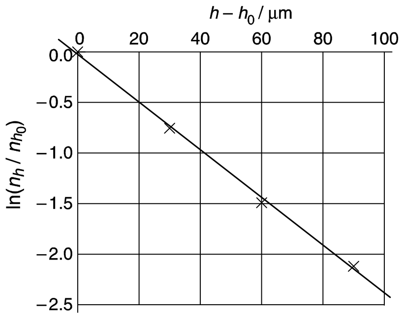
1.10 Derive an expression for the gradient (slope) of the graph.
1.11 Determine the Avogadro constant from these data.
Official English text of the problem.
Problem 2: Interstellar production of H2
Interstellar production of \(\ce{H2}\)
If two atoms collide in interstellar space the energy of the resulting molecule is so great that it rapidly dissociates. Hydrogen atoms only react to give stable \(\ce{H2}\) molecules on the surface of dust particles. The dust particles absorb most of the excess energy and the newly formed \(\ce{H2}\) rapidly desorbs. This question examines two kinetic models for \(\ce{H2}\) formation on the surface of a dust particle.
In both models, the rate constant for adsorption of \(\ce{H}\) atoms onto the surface of dust particles is \(k_a = 1.4 \cdot 10^{-5}\ \mathrm{cm^3\,s^{-1}}\). The typical number density of \(\ce{H}\) atoms (number of \(\ce{H}\) atoms per unit volume) in interstellar space is \([\ce{H}] = 10\ \mathrm{cm^{-3}}\).
[Note: In the following, you may treat numbers of surface-adsorbed atoms and number densities of gas-phase atoms in the same way as you would normally use concentrations in the rate equations. As a result, the units of the rate constants may be unfamiliar to you. Reaction rates have units of numbers of atoms or molecules per unit time.]
2.1 Calculate the rate at which \(\ce{H}\) atoms adsorb onto a dust particle. You may assume that this rate is constant throughout.
Desorption of \(\ce{H}\) atoms is first order with respect to the number of adsorbed atoms. The rate constant for the desorption step is \(k_d = 1.9 \cdot 10^{-3}\ \mathrm{s^{-1}}\).
2.2 Assuming that only adsorption and desorption take place, calculate the steady-state number, \(N\), of \(\ce{H}\) atoms on the surface of a dust particle.
The \(\ce{H}\) atoms are mobile on the surface. When they meet they react to form \(\ce{H2}\), which then desorbs. The two kinetic models under consideration differ in the way the reaction is modelled, but share the same rate constants \(k_a\), \(k_d\), and \(k_r\), for adsorption, desorption, and bimolecular reaction, as given below.
\[ \begin{aligned} k_a &= 1.4 \cdot 10^{-5}\ \mathrm{cm^3\,s^{-1}}\\ k_d &= 1.9 \cdot 10^{-3}\ \mathrm{s^{-1}}\\ k_r &= 5.1 \cdot 10^4\ \mathrm{s^{-1}} \end{aligned} \]
Model A
Reaction to form \(\ce{H2}\) is assumed to be second order. On a dust particle the rate of removal of \(\ce{H}\) atoms by reaction is \(k_rN^2\).
2.3 Write down an equation for the rate of change of \(N\), including adsorption, desorption and reaction. Assuming steady state conditions, determine the value of \(N\).
2.4 Calculate the rate of production of \(\ce{H2}\) per dust particle in this model.
Model B
Model B attempts to analyse the probability that the dust particles carry 0, 1 or 2 \(\ce{H}\) atoms. The three states are linked by the following reaction scheme. The assumption is made that no more than 2 atoms may be adsorbed simultaneously.
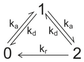
\(x_0\), \(x_1\) and \(x_2\) are the fractions of dust particles existing in state 0, 1 or 2, respectively. These fractions may be treated in the same way as concentrations in the following kinetic analysis. For a system in state \(m\) with fraction \(x_m\), the rates of the three possible processes are
Adsorption \((m\rightarrow m+1)\) : rate \(=k_a[\ce{H}]x_m\)
Desorption \((m\rightarrow m-1)\) : rate \(=k_dmx_m\)
Reaction \((m\rightarrow m-2)\) : rate \(=\frac12 k_rm(m-1)x_m\)
2.5 Write down equations for the rates of change, \(\mathrm{d}x_m/\mathrm{d}t\), of the fractions \(x_0\), \(x_1\) and \(x_2\).
2.6 Assuming steady-state conditions, use the above rate equations to find expressions for the ratios \(x_2/x_1\) and \(x_1/x_0\), and evaluate these ratios.
2.7 Evaluate the steady state fractions \(x_0\), \(x_1\) and \(x_2\).
[If you were unable to determine the ratios in 2.6, use \(x_2/x_1=a\) and \(x_1/x_0=b\) and give the result algebraically.]
2.8 Evaluate the rate of production of \(\ce{H2}\) per dust particle in this model.
2.9 It is currently not possible to measure the rate of this reaction experimentally, but the most recent computer simulations of the rate give a value of \(9.4 \cdot 10^{-6}\ \mathrm{s^{-1}}\). Which of the following statements apply to each model under these conditions? Mark any box you consider to be appropriate.
Statement Model A Model B Neither model The rate determining step is adsorption of \(\ce{H}\) atoms. The rate-determining step is desorption of \(\ce{H2}\) molecules. The rate determining step is the bimolecular reaction of \(\ce{H}\) atoms on the surface. The rate determining step is adsorption of the second \(\ce{H}\) atom. The implicit assumption that reaction can take place regardless of the number of atoms adsorbed leads to substantial error (at least a factor of two). Limiting the number of atoms adsorbed on the particle to 2 leads to substantial error (at least a factor of two). Official English text of the problem.
Problem 3: Protein Folding
The unfolding reaction for many small proteins can be represented by the equilibrium:

You may assume that the protein folding reaction takes place in a single step. The position of this equilibrium changes with temperature; the melting temperature \(T_m\) is defined as the temperature at which half of the molecules are unfolded and half are folded.
The intensity of the fluorescence signal at a wavelength of 356 nm of a \(1.0\ \mu\mathrm{M}\) (\(\mathrm{M}=\mathrm{mol\,dm^{-3}}\)) sample of the protein Chymotrypsin Inhibitor 2 (CI2) was measured as a function of temperature over the range 58 to 66 °C:
Temperature / °C 58 60 62 64 66 Fluorescence intensity (arbitrary units) 27 30 34 37 40 A \(1.0\ \mu\mathrm{M}\) sample in which all of the protein molecules are folded gives a fluorescence signal of 21 units at 356 nm. A \(1.0\ \mu\mathrm{M}\) sample in which all of the protein molecules are unfolded gives a fluorescence signal of 43 units.
3.1 Assuming that the fluorescence intensity from each species is directly proportional to its concentration, calculate the fraction, \(x\), of unfolded molecules present at each temperature.
3.2 Give an expression for the equilibrium constant, \(K\), in terms of \(x\), and hence calculate the value of \(K\) at each temperature.
3.3 Estimate the value of \(T_m\) for this protein (to the nearest 1 °C).
Assuming that the values of \(\Delta H^\circ\) and \(\Delta S^\circ\) for the protein unfolding reaction are constant with temperature then:
\[ \ln K=-\frac{\Delta H^\circ}{RT}+C \]
where \(C\) is a constant.
3.4 Plot a suitable graph and hence determine the values of \(\Delta H^\circ\) and \(\Delta S^\circ\) for the protein unfolding reaction.
[If you have been unable to calculate values for \(\Delta H^\circ\) and \(\Delta S^\circ\), you should use the following incorrect values for the subsequent parts of the problem:
\(\Delta H^\circ=130\ \mathrm{kJ\,mol^{-1}}\); \(\Delta S^\circ=250\ \mathrm{J\,K^{-1}\,mol^{-1}}\).]
3.5 Calculate the equilibrium constant for the unfolding reaction at 25 °C.
[If you have been unable to calculate a value for \(K\), you should use the following incorrect value for the subsequent parts of the problem: \(K=3.6\cdot10^{-6}\)]
The first order rate constant for the CI2 protein folding reaction can be determined by following the fluorescence intensity when a sample of unfolded protein is allowed to refold (typically the pH of the solution is changed). The concentration of protein when a \(1.0\ \mu\mathrm{M}\) sample of unfolded CI2 was allowed to refold was measured at a temperature of 25 °C:
time / ms 0 10 20 30 40 concentration / \(\mu\mathrm{M}\) 1 0.64 0.36 0.23 0.14 3.6 Plot a suitable graph and hence determine the value of the rate constant for the protein folding reaction, \(k_f\), at 25 °C.
[If you have been unable to calculate the value for \(k_f\), you should use the following incorrect value for the subsequent parts of the question: \(k_f=60\ \mathrm{s^{-1}}\).]
3.7 Determine the value of the rate constant for the protein unfolding reaction, \(k_u\), at 25 °C.
3.8 At 20 °C the rate constant for the protein folding reaction is \(33\ \mathrm{s^{-1}}\). Calculate the activation energy for the protein folding reaction.
Official English text of the problem.
Problem 4: Synthesis of Amprenavir
One class of anti-HIV drugs, known as protease inhibitors, works by blocking the active site of one of the enzymes used in assembly of the viruses within the host cell. Two successful drugs, saquinavir and amprenavir, contain the structural unit shown below which mimics the transition state within the enzyme. In the structure, \(R^1\), \(R^2\) and \(R^3\) may represent any atom or group other than hydrogen.
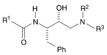
Amprenavir may be synthesised as shown in the convergent scheme below.
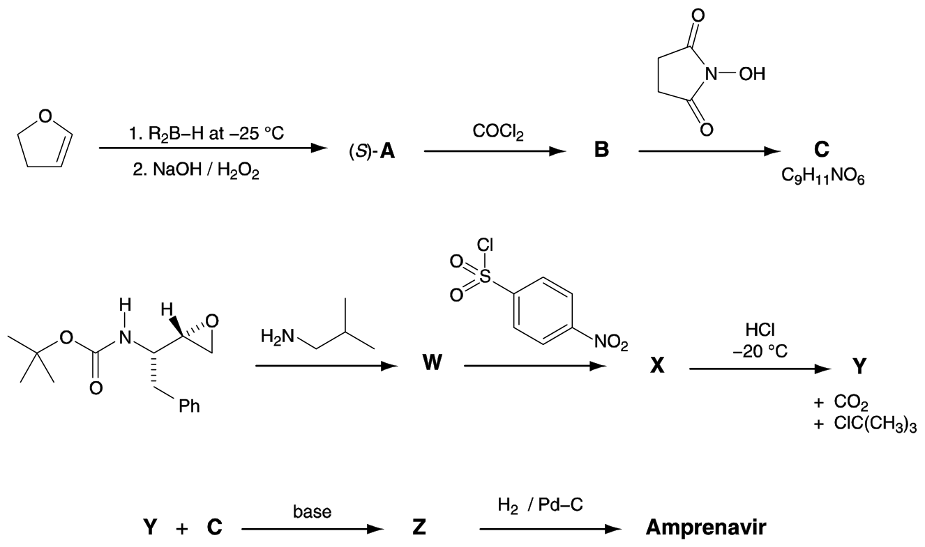
The reagent \(\ce{R2B-H}\) used in the first step is chiral.
Product A is formed as the \((S)\)-enantiomer.
Three of the signals in the \({}^{1}\mathrm{H}\) NMR spectrum of Amprenavir disappear on shaking with \(\ce{D2O}\): \(\delta\) 4.2 (2H), \(\delta\) 4.9 (1H) and \(\delta\) 5.1 (1H).
4.1 Suggest structures for:
a) the intermediates A, B, C, W, X, Y and Z,
b) Amprenavir.
Your answers should clearly show the stereochemistry at each centre.
Official English text of the problem.
Problem 5: Epoxy resins
The synthesis of epoxy resins is a multi-billion dollar industry worldwide. Epoxy resins are high performance adhesives synthesised from the reaction of a bis-epoxide with a diamine. The bis-epoxide is made from H and epichlorohydrin, C. Compounds C and H can be synthesised according to the schemes below.

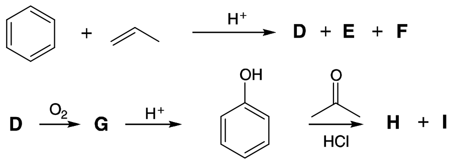
The synthesis of epichlorohydrin C begins with the reaction of propene with chlorine in the presence of light.
5.1 Draw the structures of A and B.
5.2 Give the formula of a suitable reagent for the conversion of B into epichlorohydrin C.
The synthesis of H commences with the reaction of benzene with propene in the presence of an acid catalyst which gives D as the major product and E and F as minor products.
5.3 Draw the structures of D, E, and F from the following data:
D: Elemental composition: C 89.94 %, H 10.06 %; 6 signals in the \(^{13}\mathrm{C}\) NMR spectrum.
E: Elemental composition: C 88.82 %, H 11.18 %; 4 signals in the \(^{13}\mathrm{C}\) NMR spectrum.
F: Elemental composition: C 88.82 %, H 11.18 %; 5 signals in the \(^{13}\mathrm{C}\) NMR spectrum.
Bubbling oxygen through a hot solution of D gives G which on exposure to acid gives phenol (hydroxybenzene) and acetone (propanone). G turns starch iodide paper from white to dark blue. G has 6 signals in the \(^{13}\mathrm{C}\) NMR spectrum and the following signals in the \(^{1}\mathrm{H}\) NMR spectrum: \(\delta\) 7.78 (1H, s), 7.45 – 7.22 (5H, m), 1.56 (6H, s); addition of \(\ce{D2O}\) results in the disappearance of the signal at \(\delta = 7.78\).
5.4 Draw the structure of G.
Exposure of phenol and acetone to hydrochloric acid gives compound H. The \(^{13}\mathrm{C}\) NMR spectrum for H is shown in Fig. 1. The \(^{1}\mathrm{H}\) NMR spectrum is shown in Fig. 2 together with a four-fold expansion of the region 6.5 – 7.1 ppm. The \(^{1}\mathrm{H}\) NMR spectrum after the addition of a drop of \(\ce{D2O}\), is shown in Fig. 3. Peaks due to the solvent are marked with an asterisk (*).
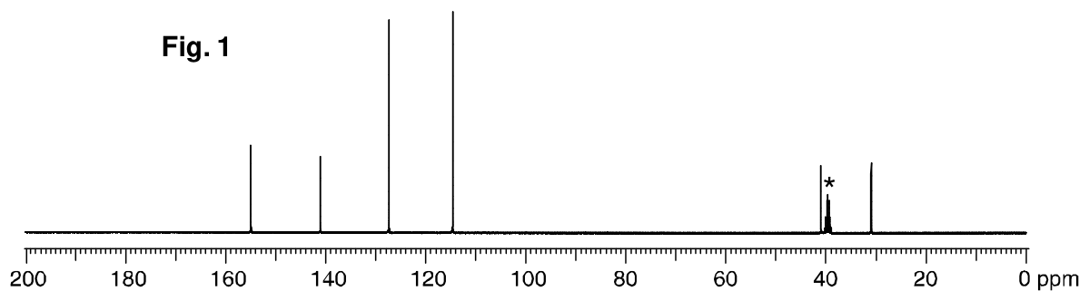
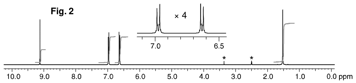
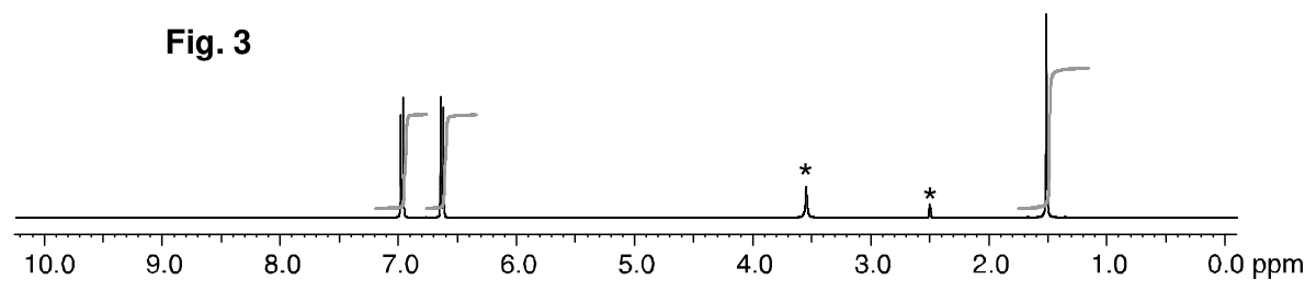
5.5 Draw the structure of H.
5.6 Draw one resonance structure of phenol which explains the regioselective formation of H.
A second compound, I, is also formed in the reaction of phenol with acetone. The \(^{13}\mathrm{C}\) NMR spectrum of I has 12 signals. The \(^{1}\mathrm{H}\) NMR spectrum has the following signals:
\(\delta\) 7.50 – 6.51 (8H, m), 5.19 (1H, s), 4.45 (1H, s), 1.67 (6H, s); addition of \(\ce{D2O}\) results in the disappearance of the signals at \(\delta = 5.19\) and 4.45
5.7 Draw a structure for I.
Excess phenol reacts with epichlorohydrin C in the presence of base to give compound L which has 6 signals in its \(^{13}\mathrm{C}\) NMR spectrum. If the reaction is stopped before completion compounds J and K can also be isolated. Compound L is formed from compound K and compound K is formed from compound J.

5.8 Draw the structures of J, K and L.
Treatment of H with a large excess of epichlorohydrin C and base gives a monomeric bis-epoxide M. M contains no chlorine atoms or OH groups.
5.9 Draw the structure of M.
Treatment of H with a small excess of epichlorohydrin and base gives N. N has the form: endgroup 1-[repeat unit]\(_n\)-endgroup 2 where \(n\) is approximately 10 – 15. N does not contain chlorine atoms and contains one OH group per repeat unit.
5.10 Draw the structure of N in the form indicated above:
(endgroup 1-[repeat unit]\(_n\)-endgroup 2).
5.11 Draw the repeat unit of the polymeric epoxy resin O formed from the reaction of the bis-epoxide M with ethane-1,2-diamine.
Official English text of the problem.
Problem 6: Transition metal complexes
Alfred Werner used the technique of ‘isomer counting’ to deduce the structure of metal complexes with coordination number six. Three of the shapes he considered are shown below.
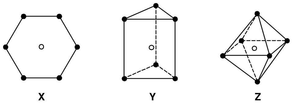
In each structure, the empty circle shows the location of the central metal atom and the filled circles show the location of the ligands. Structure X is hexagonal planar, structure Y is trigonal prismatic and structure Z is octahedral.
For each of the three shapes, there is just one structure when all of the ligands are the same, i.e. when the complex has the general formula \(\ce{MA6}\) where A is the ligand. However, when achiral ligands A are substituted by one or more achiral ligands, it may be possible for each structure to form geometrical isomers. It might also be possible for one or more of the geometrical isomers to be optically active and exist as pairs of enantiomers.
6.1 Fill in the table below to indicate how many geometrical isomers may be formed for each structure X, Y, and Z as the monodentate ligands A are substituted by monodentate ligands B or by symmetrical bidentate ligands, denoted C–C. Bidentate ligand C–C can only link between two atoms on adjacent positions, i.e. those positions connected by a line in the structures X, Y, and Z.
In each case write the number of geometrical isomers in the space provided. If one of the isomers exists as a pair of enantiomers, include an asterisk, *, in the box. If two exist as two pairs of enantiomers, include two asterisks and so on. For example, if you think there are five geometrical isomers of a particular structure, three of which exist as pairs of enantiomers, write 5 ***
Number of predicted geometrical isomers
Hexagonal planar X Trigonal Prismatic Y Octahedral Z \(\ce{MA6}\) 1 1 1 \(\ce{MA5B}\) \(\ce{MA4B2}\) \(\ce{MA3B3}\) \(\ce{MA4(C-C)}\) \(\ce{MA2(C-C)2}\) \(\ce{M(C-C)3}\) There are no known complexes that adopt the hexagonal planar geometry X, but structures are known for both the trigonal prismatic geometry Y and for the octahedral geometry Z. In these complexes, the orbitals derived from the metal d orbitals have different energies depending on the geometry of the complex. The splitting patterns for the trigonal prismatic geometry and for the octahedral geometry are shown below.
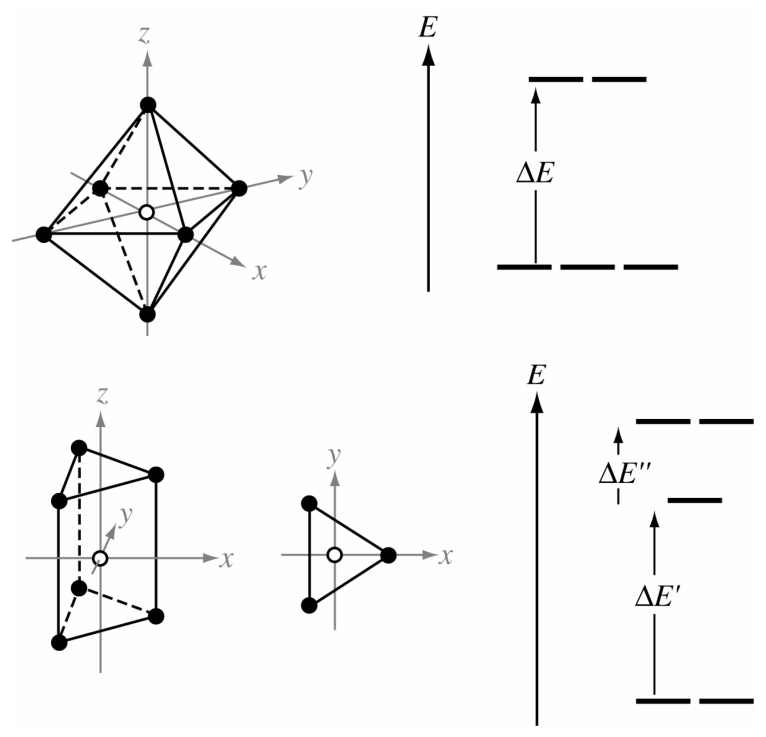
The separations in energy, \(\Delta E\), \(\Delta E'\) and \(\Delta E''\) depend on the particular complex.
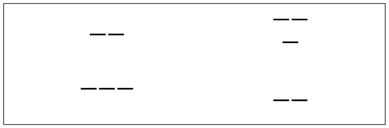
6.2 For each of the splitting patterns shown below label which d orbitals are which.
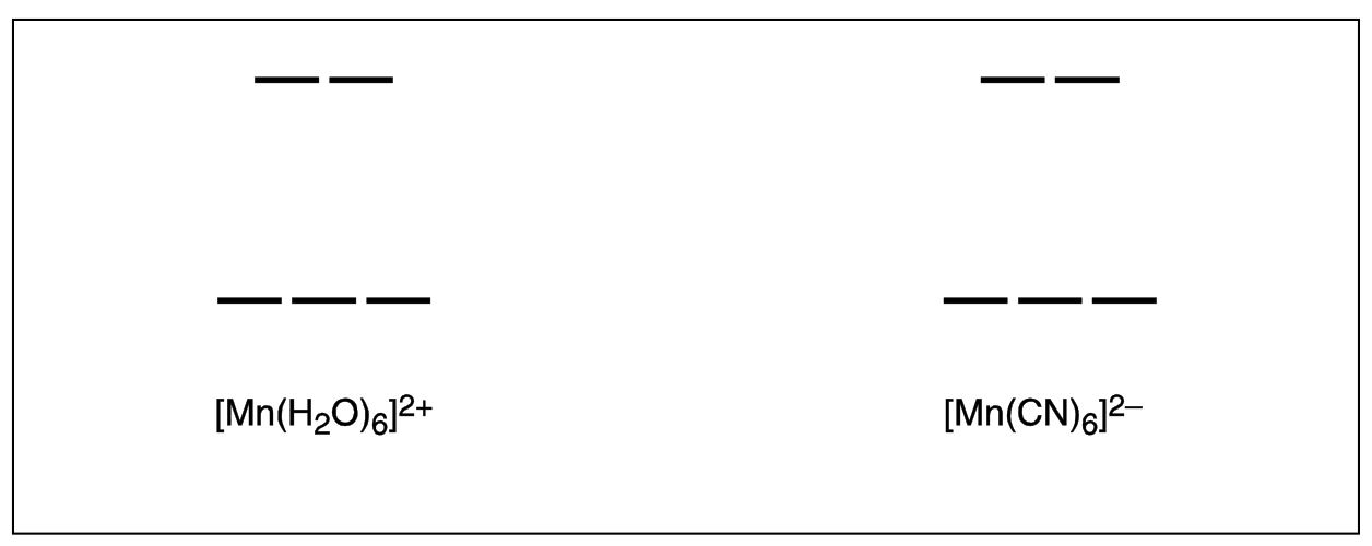
The two complexes \(\ce{[Mn(H2O)6]^2+}\) and \(\ce{[Mn(CN)6]^2-}\) are both octahedral. One has a magnetic moment of 5.9 BM, the other has a magnetic moment of 3.8 BM but you must decide which is which.
6.3 On the diagram below, draw the electronic arrangements for each of the complexes.
The magnetic moments of complexes A and B shown below have been measured and found to be 1.9 and 2.7 BM but you must decide which is which.
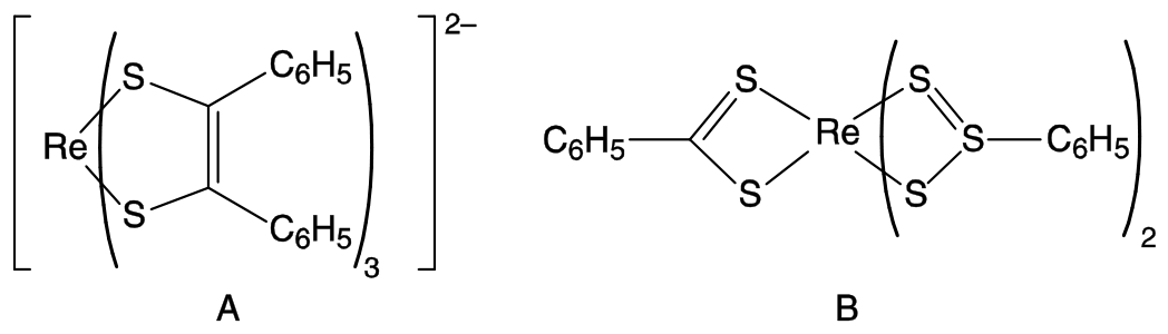
6.4 Draw the orbital splitting diagrams for the two complexes, including the arrangements of the electrons.
Octahedral complexes are far more common than trigonal prismatic. Werner isolated five compounds C – G containing Co(III), Cl, and \(\ce{NH3}\) only, each of which contained one octahedral complex. (There is actually a sixth compound but Werner could not isolate it.) Werner’s five compounds had the molar conductivities shown below. The conductivities are extrapolated to infinite dilution and are expressed in arbitrary units. Compound G does not react with aqueous \(\ce{AgNO3}\); compounds C, D, and E react with different stoichiometric ratios of aqueous \(\ce{AgNO3}\); E and F react with the same stoichiometric ratio of aqueous \(\ce{AgNO3}\).
C D E F G molar conductivity 510 372 249 249 \(\sim0\) 6.5 As far as you are able, suggest a structure for each of the compounds C – G.
Werner was also the first person to separate the enantiomers of an octahedral compound, H, which contained no carbon atoms. The compound, H, is composed of only cobalt, ammonia, chloride and an oxygen species which could be either \(\ce{H2O}\), or \(\ce{HO^-}\) or \(\ce{O^2-}\). The compound contains octahedrally coordinated cobalt ions. All of the chloride is easily removed from the compound by titration with aqueous silver nitrate. A 0.2872 g sample of H (containing no water of crystallization) required 22.8 \(\mathrm{cm^3}\) of a silver nitrate solution (\(c=0.100\ \mathrm{mol\,dm^{-3}}\)) to exchange all the chloride.
6.6 Calculate the percentage, by mass, of chloride in H.
H is stable to acids, but is hydrolysed in alkali. A 0.7934 g sample of H (containing no water of crystallization) was heated with excess aqueous sodium hydroxide. Cobalt(III) oxide was formed and ammonia gas given off. The ammonia produced was distilled off and absorbed into 50.0 \(\mathrm{cm^3}\) of aqueous \(\ce{HCl}\) (\(c_{\ce{HCl}}=0.500\ \mathrm{mol\,dm^{-3}}\)). The residual \(\ce{HCl}\) required 24.8 \(\mathrm{cm^3}\) of aqueous \(\ce{KOH}\) solution (\(c_{\ce{KOH}}=0.500\ \mathrm{mol\,dm^{-3}}\)) to be neutralized.
The remaining suspension of cobalt(III) oxide was allowed to cool, approximately 1 g of potassium iodide was added, and then the mixture was acidified with aqueous \(\ce{HCl}\). The liberated iodine was then titrated with aqueous solution of sodium thiosulfate (\(c=0.200\ \mathrm{mol\,dm^{-3}}\)) and required 21.0 \(\mathrm{cm^3}\) for complete reaction.
6.7 Calculate the percentage, by mass, of ammonia in H.
6.8 Give the equation for the reaction of cobalt(III) oxide with potassium iodide in aqueous acid.
6.9 Calculate the percentage, by mass, of cobalt in H.
6.10 Calculate the identity of the oxygen species contained in H. Show your working.
6.11 Give the empirical formula of H.
6.12 Suggest a structure for the chiral compound H.
Official English text of the problem.
Internationale Chemie-Olympiade
IChO 2009
Cambridge, Vereinigtes Königreich · 6 Theorieaufgaben mit vollständigen Lösungen
Aufgabe 1: Abschätzung der Avogadro-Konstante
Zur Bestimmung der Avogadro-Konstante wurden viele verschiedene Methoden verwendet. Drei verschiedene Methoden sind im Folgenden beschrieben.
Methode A – anhand von Röntgenbeugungsdaten (modern)
Die Elementarzelle ist die kleinste sich wiederholende Einheit einer Kristallstruktur. Mittels Röntgenbeugung wird festgestellt, dass die Elementarzelle eines Goldkristalls eine kubisch flächenzentrierte Struktur besitzt (d. h., der Mittelpunkt eines Atoms befindet sich an jeder Ecke eines Würfels und in der Mitte jeder Fläche). Die Kantenlänge der Elementarzelle beträgt 0.408 nm.
1.1 Skizzieren Sie die Elementarzelle und berechnen Sie, wie viele \(\ce{Au}\)-Atome sie enthält.
1.2 Die Dichte von \(\ce{Au}\) beträgt \(1.93\cdot10^4\ \mathrm{kg\,m^{-3}}\). Berechnen Sie das Volumen und die Masse der würfelförmigen Elementarzelle.
1.3 Berechnen Sie daraus die Masse eines Goldatoms und die Avogadro-Konstante, wenn die relative Atommasse von \(\ce{Au}\) 196.97 beträgt.
Methode B – anhand des radioaktiven Zerfalls (Rutherford, 1911)
Die radioaktive Zerfallsreihe von \(\ce{^{226}Ra}\) lautet wie folgt:
Die angegebenen Zeiten sind Halbwertszeiten; die Einheiten sind y = Jahre, d = Tage, m = Minuten. Der erste Zerfall, oben mit \(t\) gekennzeichnet, hat eine wesentlich längere Halbwertszeit als die anderen.
1.4 Geben Sie in der folgenden Tabelle an, welche Umwandlungen \(\alpha\)-Zerfälle und welche \(\beta\)-Zerfälle sind.
\(\alpha\)-Zerfall \(\beta\)-Zerfall \(\ce{^{226}Ra -> ^{222}Rn}\) \(\ce{^{222}Rn -> ^{218}Po}\) \(\ce{^{218}Po -> ^{214}Pb}\) \(\ce{^{214}Pb -> ^{214}Bi}\) \(\ce{^{214}Bi -> ^{214}Po}\) \(\ce{^{214}Po -> ^{210}Pb}\) \(\ce{^{210}Pb -> ^{210}Bi}\) \(\ce{^{210}Bi -> ^{210}Po}\) \(\ce{^{210}Po -> ^{206}Pb}\) 1.5 Eine Probe mit 192 mg \(\ce{^{226}Ra}\) wurde gereinigt und 40 Tage stehen gelassen. Geben Sie das erste Isotop in der Reihe (mit Ausnahme von \(\ce{Ra}\)) an, das noch keinen stationären Zustand erreicht hat.
1.6 Anschließend wurde die gesamte \(\alpha\)-Zerfallsrate der Probe durch Szintillationsmessung zu 27.7 GBq bestimmt (wobei \(1\ \mathrm{Bq}=1\ \mathrm{count\,s^{-1}}\)). Danach wurde die Probe für 163 Tage dicht verschlossen. Berechnen Sie die Anzahl der erzeugten \(\alpha\)-Teilchen.
1.7 Nach Ablauf der 163 Tage wurde festgestellt, dass die Probe \(10.4\ \mathrm{mm^3}\) \(\ce{He}\) enthielt, gemessen bei 101325 Pa und 273 K. Berechnen Sie aus diesen Daten die Avogadro-Konstante.
1.8 Die mittels Massenspektrometrie gemessene relative Isotopenmasse von \(\ce{^{226}Ra}\) beträgt 226.25. Berechnen Sie mit dem Lehrbuchwert der Avogadro-Konstante (\(6.022\cdot10^{23}\ \mathrm{mol^{-1}}\)) die Anzahl der \(\ce{^{226}Ra}\)-Atome in der ursprünglichen Probe, \(n_{\ce{Ra}}\), die Zerfallskonstante, \(\lambda\), und die Halbwertszeit, \(t\), von \(\ce{^{226}Ra}\) (in Jahren). Sie müssen nur die Zerfälle bis zu dem in 1.5 bestimmten Isotop berücksichtigen, jedoch nicht den Zerfall dieses Isotops selbst.
Methode C – Verteilung von Teilchen (Perrin, 1909)
Eine der ersten genauen Bestimmungen der Avogadro-Konstante erfolgte durch Untersuchung der vertikalen Verteilung von in Wasser suspendierten kolloidalen Teilchen unter dem Einfluss der Schwerkraft. In einem solchen Experiment wurden Teilchen mit einem Radius von \(2.12\cdot10^{-7}\ \mathrm{m}\) und einer Dichte von \(1.206\cdot10^3\ \mathrm{kg\,m^{-3}}\) in einem mit Wasser gefüllten Rohr bei \(15\ ^\circ\mathrm{C}\) suspendiert. Nachdem ausreichend Zeit zur Einstellung des Gleichgewichts verstrichen war, wurden in vier Höhen über dem Boden des Rohrs folgende mittlere Teilchenzahlen pro Volumeneinheit beobachtet:
Höhe / \(10^{-6}\ \mathrm{m}\) 5 35 65 95 mittlere Anzahl pro Volumeneinheit 4.00 1.88 0.90 0.48 1.9 Berechnen Sie unter der Annahme kugelförmiger Teilchen:
i) die Masse, \(m\), eines Teilchens;
ii) die Masse, \(m_{\ce{H2O}}\), des von ihm verdrängten Wassers;
iii) die effektive Masse, \(m^*\), des Teilchens in Wasser unter Berücksichtigung des Auftriebs (d. h. unter Berücksichtigung der durch das verdrängte Wasservolumen verursachten Auftriebskraft).
Nehmen Sie für die Dichte des Wassers \(999\ \mathrm{kg\,m^{-3}}\) an.
Im Gleichgewicht lässt sich die Teilchenzahl pro Volumeneinheit in verschiedenen Höhen durch eine Boltzmann-Verteilung beschreiben:
\[ \frac{n_h}{n_{h_0}} = \exp\left[-\frac{E_h-E_{h_0}}{RT}\right] \]
wobei
\(n_h\) die Teilchenzahl pro Volumeneinheit in der Höhe \(h\) ist,
\(n_{h_0}\) die Teilchenzahl pro Volumeneinheit in der Bezugshöhe \(h_0\) ist,
\(E_h\) die potenzielle Energie im Schwerefeld pro Mol Teilchen in der Höhe \(h\), bezogen auf die Teilchen am Boden des Rohrs, ist,
\(R\) die Gaskonstante ist, \(8.3145\ \mathrm{J\,K^{-1}\,mol^{-1}}\).
Ein Diagramm von \(\ln(n_h/n_{h_0})\) gegen \((h-h_0)\), das auf den Daten der obigen Tabelle beruht, ist unten dargestellt. Als Bezugshöhe werden \(5\ \mu\mathrm{m}\) über dem Boden des Rohrs gewählt.
1.10 Leiten Sie einen Ausdruck für die Steigung des Graphen her.
1.11 Bestimmen Sie aus diesen Daten die Avogadro-Konstante.
Aufgabentext aus dem offiziellen englischen Text übersetzt.
Aufgabe 2: Interstellare Bildung von H2
Interstellare Bildung von \(\ce{H2}\)
Wenn zwei Atome im interstellaren Raum zusammenstoßen, ist die Energie des entstehenden Moleküls so groß, dass es rasch dissoziiert. Wasserstoffatome reagieren nur auf der Oberfläche von Staubteilchen zu stabilen \(\ce{H2}\)-Molekülen. Die Staubteilchen nehmen den größten Teil der überschüssigen Energie auf, und das neu gebildete \(\ce{H2}\) desorbiert rasch. In dieser Aufgabe werden zwei kinetische Modelle für die Bildung von \(\ce{H2}\) auf der Oberfläche eines Staubteilchens untersucht.
In beiden Modellen beträgt die Geschwindigkeitskonstante für die Adsorption von \(\ce{H}\)-Atomen an der Oberfläche von Staubteilchen \(k_a = 1.4 \cdot 10^{-5}\ \mathrm{cm^3\,s^{-1}}\). Die typische Teilchenzahldichte der \(\ce{H}\)-Atome (Anzahl der \(\ce{H}\)-Atome pro Volumeneinheit) im interstellaren Raum beträgt \([\ce{H}] = 10\ \mathrm{cm^{-3}}\).
[Hinweis: Im Folgenden dürfen Sie die Anzahlen der an der Oberfläche adsorbierten Atome und die Teilchenzahldichten der Atome in der Gasphase genauso behandeln, wie Sie normalerweise Konzentrationen in Geschwindigkeitsgleichungen verwenden würden. Daher können Ihnen die Einheiten der Geschwindigkeitskonstanten ungewohnt erscheinen. Reaktionsgeschwindigkeiten haben die Einheit Anzahl der Atome oder Moleküle pro Zeiteinheit.]
2.1 Berechnen Sie die Geschwindigkeit, mit der \(\ce{H}\)-Atome an einem Staubteilchen adsorbieren. Sie dürfen durchgehend annehmen, dass diese Geschwindigkeit konstant ist.
Die Desorption von \(\ce{H}\)-Atomen ist bezüglich der Anzahl der adsorbierten Atome erster Ordnung. Die Geschwindigkeitskonstante des Desorptionsschritts beträgt \(k_d = 1.9 \cdot 10^{-3}\ \mathrm{s^{-1}}\).
2.2 Berechnen Sie unter der Annahme, dass nur Adsorption und Desorption stattfinden, die Anzahl \(N\) der \(\ce{H}\)-Atome auf der Oberfläche eines Staubteilchens im stationären Zustand.
Die \(\ce{H}\)-Atome sind auf der Oberfläche beweglich. Wenn sie aufeinandertreffen, reagieren sie zu \(\ce{H2}\), das anschließend desorbiert. Die beiden betrachteten kinetischen Modelle unterscheiden sich in der Modellierung der Reaktion, verwenden jedoch dieselben unten angegebenen Geschwindigkeitskonstanten \(k_a\), \(k_d\) und \(k_r\) für Adsorption, Desorption beziehungsweise bimolekulare Reaktion.
\[ \begin{aligned} k_a &= 1.4 \cdot 10^{-5}\ \mathrm{cm^3\,s^{-1}}\\ k_d &= 1.9 \cdot 10^{-3}\ \mathrm{s^{-1}}\\ k_r &= 5.1 \cdot 10^4\ \mathrm{s^{-1}} \end{aligned} \]
Modell A
Es wird angenommen, dass die Reaktion zur Bildung von \(\ce{H2}\) zweiter Ordnung ist. Auf einem Staubteilchen beträgt die Geschwindigkeit des Verbrauchs von \(\ce{H}\)-Atomen durch die Reaktion \(k_rN^2\).
2.3 Stellen Sie eine Gleichung für die zeitliche Änderung von \(N\) auf, die Adsorption, Desorption und Reaktion berücksichtigt. Bestimmen Sie den Wert von \(N\) unter der Annahme stationärer Bedingungen.
2.4 Berechnen Sie die Bildungsgeschwindigkeit von \(\ce{H2}\) pro Staubteilchen in diesem Modell.
Modell B
Modell B versucht, die Wahrscheinlichkeit dafür zu analysieren, dass die Staubteilchen 0, 1 oder 2 \(\ce{H}\)-Atome tragen. Die drei Zustände sind durch das folgende Reaktionsschema miteinander verknüpft. Es wird angenommen, dass höchstens 2 Atome gleichzeitig adsorbiert sein können.
\(x_0\), \(x_1\) und \(x_2\) sind die Anteile der Staubteilchen, die sich im Zustand 0, 1 beziehungsweise 2 befinden. Diese Anteile dürfen in der folgenden kinetischen Analyse genauso wie Konzentrationen behandelt werden. Für ein System im Zustand \(m\) mit dem Anteil \(x_m\) lauten die Geschwindigkeiten der drei möglichen Prozesse
Adsorption \((m\rightarrow m+1)\) : Geschwindigkeit \(=k_a[\ce{H}]x_m\)
Desorption \((m\rightarrow m-1)\) : Geschwindigkeit \(=k_dmx_m\)
Reaktion \((m\rightarrow m-2)\) : Geschwindigkeit \(=\frac12 k_rm(m-1)x_m\)
2.5 Stellen Sie Gleichungen für die zeitlichen Änderungen \(\mathrm{d}x_m/\mathrm{d}t\) der Anteile \(x_0\), \(x_1\) und \(x_2\) auf.
2.6 Leiten Sie unter der Annahme stationärer Bedingungen mithilfe der obigen Geschwindigkeitsgleichungen Ausdrücke für die Verhältnisse \(x_2/x_1\) und \(x_1/x_0\) her und berechnen Sie diese Verhältnisse.
2.7 Berechnen Sie die Anteile \(x_0\), \(x_1\) und \(x_2\) im stationären Zustand.
[Falls Sie die Verhältnisse in 2.6 nicht bestimmen konnten, verwenden Sie \(x_2/x_1=a\) und \(x_1/x_0=b\) und geben Sie das Ergebnis algebraisch an.]
2.8 Berechnen Sie die Bildungsgeschwindigkeit von \(\ce{H2}\) pro Staubteilchen in diesem Modell.
2.9 Derzeit ist es nicht möglich, die Geschwindigkeit dieser Reaktion experimentell zu messen; die neuesten Computersimulationen der Geschwindigkeit ergeben jedoch einen Wert von \(9.4 \cdot 10^{-6}\ \mathrm{s^{-1}}\). Welche der folgenden Aussagen treffen unter diesen Bedingungen auf das jeweilige Modell zu? Kreuzen Sie alle Kästchen an, die Sie für zutreffend halten.
Aussage Modell A Modell B Keines der beiden Modelle Der geschwindigkeitsbestimmende Schritt ist die Adsorption von \(\ce{H}\)-Atomen. Der geschwindigkeitsbestimmende Schritt ist die Desorption von \(\ce{H2}\)-Molekülen. Der geschwindigkeitsbestimmende Schritt ist die bimolekulare Reaktion von \(\ce{H}\)-Atomen auf der Oberfläche. Der geschwindigkeitsbestimmende Schritt ist die Adsorption des zweiten \(\ce{H}\)-Atoms. Die implizite Annahme, dass die Reaktion unabhängig von der Anzahl adsorbierter Atome stattfinden kann, führt zu einem erheblichen Fehler (mindestens um einen Faktor zwei). Die Begrenzung der Anzahl der am Teilchen adsorbierten Atome auf 2 führt zu einem erheblichen Fehler (mindestens um einen Faktor zwei). Aufgabentext aus dem offiziellen englischen Text übersetzt.
Aufgabe 3: Proteinfaltung
Die Entfaltungsreaktion vieler kleiner Proteine lässt sich durch das folgende Gleichgewicht darstellen:
Sie dürfen annehmen, dass die Proteinfaltungsreaktion in einem einzigen Schritt abläuft. Die Lage dieses Gleichgewichts ändert sich mit der Temperatur; die Schmelztemperatur \(T_m\) ist als die Temperatur definiert, bei der die Hälfte der Moleküle entfaltet und die andere Hälfte gefaltet ist.
Die Intensität des Fluoreszenzsignals bei einer Wellenlänge von 356 nm einer \(1.0\ \mu\mathrm{M}\) (\(\mathrm{M}=\mathrm{mol\,dm^{-3}}\)) Lösung des Proteins Chymotrypsin-Inhibitor 2 (CI2) wurde im Bereich von 58 bis 66 °C in Abhängigkeit von der Temperatur gemessen:
Temperatur / °C 58 60 62 64 66 Fluoreszenzintensität (willkürliche Einheiten) 27 30 34 37 40 Eine \(1.0\ \mu\mathrm{M}\) Lösung, in der alle Proteinmoleküle gefaltet sind, liefert bei 356 nm ein Fluoreszenzsignal von 21 Einheiten. Eine \(1.0\ \mu\mathrm{M}\) Lösung, in der alle Proteinmoleküle entfaltet sind, liefert ein Fluoreszenzsignal von 43 Einheiten.
3.1 Berechnen Sie unter der Annahme, dass die Fluoreszenzintensität jeder Spezies direkt proportional zu ihrer Konzentration ist, den Anteil \(x\) der entfalteten Moleküle bei jeder Temperatur.
3.2 Geben Sie einen Ausdruck für die Gleichgewichtskonstante \(K\) in Abhängigkeit von \(x\) an und berechnen Sie damit den Wert von \(K\) bei jeder Temperatur.
3.3 Schätzen Sie den Wert von \(T_m\) für dieses Protein (auf 1 °C gerundet).
Unter der Annahme, dass die Werte von \(\Delta H^\circ\) und \(\Delta S^\circ\) für die Proteinentfaltungsreaktion temperaturunabhängig sind, gilt:
\[ \ln K=-\frac{\Delta H^\circ}{RT}+C \]
wobei \(C\) eine Konstante ist.
3.4 Zeichnen Sie ein geeignetes Diagramm und bestimmen Sie daraus die Werte von \(\Delta H^\circ\) und \(\Delta S^\circ\) für die Proteinentfaltungsreaktion.
[Falls Sie keine Werte für \(\Delta H^\circ\) und \(\Delta S^\circ\) berechnen konnten, verwenden Sie für die folgenden Teilaufgaben die nachstehenden falschen Werte:
\(\Delta H^\circ=130\ \mathrm{kJ\,mol^{-1}}\); \(\Delta S^\circ=250\ \mathrm{J\,K^{-1}\,mol^{-1}}\).]
3.5 Berechnen Sie die Gleichgewichtskonstante der Entfaltungsreaktion bei 25 °C.
[Falls Sie keinen Wert für \(K\) berechnen konnten, verwenden Sie für die folgenden Teilaufgaben den nachstehenden falschen Wert: \(K=3.6\cdot10^{-6}\)]
Die Geschwindigkeitskonstante erster Ordnung der Faltungsreaktion des Proteins CI2 lässt sich bestimmen, indem man die Fluoreszenzintensität verfolgt, während sich eine Probe des entfalteten Proteins wieder faltet (üblicherweise wird dazu der pH-Wert der Lösung geändert). Bei der Rückfaltung einer \(1.0\ \mu\mathrm{M}\) Lösung von entfaltetem CI2 wurde die Proteinkonzentration bei einer Temperatur von 25 °C gemessen:
Zeit / ms 0 10 20 30 40 Konzentration / \(\mu\mathrm{M}\) 1 0.64 0.36 0.23 0.14 3.6 Zeichnen Sie ein geeignetes Diagramm und bestimmen Sie daraus den Wert der Geschwindigkeitskonstante der Proteinfaltungsreaktion \(k_f\) bei 25 °C.
[Falls Sie den Wert für \(k_f\) nicht berechnen konnten, verwenden Sie für die folgenden Teilaufgaben den nachstehenden falschen Wert: \(k_f=60\ \mathrm{s^{-1}}\).]
3.7 Bestimmen Sie den Wert der Geschwindigkeitskonstante der Proteinentfaltungsreaktion \(k_u\) bei 25 °C.
3.8 Bei 20 °C beträgt die Geschwindigkeitskonstante der Proteinfaltungsreaktion \(33\ \mathrm{s^{-1}}\). Berechnen Sie die Aktivierungsenergie der Proteinfaltungsreaktion.
Aufgabentext aus dem offiziellen englischen Text übersetzt.
Aufgabe 4: Synthese von Amprenavir
Eine Klasse von Arzneistoffen gegen HIV, die als Proteaseinhibitoren bezeichnet werden, wirkt durch Blockierung des aktiven Zentrums eines der Enzyme, die am Zusammenbau der Viren innerhalb der Wirtszelle beteiligt sind. Zwei erfolgreiche Arzneistoffe, Saquinavir und Amprenavir, enthalten die unten gezeigte Struktureinheit, die den Übergangszustand im Enzym nachahmt. In der Struktur können \(R^1\), \(R^2\) und \(R^3\) für beliebige Atome oder Gruppen außer Wasserstoff stehen.
Amprenavir lässt sich nach dem unten gezeigten konvergenten Syntheseschema herstellen.
Das im ersten Schritt verwendete Reagenz \(\ce{R2B-H}\) ist chiral.
Produkt A entsteht als \((S)\)-Enantiomer.
Drei der Signale im \({}^{1}\mathrm{H}\)-NMR-Spektrum von Amprenavir verschwinden beim Schütteln mit \(\ce{D2O}\): \(\delta\) 4.2 (2H), \(\delta\) 4.9 (1H) und \(\delta\) 5.1 (1H).
4.1 Schlagen Sie Strukturen vor für:
a) die Zwischenprodukte A, B, C, W, X, Y und Z,
b) Amprenavir.
Ihre Antworten sollen die Stereochemie an jedem Zentrum eindeutig zeigen.
Aufgabentext aus dem offiziellen englischen Text übersetzt.
Aufgabe 5: Epoxidharze
Die Synthese von Epoxidharzen ist weltweit ein Industriezweig mit Milliardenumsätzen in US-Dollar. Epoxidharze sind Hochleistungsklebstoffe, die durch die Reaktion eines Bisepoxids mit einem Diamin hergestellt werden. Das Bisepoxid wird aus H und Epichlorhydrin, C, hergestellt. Die Verbindungen C und H können nach den folgenden Schemata synthetisiert werden.
Die Synthese von Epichlorhydrin C beginnt mit der Reaktion von Propen mit Chlor unter Lichteinwirkung.
5.1 Zeichnen Sie die Strukturen von A und B.
5.2 Geben Sie die Formel eines geeigneten Reagenzes für die Umwandlung von B in Epichlorhydrin C an.
Die Synthese von H beginnt mit der Reaktion von Benzol mit Propen in Gegenwart eines Säurekatalysators, wobei D als Hauptprodukt und E und F als Nebenprodukte entstehen.
5.3 Zeichnen Sie anhand der folgenden Daten die Strukturen von D, E und F:
D: Elementarzusammensetzung: C 89.94 %, H 10.06 %; 6 Signale im \(^{13}\mathrm{C}\)-NMR-Spektrum.
E: Elementarzusammensetzung: C 88.82 %, H 11.18 %; 4 Signale im \(^{13}\mathrm{C}\)-NMR-Spektrum.
F: Elementarzusammensetzung: C 88.82 %, H 11.18 %; 5 Signale im \(^{13}\mathrm{C}\)-NMR-Spektrum.
Beim Durchleiten von Sauerstoff durch eine heiße Lösung von D entsteht G, das unter Säureeinwirkung Phenol (Hydroxybenzol) und Aceton (Propanon) ergibt. G färbt weißes Iodidstärkepapier dunkelblau. G weist 6 Signale im \(^{13}\mathrm{C}\)-NMR-Spektrum und die folgenden Signale im \(^{1}\mathrm{H}\)-NMR-Spektrum auf: \(\delta\) 7.78 (1H, s), 7.45 – 7.22 (5H, m), 1.56 (6H, s); die Zugabe von \(\ce{D2O}\) führt zum Verschwinden des Signals bei \(\delta = 7.78\).
5.4 Zeichnen Sie die Struktur von G.
Die Einwirkung von Salzsäure auf Phenol und Aceton ergibt die Verbindung H. Das \(^{13}\mathrm{C}\)-NMR-Spektrum von H ist in Abb. 1 dargestellt. Das \(^{1}\mathrm{H}\)-NMR-Spektrum ist in Abb. 2 zusammen mit einer vierfachen Vergrößerung des Bereichs 6.5 – 7.1 ppm dargestellt. Das \(^{1}\mathrm{H}\)-NMR-Spektrum nach Zugabe eines Tropfens \(\ce{D2O}\) ist in Abb. 3 dargestellt. Die vom Lösungsmittel verursachten Peaks sind mit einem Sternchen (*) gekennzeichnet.
5.5 Zeichnen Sie die Struktur von H.
5.6 Zeichnen Sie eine mesomere Grenzstruktur von Phenol, die die regioselektive Bildung von H erklärt.
Bei der Reaktion von Phenol mit Aceton entsteht auch eine zweite Verbindung, I. Das \(^{13}\mathrm{C}\)-NMR-Spektrum von I weist 12 Signale auf. Das \(^{1}\mathrm{H}\)-NMR-Spektrum weist die folgenden Signale auf:
\(\delta\) 7.50 – 6.51 (8H, m), 5.19 (1H, s), 4.45 (1H, s), 1.67 (6H, s); die Zugabe von \(\ce{D2O}\) führt zum Verschwinden der Signale bei \(\delta = 5.19\) und 4.45
5.7 Zeichnen Sie eine Struktur für I.
Überschüssiges Phenol reagiert mit Epichlorhydrin C in Gegenwart einer Base zur Verbindung L, die 6 Signale in ihrem \(^{13}\mathrm{C}\)-NMR-Spektrum aufweist. Wird die Reaktion vor ihrem vollständigen Ablauf abgebrochen, können auch die Verbindungen J und K isoliert werden. Verbindung L entsteht aus Verbindung K, und Verbindung K entsteht aus Verbindung J.
5.8 Zeichnen Sie die Strukturen von J, K und L.
Die Umsetzung von H mit einem großen Überschuss an Epichlorhydrin C und einer Base ergibt ein monomeres Bisepoxid M. M enthält weder Chloratome noch OH-Gruppen.
5.9 Zeichnen Sie die Struktur von M.
Die Umsetzung von H mit einem geringen Überschuss an Epichlorhydrin und einer Base ergibt N. N hat die Form: Endgruppe 1-[Wiederholungseinheit]\(_n\)-Endgruppe 2, wobei \(n\) ungefähr 10 – 15 beträgt. N enthält keine Chloratome und enthält eine OH-Gruppe pro Wiederholungseinheit.
5.10 Zeichnen Sie die Struktur von N in der oben angegebenen Form:
(Endgruppe 1-[Wiederholungseinheit]\(_n\)-Endgruppe 2).
5.11 Zeichnen Sie die Wiederholungseinheit des polymeren Epoxidharzes O, das bei der Reaktion des Bisepoxids M mit Ethan-1,2-diamin entsteht.
Aufgabentext aus dem offiziellen englischen Text übersetzt.
Aufgabe 6: Übergangsmetallkomplexe
Alfred Werner nutzte die Methode der „Isomerenzählung“, um die Struktur von Metallkomplexen mit der Koordinationszahl sechs abzuleiten. Drei der von ihm betrachteten Geometrien sind unten dargestellt.
In jeder Struktur zeigt der leere Kreis die Position des zentralen Metallatoms und die ausgefüllten Kreise zeigen die Positionen der Liganden. Struktur X ist hexagonal-planar, Struktur Y trigonal-prismatisch und Struktur Z oktaedrisch.
Für jede der drei Geometrien gibt es nur eine Struktur, wenn alle Liganden gleich sind, d. h., wenn der Komplex die allgemeine Formel \(\ce{MA6}\) hat, wobei A der Ligand ist. Werden jedoch achirale Liganden A durch einen oder mehrere achirale Liganden ersetzt, kann jede Struktur möglicherweise geometrische Isomere bilden. Außerdem können eines oder mehrere der geometrischen Isomere optisch aktiv sein und als Enantiomerenpaare vorliegen.
6.1 Tragen Sie in die folgende Tabelle ein, wie viele geometrische Isomere für jede der Strukturen X, Y und Z entstehen können, wenn die einzähnigen Liganden A durch einzähnige Liganden B oder durch symmetrische zweizähnige Liganden, bezeichnet als C–C, ersetzt werden. Der zweizähnige Ligand C–C kann nur zwei Atome an benachbarten Positionen miteinander verbinden, d. h. an Positionen, die in den Strukturen X, Y und Z durch eine Linie verbunden sind.
Schreiben Sie jeweils die Anzahl der geometrischen Isomere in das dafür vorgesehene Feld. Wenn eines der Isomere als Enantiomerenpaar vorliegt, fügen Sie im Feld ein Sternchen, *, hinzu. Wenn zwei als zwei Enantiomerenpaare vorliegen, fügen Sie zwei Sternchen hinzu und so weiter. Wenn Sie beispielsweise annehmen, dass es fünf geometrische Isomere einer bestimmten Struktur gibt, von denen drei als Enantiomerenpaare vorliegen, schreiben Sie 5 ***
Anzahl der vorhergesagten geometrischen Isomere
Hexagonal-planar X Trigonal-prismatisch Y Oktaedrisch Z \(\ce{MA6}\) 1 1 1 \(\ce{MA5B}\) \(\ce{MA4B2}\) \(\ce{MA3B3}\) \(\ce{MA4(C-C)}\) \(\ce{MA2(C-C)2}\) \(\ce{M(C-C)3}\) Es sind keine Komplexe bekannt, die die hexagonal-planare Geometrie X annehmen; es sind jedoch Strukturen sowohl mit der trigonal-prismatischen Geometrie Y als auch mit der oktaedrischen Geometrie Z bekannt. In diesen Komplexen haben die von den d-Orbitalen des Metalls abgeleiteten Orbitale je nach Geometrie des Komplexes unterschiedliche Energien. Die Aufspaltungsmuster für die trigonal-prismatische und die oktaedrische Geometrie sind unten dargestellt.
Die Energieabstände \(\Delta E\), \(\Delta E'\) und \(\Delta E''\) hängen vom jeweiligen Komplex ab.
6.2 Beschriften Sie für jedes der unten dargestellten Aufspaltungsmuster die Niveaus mit den zugehörigen d-Orbitalen.
Die beiden Komplexe \(\ce{[Mn(H2O)6]^2+}\) und \(\ce{[Mn(CN)6]^2-}\) sind beide oktaedrisch. Einer hat ein magnetisches Moment von 5.9 BM, der andere ein magnetisches Moment von 3.8 BM. Sie müssen entscheiden, welcher Wert zu welchem Komplex gehört.
6.3 Zeichnen Sie im folgenden Diagramm die Elektronenbesetzung für jeden der Komplexe ein.
Die magnetischen Momente der unten dargestellten Komplexe A und B wurden gemessen und betragen 1.9 und 2.7 BM. Sie müssen entscheiden, welcher Wert zu welchem Komplex gehört.
6.4 Zeichnen Sie die Orbitalaufspaltungsdiagramme für die beiden Komplexe einschließlich der Elektronenbesetzung.
Oktaedrische Komplexe sind weitaus häufiger als trigonal-prismatische. Werner isolierte fünf Verbindungen C – G, die ausschließlich Co(III), Cl und \(\ce{NH3}\) enthielten und jeweils einen oktaedrischen Komplex aufwiesen. (Tatsächlich gibt es eine sechste Verbindung, doch Werner konnte sie nicht isolieren.) Werners fünf Verbindungen hatten die unten angegebenen molaren Leitfähigkeiten. Die Leitfähigkeiten sind auf unendliche Verdünnung extrapoliert und in willkürlichen Einheiten angegeben. Verbindung G reagiert nicht mit wässrigem \(\ce{AgNO3}\); die Verbindungen C, D und E reagieren in unterschiedlichen stöchiometrischen Verhältnissen mit wässrigem \(\ce{AgNO3}\); E und F reagieren im gleichen stöchiometrischen Verhältnis mit wässrigem \(\ce{AgNO3}\).
C D E F G Molare Leitfähigkeit 510 372 249 249 \(\sim0\) 6.5 Schlagen Sie, soweit möglich, für jede der Verbindungen C – G eine Struktur vor.
Werner war außerdem der Erste, der die Enantiomere einer oktaedrischen Verbindung H trennte, die keine Kohlenstoffatome enthielt. Die Verbindung H besteht nur aus Cobalt, Ammoniak, Chlorid und einer Sauerstoffspezies, bei der es sich um \(\ce{H2O}\), \(\ce{HO^-}\) oder \(\ce{O^2-}\) handeln könnte. Die Verbindung enthält oktaedrisch koordinierte Cobaltionen. Das gesamte Chlorid lässt sich durch Titration mit wässriger Silbernitratlösung leicht aus der Verbindung entfernen. Eine 0.2872 g schwere Probe von H (ohne Kristallwasser) benötigte 22.8 \(\mathrm{cm^3}\) einer Silbernitratlösung (\(c=0.100\ \mathrm{mol\,dm^{-3}}\)), um das gesamte Chlorid auszutauschen.
6.6 Berechnen Sie den Massenanteil von Chlorid in H in Prozent.
H ist gegenüber Säuren stabil, wird aber in alkalischer Lösung hydrolysiert. Eine 0.7934 g schwere Probe von H (ohne Kristallwasser) wurde mit überschüssiger wässriger Natriumhydroxidlösung erhitzt. Dabei bildete sich Cobalt(III)-oxid und gasförmiges Ammoniak wurde freigesetzt. Das entstandene Ammoniak wurde abdestilliert und in 50.0 \(\mathrm{cm^3}\) wässriger \(\ce{HCl}\) (\(c_{\ce{HCl}}=0.500\ \mathrm{mol\,dm^{-3}}\)) absorbiert. Zur Neutralisation der verbleibenden \(\ce{HCl}\) wurden 24.8 \(\mathrm{cm^3}\) einer wässrigen \(\ce{KOH}\)-Lösung (\(c_{\ce{KOH}}=0.500\ \mathrm{mol\,dm^{-3}}\)) benötigt.
Die verbleibende Suspension von Cobalt(III)-oxid wurde abkühlen gelassen, etwa 1 g Kaliumiodid wurde zugegeben, und anschließend wurde das Gemisch mit wässriger \(\ce{HCl}\) angesäuert. Das freigesetzte Iod wurde anschließend mit einer wässrigen Natriumthiosulfatlösung (\(c=0.200\ \mathrm{mol\,dm^{-3}}\)) titriert; für die vollständige Reaktion wurden 21.0 \(\mathrm{cm^3}\) benötigt.
6.7 Berechnen Sie den Massenanteil von Ammoniak in H in Prozent.
6.8 Geben Sie die Reaktionsgleichung für die Reaktion von Cobalt(III)-oxid mit Kaliumiodid in wässriger Säure an.
6.9 Berechnen Sie den Massenanteil von Cobalt in H in Prozent.
6.10 Bestimmen Sie rechnerisch die Identität der in H enthaltenen Sauerstoffspezies. Geben Sie Ihren Rechenweg an.
6.11 Geben Sie die Verhältnisformel von H an.
6.12 Schlagen Sie eine Struktur für die chirale Verbindung H vor.
Aufgabentext aus dem offiziellen englischen Text übersetzt.
Uluslararası Kimya Olimpiyatı
IChO 2009
Cambridge, Birleşik Krallık · 6 teori sorusu, tam çözümleriyle
Soru 1: Avogadro sabitinin tahmin edilmesi
Avogadro sabitini belirlemek için pek çok farklı yöntem kullanılmıştır. Aşağıda üç farklı yöntem verilmiştir.
Yöntem A – X-ışını kırınımı verilerinden (modern)
Birim hücre, bir kristal yapıda tekrarlanan en küçük birimdir. X-ışını kırınımıyla, bir altın kristalinin birim hücresinin yüzey merkezli kübik yapıda olduğu (yani bir küpün her köşesinde ve her yüzünün ortasında bir atom merkezinin bulunduğu) belirlenmiştir. Birim hücrenin kenar uzunluğu 0.408 nm olarak bulunmuştur.
1.1 Birim hücrenin bir çizimini yapınız ve hücrenin kaç tane \(\ce{Au}\) atomu içerdiğini hesaplayınız.
1.2 \(\ce{Au}\)'nun yoğunluğu \(1.93\cdot10^4\ \mathrm{kg\,m^{-3}}\)'tür. Kübik birim hücrenin hacmini ve kütlesini hesaplayınız.
1.3 Buradan, \(\ce{Au}\)'nun bağıl atom kütlesinin 196.97 olduğunu kullanarak bir altın atomunun kütlesini ve Avogadro sabitini hesaplayınız.
Yöntem B – radyoaktif bozunmadan (Rutherford, 1911)
\(\ce{^{226}Ra}\)'nın radyoaktif bozunma serisi şöyledir:
Belirtilen süreler yarı ömürlerdir; birimler y = yıl, d = gün, m = dakikadır. Yukarıda \(t\) ile gösterilen ilk bozunmanın yarı ömrü diğerlerininkinden çok daha uzundur.
1.4 Aşağıdaki tabloda hangi dönüşümlerin \(\alpha\) bozunması, hangilerinin \(\beta\) bozunması olduğunu belirleyiniz.
\(\alpha\) bozunması \(\beta\) bozunması \(\ce{^{226}Ra -> ^{222}Rn}\) \(\ce{^{222}Rn -> ^{218}Po}\) \(\ce{^{218}Po -> ^{214}Pb}\) \(\ce{^{214}Pb -> ^{214}Bi}\) \(\ce{^{214}Bi -> ^{214}Po}\) \(\ce{^{214}Po -> ^{210}Pb}\) \(\ce{^{210}Pb -> ^{210}Bi}\) \(\ce{^{210}Bi -> ^{210}Po}\) \(\ce{^{210}Po -> ^{206}Pb}\) 1.5 192 mg \(\ce{^{226}Ra}\) içeren bir numune saflaştırılmış ve 40 gün bekletilmiştir. Seride (\(\ce{Ra}\) hariç) kararlı duruma ulaşmamış ilk izotopu belirleyiniz.
1.6 Daha sonra numunenin toplam \(\alpha\) bozunma hızı sintilasyon yöntemiyle 27.7 GBq olarak belirlenmiştir (burada \(1\ \mathrm{Bq}=1\ \mathrm{count\,s^{-1}}\)). Ardından numune sızdırmaz biçimde kapatılarak 163 gün bekletilmiştir. Oluşan \(\alpha\) parçacıklarının sayısını hesaplayınız.
1.7 163 günün sonunda numunenin, 101325 Pa ve 273 K'de ölçülen \(10.4\ \mathrm{mm^3}\) \(\ce{He}\) içerdiği bulunmuştur. Bu verilerden Avogadro sabitini hesaplayınız.
1.8 \(\ce{^{226}Ra}\)'nın kütle spektrometrisiyle ölçülen bağıl izotop kütlesinin 226.25 olduğu verilmektedir. Avogadro sabitinin ders kitaplarında verilen değerini (\(6.022\cdot10^{23}\ \mathrm{mol^{-1}}\)) kullanarak başlangıçtaki numunede bulunan \(\ce{^{226}Ra}\) atomlarının sayısını, \(n_{\ce{Ra}}\), bozunma hız sabitini, \(\lambda\), ve \(\ce{^{226}Ra}\)'nın yarı ömrünü, \(t\), (yıl cinsinden) hesaplayınız. Yalnızca 1.5'te belirlediğiniz izotopa kadar olan bozunmaları, bu izotopun bozunması hariç, dikkate almanız yeterlidir.
Yöntem C – parçacıkların dağılımı (Perrin, 1909)
Avogadro sabitinin ilk doğru belirlemelerinden biri, suda asılı duran kolloidal parçacıkların yerçekimi etkisi altındaki düşey dağılımının incelenmesiyle gerçekleştirilmiştir. Bu tür bir deneyde, yarıçapı \(2.12\cdot10^{-7}\ \mathrm{m}\) ve yoğunluğu \(1.206\cdot10^3\ \mathrm{kg\,m^{-3}}\) olan parçacıklar, \(15\ ^\circ\mathrm{C}\)'de su içeren bir tüpte süspansiyon hâline getirilmiştir. Dengeye ulaşılması için yeterince beklendikten sonra, tüpün tabanından itibaren dört farklı yükseklikte gözlenen birim hacim başına ortalama parçacık sayıları şöyledir:
yükseklik / \(10^{-6}\ \mathrm{m}\) 5 35 65 95 birim hacim başına ortalama sayı 4.00 1.88 0.90 0.48 1.9 Parçacıkların küresel olduğunu varsayarak aşağıdakileri hesaplayınız:
i) bir parçacığın kütlesi, \(m\);
ii) parçacığın yer değiştirdiği suyun kütlesi, \(m_{\ce{H2O}}\);
iii) kaldırma kuvvetini hesaba katarak (yani yer değiştirilen su hacminden kaynaklanan yukarı yönlü kuvveti dikkate alarak) parçacığın sudaki etkin kütlesi, \(m^*\).
Suyun yoğunluğunu \(999\ \mathrm{kg\,m^{-3}}\) alınız.
Dengede, farklı yüksekliklerdeki birim hacim başına parçacık sayısı Boltzmann dağılımına göre modellenebilir:
\[ \frac{n_h}{n_{h_0}} = \exp\left[-\frac{E_h-E_{h_0}}{RT}\right] \]
burada
\(n_h\), \(h\) yüksekliğinde birim hacim başına parçacık sayısıdır,
\(n_{h_0}\), referans yüksekliği \(h_0\)'da birim hacim başına parçacık sayısıdır,
\(E_h\), \(h\) yüksekliğindeki parçacıkların, tüpün tabanındaki parçacıklara göre mol başına kütleçekimsel potansiyel enerjisidir,
\(R\), gaz sabitidir ve değeri \(8.3145\ \mathrm{J\,K^{-1}\,mol^{-1}}\)'dir.
Yukarıdaki tabloda verilen verilere dayanan, \(\ln(n_h/n_{h_0})\)'ın \((h-h_0)\)'a karşı grafiği aşağıda gösterilmiştir. Referans yüksekliği, tüpün tabanından itibaren \(5\ \mu\mathrm{m}\) olarak alınmıştır.
1.10 Grafiğin eğimi için bir ifade türetiniz.
1.11 Bu verilerden Avogadro sabitini belirleyiniz.
Soru metni resmi İngilizce metinden çevrilmiştir.
Soru 2: Yıldızlararası ortamda H2 oluşumu
Yıldızlararası ortamda \(\ce{H2}\) oluşumu
Yıldızlararası uzayda iki atom çarpıştığında oluşan molekülün enerjisi o kadar büyüktür ki molekül hızla ayrışır. Hidrojen atomları, kararlı \(\ce{H2}\) molekülleri oluşturmak üzere yalnızca toz taneciklerinin yüzeyinde tepkimeye girer. Toz tanecikleri fazla enerjinin büyük bölümünü soğurur ve yeni oluşan \(\ce{H2}\) hızla desorbe olur. Bu soru, bir toz taneciğinin yüzeyinde \(\ce{H2}\) oluşumuna ilişkin iki kinetik modeli incelemektedir.
Her iki modelde de \(\ce{H}\) atomlarının toz taneciklerinin yüzeyine adsorpsiyonu için hız sabiti \(k_a = 1.4 \cdot 10^{-5}\ \mathrm{cm^3\,s^{-1}}\) değerindedir. Yıldızlararası uzayda \(\ce{H}\) atomlarının tipik sayı yoğunluğu (birim hacim başına \(\ce{H}\) atomu sayısı) \([\ce{H}] = 10\ \mathrm{cm^{-3}}\) değerindedir.
[Not: Aşağıda, yüzeye adsorplanmış atom sayılarını ve gaz fazındaki atomların sayı yoğunluklarını, hız denklemlerinde normalde derişimleri kullandığınız şekilde kullanabilirsiniz. Bu nedenle hız sabitlerinin birimleri size alışılmadık gelebilir. Tepkime hızlarının birimi, birim zaman başına atom veya molekül sayısıdır.]
2.1 \(\ce{H}\) atomlarının bir toz taneciğine adsorplanma hızını hesaplayınız. Bu hızın sorunun tamamında sabit olduğunu varsayabilirsiniz.
\(\ce{H}\) atomlarının desorpsiyonu, adsorplanmış atom sayısına göre birinci derecedendir. Desorpsiyon basamağının hız sabiti \(k_d = 1.9 \cdot 10^{-3}\ \mathrm{s^{-1}}\) değerindedir.
2.2 Yalnızca adsorpsiyon ve desorpsiyonun gerçekleştiğini varsayarak, bir toz taneciğinin yüzeyindeki \(\ce{H}\) atomlarının kararlı durumdaki sayısını, \(N\), hesaplayınız.
\(\ce{H}\) atomları yüzey üzerinde hareketlidir. Karşılaştıklarında tepkimeye girerek \(\ce{H2}\) oluştururlar; oluşan \(\ce{H2}\) daha sonra desorbe olur. Ele alınan iki kinetik model, tepkimenin modellenme biçimi bakımından farklılık gösterir; ancak adsorpsiyon, desorpsiyon ve bimoleküler tepkime için aşağıda verilen aynı \(k_a\), \(k_d\) ve \(k_r\) hız sabitlerini kullanır.
\[ \begin{aligned} k_a &= 1.4 \cdot 10^{-5}\ \mathrm{cm^3\,s^{-1}}\\ k_d &= 1.9 \cdot 10^{-3}\ \mathrm{s^{-1}}\\ k_r &= 5.1 \cdot 10^4\ \mathrm{s^{-1}} \end{aligned} \]
Model A
\(\ce{H2}\) oluşum tepkimesinin ikinci dereceden olduğu varsayılmaktadır. Bir toz taneciğinde \(\ce{H}\) atomlarının tepkime yoluyla tüketilme hızı \(k_rN^2\) olarak verilir.
2.3 Adsorpsiyon, desorpsiyon ve tepkimeyi içerecek şekilde \(N\)'nin değişim hızı için bir denklem yazınız. Kararlı durum koşullarını varsayarak \(N\)'nin değerini belirleyiniz.
2.4 Bu modelde toz taneciği başına \(\ce{H2}\) oluşum hızını hesaplayınız.
Model B
Model B, toz taneciklerinin 0, 1 veya 2 \(\ce{H}\) atomu taşıma olasılığını incelemeyi amaçlar. Bu üç durum, aşağıdaki tepkime şemasıyla birbirine bağlıdır. Aynı anda en fazla 2 atomun adsorplanabileceği varsayılmaktadır.
\(x_0\), \(x_1\) ve \(x_2\), sırasıyla 0, 1 veya 2 durumunda bulunan toz taneciklerinin kesirleridir. Aşağıdaki kinetik analizde bu kesirler derişimlerle aynı şekilde ele alınabilir. Kesri \(x_m\) olan \(m\) durumundaki bir sistem için olası üç sürecin hızları şöyledir:
Adsorpsiyon \((m\rightarrow m+1)\) : hız \(=k_a[\ce{H}]x_m\)
Desorpsiyon \((m\rightarrow m-1)\) : hız \(=k_dmx_m\)
Tepkime \((m\rightarrow m-2)\) : hız \(=\frac12 k_rm(m-1)x_m\)
2.5 \(x_0\), \(x_1\) ve \(x_2\) kesirlerinin değişim hızları, \(\mathrm{d}x_m/\mathrm{d}t\), için denklemleri yazınız.
2.6 Kararlı durum koşullarını varsayarak, yukarıdaki hız denklemlerini kullanıp \(x_2/x_1\) ve \(x_1/x_0\) oranları için ifadeler bulunuz ve bu oranların değerlerini hesaplayınız.
2.7 Kararlı durum kesirleri \(x_0\), \(x_1\) ve \(x_2\)'nin değerlerini hesaplayınız.
[2.6'daki oranları belirleyemediyseniz, \(x_2/x_1=a\) ve \(x_1/x_0=b\) kullanarak sonucu cebirsel olarak veriniz.]
2.8 Bu modelde toz taneciği başına \(\ce{H2}\) oluşum hızını hesaplayınız.
2.9 Bu tepkimenin hızını deneysel olarak ölçmek şu anda mümkün değildir; ancak hıza ilişkin en son bilgisayar benzetimleri \(9.4 \cdot 10^{-6}\ \mathrm{s^{-1}}\) değerini vermektedir. Bu koşullar altında aşağıdaki ifadelerden hangileri her bir model için geçerlidir? Uygun bulduğunuz tüm kutuları işaretleyiniz.
İfade Model A Model B Hiçbir model Hız belirleyen basamak, \(\ce{H}\) atomlarının adsorpsiyonudur. Hız belirleyen basamak, \(\ce{H2}\) moleküllerinin desorpsiyonudur. Hız belirleyen basamak, yüzeydeki \(\ce{H}\) atomlarının bimoleküler tepkimesidir. Hız belirleyen basamak, ikinci \(\ce{H}\) atomunun adsorpsiyonudur. Adsorplanmış atom sayısından bağımsız olarak tepkimenin gerçekleşebileceği yönündeki örtük varsayım, önemli bir hataya (en az iki katlık bir farka) yol açar. Tanecik üzerinde adsorplanmış atom sayısını 2 ile sınırlamak, önemli bir hataya (en az iki katlık bir farka) yol açar. Soru metni resmi İngilizce metinden çevrilmiştir.
Soru 3: Protein Katlanması
Birçok küçük proteinin açılma tepkimesi aşağıdaki denge ile gösterilebilir:
Protein katlanma tepkimesinin tek basamakta gerçekleştiğini varsayabilirsiniz. Bu dengenin konumu sıcaklıkla değişir; erime sıcaklığı \(T_m\), moleküllerin yarısının açılmış, yarısının ise katlanmış olduğu sıcaklık olarak tanımlanır.
Kimotripsin İnhibitörü 2 (CI2) proteininin \(1.0\ \mu\mathrm{M}\) (\(\mathrm{M}=\mathrm{mol\,dm^{-3}}\)) derişimli bir örneğinin 356 nm dalga boyundaki floresans sinyalinin şiddeti, 58 ile 66 °C aralığında sıcaklığın bir fonksiyonu olarak ölçülmüştür:
Sıcaklık / °C 58 60 62 64 66 Floresans şiddeti (keyfî birimler) 27 30 34 37 40 Protein moleküllerinin tamamının katlanmış olduğu \(1.0\ \mu\mathrm{M}\) derişimli bir örnek, 356 nm'de 21 birimlik bir floresans sinyali verir. Protein moleküllerinin tamamının açılmış olduğu \(1.0\ \mu\mathrm{M}\) derişimli bir örnek ise 43 birimlik bir floresans sinyali verir.
3.1 Her bir türden kaynaklanan floresans şiddetinin o türün derişimiyle doğru orantılı olduğunu varsayarak, her sıcaklıkta bulunan açılmış moleküllerin kesrini, \(x\), hesaplayınız.
3.2 Denge sabiti \(K\) için \(x\) cinsinden bir ifade yazınız ve buradan her sıcaklıktaki \(K\) değerini hesaplayınız.
3.3 Bu protein için \(T_m\) değerini tahmin ediniz (en yakın 1 °C'ye yuvarlayınız).
Proteinin açılma tepkimesi için \(\Delta H^\circ\) ve \(\Delta S^\circ\) değerlerinin sıcaklıkla değişmediği varsayılırsa:
\[ \ln K=-\frac{\Delta H^\circ}{RT}+C \]
Burada \(C\) bir sabittir.
3.4 Uygun bir grafik çiziniz ve bu grafikten proteinin açılma tepkimesi için \(\Delta H^\circ\) ve \(\Delta S^\circ\) değerlerini belirleyiniz.
[\(\Delta H^\circ\) ve \(\Delta S^\circ\) değerlerini hesaplayamadıysanız, sorunun sonraki kısımlarında aşağıdaki yanlış değerleri kullanınız:
\(\Delta H^\circ=130\ \mathrm{kJ\,mol^{-1}}\); \(\Delta S^\circ=250\ \mathrm{J\,K^{-1}\,mol^{-1}}\).]
3.5 Açılma tepkimesinin 25 °C'deki denge sabitini hesaplayınız.
[\(K\) değerini hesaplayamadıysanız, sorunun sonraki kısımlarında aşağıdaki yanlış değeri kullanınız: \(K=3.6\cdot10^{-6}\)]
CI2 proteininin katlanma tepkimesinin birinci dereceden hız sabiti, açılmış protein içeren bir örneğin yeniden katlanmasına izin verildiğinde floresans şiddeti izlenerek belirlenebilir (genellikle çözeltinin pH'ı değiştirilir). Açılmış CI2'nin \(1.0\ \mu\mathrm{M}\) derişimli bir örneğinin yeniden katlanmasına izin verildiğinde, protein derişimi 25 °C sıcaklıkta ölçülmüştür:
zaman / ms 0 10 20 30 40 derişim / \(\mu\mathrm{M}\) 1 0.64 0.36 0.23 0.14 3.6 Uygun bir grafik çiziniz ve bu grafikten proteinin katlanma tepkimesinin 25 °C'deki hız sabitini, \(k_f\), belirleyiniz.
[\(k_f\) değerini hesaplayamadıysanız, sorunun sonraki kısımlarında aşağıdaki yanlış değeri kullanınız: \(k_f=60\ \mathrm{s^{-1}}\).]
3.7 Proteinin açılma tepkimesinin 25 °C'deki hız sabitini, \(k_u\), belirleyiniz.
3.8 Proteinin katlanma tepkimesinin 20 °C'deki hız sabiti \(33\ \mathrm{s^{-1}}\)'dir. Proteinin katlanma tepkimesinin aktivasyon enerjisini hesaplayınız.
Soru metni resmi İngilizce metinden çevrilmiştir.
Soru 4: Amprenavir Sentezi
Proteaz inhibitörleri olarak bilinen bir anti-HIV ilaç sınıfı, konak hücre içinde virüslerin bir araya getirilmesinde kullanılan enzimlerden birinin aktif bölgesini bloke ederek etki gösterir. Başarıyla kullanılan iki ilaç, sakinavir ve amprenavir, enzim içindeki geçiş durumunu taklit eden ve aşağıda gösterilen yapısal birimi içerir. Bu yapıda \(R^1\), \(R^2\) ve \(R^3\), hidrojen dışındaki herhangi bir atomu veya grubu temsil edebilir.
Amprenavir, aşağıdaki yakınsak sentez şemasında gösterildiği gibi sentezlenebilir.
İlk basamakta kullanılan \(\ce{R2B-H}\) reaktifi kiraldir.
A ürünü, \((S)\)-enantiyomeri olarak oluşur.
Amprenavirin \({}^{1}\mathrm{H}\) NMR spektrumundaki sinyallerden üçü, \(\ce{D2O}\) ile çalkalandığında kaybolur: \(\delta\) 4.2 (2H), \(\delta\) 4.9 (1H) ve \(\delta\) 5.1 (1H).
4.1 Aşağıdakiler için yapılar öneriniz:
a) A, B, C, W, X, Y ve Z ara ürünleri,
b) Amprenavir.
Yanıtlarınız her bir merkezdeki stereokimyayı açıkça göstermelidir.
Soru metni resmi İngilizce metinden çevrilmiştir.
Soru 5: Epoksi reçineleri
Epoksi reçinelerinin sentezi, dünya çapında milyarlarca dolarlık bir endüstridir. Epoksi reçineleri, bir bis-epoksidin bir diaminle tepkimesinden sentezlenen yüksek performanslı yapıştırıcılardır. Bis-epoksit, H ve epiklorohidrin C kullanılarak hazırlanır. C ve H bileşikleri aşağıdaki şemalara göre sentezlenebilir.
Epiklorohidrin C'nin sentezi, propenin ışık varlığında klorla tepkimesiyle başlar.
5.1 A ve B'nin yapılarını çiziniz.
5.2 B'nin epiklorohidrin C'ye dönüştürülmesi için uygun bir reaktifin formülünü veriniz.
H'nin sentezi, benzenin bir asit katalizörü varlığında propenle tepkimesiyle başlar; bu tepkimede ana ürün olarak D, yan ürünler olarak E ve F oluşur.
5.3 Aşağıdaki verilerden yararlanarak D, E ve F'nin yapılarını çiziniz:
D: Elementel bileşim: C %89.94, H %10.06; \(^{13}\mathrm{C}\) NMR spektrumunda 6 sinyal.
E: Elementel bileşim: C %88.82, H %11.18; \(^{13}\mathrm{C}\) NMR spektrumunda 4 sinyal.
F: Elementel bileşim: C %88.82, H %11.18; \(^{13}\mathrm{C}\) NMR spektrumunda 5 sinyal.
D'nin sıcak bir çözeltisinden oksijen gazı geçirilmesiyle G elde edilir; G, asitle muamele edildiğinde fenol (hidroksibenzen) ve aseton (propanon) verir. G, nişastalı iyodür kâğıdının rengini beyazdan koyu maviye dönüştürür. G'nin \(^{13}\mathrm{C}\) NMR spektrumunda 6 sinyal ve \(^{1}\mathrm{H}\) NMR spektrumunda şu sinyaller vardır: \(\delta\) 7.78 (1H, s), 7.45 – 7.22 (5H, m), 1.56 (6H, s); \(\ce{D2O}\) eklenmesi, \(\delta = 7.78\) konumundaki sinyalin kaybolmasına yol açar.
5.4 G'nin yapısını çiziniz.
Fenol ve asetonun hidroklorik asitle muamele edilmesiyle H bileşiği elde edilir. H'nin \(^{13}\mathrm{C}\) NMR spektrumu Şekil 1'de gösterilmiştir. \(^{1}\mathrm{H}\) NMR spektrumu, 6.5 – 7.1 ppm bölgesinin dört kat büyütülmüş hâliyle birlikte Şekil 2'de gösterilmiştir. Bir damla \(\ce{D2O}\) eklendikten sonraki \(^{1}\mathrm{H}\) NMR spektrumu Şekil 3'te gösterilmiştir. Çözücüden kaynaklanan pikler yıldız işareti (*) ile belirtilmiştir.
5.5 H'nin yapısını çiziniz.
5.6 H'nin bölge seçici oluşumunu açıklayan bir fenol rezonans yapısı çiziniz.
Fenolün asetonla tepkimesinde ikinci bir bileşik olan I de oluşur. I'nın \(^{13}\mathrm{C}\) NMR spektrumunda 12 sinyal vardır. \(^{1}\mathrm{H}\) NMR spektrumunda şu sinyaller bulunur:
\(\delta\) 7.50 – 6.51 (8H, m), 5.19 (1H, s), 4.45 (1H, s), 1.67 (6H, s); \(\ce{D2O}\) eklenmesi, \(\delta = 5.19\) ve 4.45 konumlarındaki sinyallerin kaybolmasına yol açar
5.7 I için bir yapı çiziniz.
Fazla miktarda fenol, baz varlığında epiklorohidrin C ile tepkimeye girerek \(^{13}\mathrm{C}\) NMR spektrumunda 6 sinyal bulunan L bileşiğini verir. Tepkime tamamlanmadan durdurulursa J ve K bileşikleri de izole edilebilir. L bileşiği K bileşiğinden, K bileşiği ise J bileşiğinden oluşur.
5.8 J, K ve L'nin yapılarını çiziniz.
H'nin çok fazla miktarda epiklorohidrin C ve baz ile muamele edilmesiyle monomerik bir bis-epoksit olan M elde edilir. M, klor atomu veya OH grubu içermez.
5.9 M'nin yapısını çiziniz.
H'nin az miktarda fazla epiklorohidrin ve baz ile muamele edilmesiyle N elde edilir. N şu biçimdedir: uç grup 1-[tekrarlanan birim]\(_n\)-uç grup 2; burada \(n\) yaklaşık 10 – 15'tir. N, klor atomu içermez ve tekrarlanan birim başına bir OH grubu içerir.
5.10 N'nin yapısını yukarıda belirtilen biçimde çiziniz:
(uç grup 1-[tekrarlanan birim]\(_n\)-uç grup 2).
5.11 Bis-epoksit M'nin etan-1,2-diamin ile tepkimesinden oluşan polimerik epoksi reçinesi O'nun tekrarlanan birimini çiziniz.
Soru metni resmi İngilizce metinden çevrilmiştir.
Soru 6: Geçiş metali kompleksleri
Alfred Werner, koordinasyon sayısı altı olan metal komplekslerinin yapısını belirlemek için ‘izomer sayma’ tekniğini kullanmıştır. Ele aldığı geometrilerden üçü aşağıda gösterilmiştir.
Her yapıda boş daire merkez metal atomunun konumunu, dolu daireler ise ligandların konumlarını göstermektedir. X yapısı altıgen düzlemsel, Y yapısı üçgen prizmatik ve Z yapısı oktahedraldir.
Bu üç geometrinin her biri için, ligandların tamamı aynı olduğunda, yani kompleksin genel formülü A'nın ligandı gösterdiği \(\ce{MA6}\) olduğunda, yalnızca bir yapı vardır. Ancak akiral A ligandları bir veya daha fazla akiral ligandla yer değiştirdiğinde, her yapının geometrik izomerler oluşturması mümkün olabilir. Geometrik izomerlerden bir veya daha fazlasının optikçe aktif olması ve enantiyomer çiftleri hâlinde bulunması da mümkündür.
6.1 Tek dişli A ligandları tek dişli B ligandlarıyla veya C–C ile gösterilen simetrik iki dişli ligandlarla yer değiştirdiğinde, X, Y ve Z yapılarının her biri için kaç geometrik izomer oluşabileceğini belirtmek üzere aşağıdaki tabloyu doldurunuz. İki dişli C–C ligandı yalnızca komşu konumlardaki iki atom arasında, yani X, Y ve Z yapılarında bir çizgiyle bağlanmış konumlar arasında bağlantı kurabilir.
Her durumda geometrik izomer sayısını ayrılan boşluğa yazınız. İzomerlerden biri bir enantiyomer çifti hâlinde bulunuyorsa kutuya bir yıldız, *, ekleyiniz. İkisi iki enantiyomer çifti hâlinde bulunuyorsa iki yıldız ekleyiniz ve bu şekilde devam ediniz. Örneğin, belirli bir yapının beş geometrik izomeri olduğunu ve bunlardan üçünün enantiyomer çiftleri hâlinde bulunduğunu düşünüyorsanız 5 *** yazınız.
Öngörülen geometrik izomer sayısı
Altıgen düzlemsel X Üçgen prizmatik Y Oktahedral Z \(\ce{MA6}\) 1 1 1 \(\ce{MA5B}\) \(\ce{MA4B2}\) \(\ce{MA3B3}\) \(\ce{MA4(C-C)}\) \(\ce{MA2(C-C)2}\) \(\ce{M(C-C)3}\) Altıgen düzlemsel X geometrisini benimseyen bilinen bir kompleks yoktur; ancak hem üçgen prizmatik Y geometrisine hem de oktahedral Z geometrisine sahip yapılar bilinmektedir. Bu komplekslerde metalin d orbitallerinden türeyen orbitaller, kompleksin geometrisine bağlı olarak farklı enerjilere sahiptir. Üçgen prizmatik geometri ve oktahedral geometri için yarılma düzenleri aşağıda gösterilmiştir.
Enerji farkları \(\Delta E\), \(\Delta E'\) ve \(\Delta E''\), söz konusu komplekse bağlıdır.
6.2 Aşağıda gösterilen yarılma düzenlerinin her birinde hangi d orbitalinin hangisi olduğunu etiketleyiniz.
\(\ce{[Mn(H2O)6]^2+}\) ve \(\ce{[Mn(CN)6]^2-}\) komplekslerinin ikisi de oktahedraldir. Birinin manyetik momenti 5.9 BM, diğerinin manyetik momenti ise 3.8 BM'dir; ancak hangisinin hangisi olduğuna sizin karar vermeniz gerekmektedir.
6.3 Aşağıdaki diyagramda her bir kompleksin elektron yerleşimini çiziniz.
Aşağıda gösterilen A ve B komplekslerinin manyetik momentleri ölçülmüş ve 1.9 ile 2.7 BM olarak bulunmuştur; ancak hangisinin hangisi olduğuna sizin karar vermeniz gerekmektedir.
6.4 Elektronların yerleşimlerini de içerecek şekilde iki kompleksin orbital yarılma diyagramlarını çiziniz.
Oktahedral kompleksler üçgen prizmatik komplekslerden çok daha yaygındır. Werner, yalnızca Co(III), Cl ve \(\ce{NH3}\) içeren ve her birinde bir oktahedral kompleks bulunan beş bileşiği, C – G, izole etmiştir. (Aslında altıncı bir bileşik daha vardır, ancak Werner bunu izole edememiştir.) Werner'in beş bileşiğinin molar iletkenlikleri aşağıda verilmiştir. İletkenlikler sonsuz seyreltikliğe ekstrapole edilmiş olup keyfî birimlerle ifade edilmiştir. G bileşiği sulu \(\ce{AgNO3}\) ile tepkime vermez; C, D ve E bileşikleri sulu \(\ce{AgNO3}\) ile farklı stokiyometrik oranlarda tepkime verir; E ve F ise sulu \(\ce{AgNO3}\) ile aynı stokiyometrik oranda tepkime verir.
C D E F G molar iletkenlik 510 372 249 249 \(\sim0\) 6.5 Belirleyebildiğiniz ölçüde, C – G bileşiklerinin her biri için bir yapı öneriniz.
Werner, karbon atomu içermeyen oktahedral bir bileşiğin, H'nin, enantiyomerlerini birbirinden ayıran ilk kişidir. H bileşiği yalnızca kobalt, amonyak, klorür ve \(\ce{H2O}\), \(\ce{HO^-}\) veya \(\ce{O^2-}\) olabilecek bir oksijen türünden oluşmaktadır. Bileşik, oktahedral koordinasyona sahip kobalt iyonları içerir. Klorürün tamamı, sulu gümüş nitratla titrasyon yoluyla bileşikten kolayca uzaklaştırılır. H'nin (kristal suyu içermeyen) 0.2872 g'lık bir örneğindeki klorürün tamamının yer değiştirmesi için 22.8 \(\mathrm{cm^3}\) gümüş nitrat çözeltisi (\(c=0.100\ \mathrm{mol\,dm^{-3}}\)) gerekmiştir.
6.6 H'deki klorürün kütlece yüzdesini hesaplayınız.
H asitlere karşı kararlıdır, ancak bazik ortamda hidrolize olur. H'nin (kristal suyu içermeyen) 0.7934 g'lık bir örneği, fazla miktarda sulu sodyum hidroksitle ısıtılmıştır. Kobalt(III) oksit oluşmuş ve amonyak gazı açığa çıkmıştır. Oluşan amonyak damıtılarak uzaklaştırılmış ve 50.0 \(\mathrm{cm^3}\) sulu \(\ce{HCl}\) (\(c_{\ce{HCl}}=0.500\ \mathrm{mol\,dm^{-3}}\)) içinde soğurulmuştur. Artan \(\ce{HCl}\)'nin nötralleştirilmesi için 24.8 \(\mathrm{cm^3}\) sulu \(\ce{KOH}\) çözeltisi (\(c_{\ce{KOH}}=0.500\ \mathrm{mol\,dm^{-3}}\)) gerekmiştir.
Geriye kalan kobalt(III) oksit süspansiyonu soğumaya bırakılmış, yaklaşık 1 g potasyum iyodür eklenmiş ve ardından karışım sulu \(\ce{HCl}\) ile asitlendirilmiştir. Açığa çıkan iyot daha sonra sulu sodyum tiyosülfat çözeltisi (\(c=0.200\ \mathrm{mol\,dm^{-3}}\)) ile titre edilmiş ve tepkimenin tamamlanması için 21.0 \(\mathrm{cm^3}\) gerekmiştir.
6.7 H'deki amonyağın kütlece yüzdesini hesaplayınız.
6.8 Kobalt(III) oksidin sulu asit ortamında potasyum iyodürle tepkimesinin denklemini yazınız.
6.9 H'deki kobaltın kütlece yüzdesini hesaplayınız.
6.10 H'de bulunan oksijen türünün ne olduğunu hesaplayarak belirleyiniz. İşlemlerinizi gösteriniz.
6.11 H'nin basit formülünü veriniz.
6.12 Kiral H bileşiği için bir yapı öneriniz.
Soru metni resmi İngilizce metinden çevrilmiştir.
Olimpiada Internacional de Química
IChO 2009
Cambridge, Reino Unido · 6 problemas teóricos con soluciones completas
Problema 1: Estimación de la constante de Avogadro
Se han utilizado muchos métodos distintos para determinar la constante de Avogadro. A continuación se presentan tres métodos diferentes.
Método A – a partir de datos de difracción de rayos X (moderno)
La celda unidad es la unidad repetitiva más pequeña de una estructura cristalina. Mediante difracción de rayos X se determina que la celda unidad de un cristal de oro tiene una estructura cúbica centrada en las caras (es decir, el centro de un átomo se encuentra en cada vértice de un cubo y en el centro de cada cara). Se determina que la arista de la celda unidad mide 0.408 nm.
1.1 Dibuje un esquema de la celda unidad y calcule cuántos átomos de \(\ce{Au}\) contiene.
1.2 La densidad del \(\ce{Au}\) es \(1.93\cdot10^4\ \mathrm{kg\,m^{-3}}\). Calcule el volumen y la masa de la celda unidad cúbica.
1.3 A partir de estos resultados, calcule la masa de un átomo de oro y la constante de Avogadro, sabiendo que la masa atómica relativa del \(\ce{Au}\) es 196.97.
Método B – a partir de la desintegración radiactiva (Rutherford, 1911)
La serie de desintegración radiactiva del \(\ce{^{226}Ra}\) es la siguiente:
Los tiempos indicados son períodos de semidesintegración; las unidades son y = años, d = días, m = minutos. La primera desintegración, señalada arriba con \(t\), tiene un período de semidesintegración mucho mayor que las demás.
1.4 En la tabla siguiente, identifique qué transformaciones son desintegraciones \(\alpha\) y cuáles son desintegraciones \(\beta\).
Desintegración \(\alpha\) Desintegración \(\beta\) \(\ce{^{226}Ra -> ^{222}Rn}\) \(\ce{^{222}Rn -> ^{218}Po}\) \(\ce{^{218}Po -> ^{214}Pb}\) \(\ce{^{214}Pb -> ^{214}Bi}\) \(\ce{^{214}Bi -> ^{214}Po}\) \(\ce{^{214}Po -> ^{210}Pb}\) \(\ce{^{210}Pb -> ^{210}Bi}\) \(\ce{^{210}Bi -> ^{210}Po}\) \(\ce{^{210}Po -> ^{206}Pb}\) 1.5 Una muestra que contenía 192 mg de \(\ce{^{226}Ra}\) se purificó y se dejó reposar durante 40 días. Identifique el primer isótopo de la serie (excluyendo el \(\ce{Ra}\)) que no ha alcanzado un estado estacionario.
1.6 A continuación, se determinó por centelleo que la tasa total de desintegración \(\alpha\) de la muestra era de 27.7 GBq (donde \(1\ \mathrm{Bq}=1\ \mathrm{count\,s^{-1}}\)). Después, la muestra se mantuvo sellada durante 163 días. Calcule el número de partículas \(\alpha\) producidas.
1.7 Al cabo de los 163 días, se determinó que la muestra contenía \(10.4\ \mathrm{mm^3}\) de \(\ce{He}\), medidos a 101325 Pa y 273 K. Calcule la constante de Avogadro a partir de estos datos.
1.8 Sabiendo que la masa isotópica relativa del \(\ce{^{226}Ra}\), medida mediante espectrometría de masas, es 226.25, utilice el valor de la constante de Avogadro que figura en los libros de texto (\(6.022\cdot10^{23}\ \mathrm{mol^{-1}}\)) para calcular el número de átomos de \(\ce{^{226}Ra}\) en la muestra original, \(n_{\ce{Ra}}\), la constante de desintegración, \(\lambda\), y el período de semidesintegración, \(t\), del \(\ce{^{226}Ra}\) (en años). Solo es necesario considerar las desintegraciones hasta el isótopo identificado en 1.5, sin incluirlo.
Método C – dispersión de partículas (Perrin, 1909)
Una de las primeras determinaciones precisas de la constante de Avogadro se realizó estudiando la distribución vertical, bajo la acción de la gravedad, de partículas coloidales suspendidas en agua. En uno de estos experimentos, se suspendieron partículas de radio \(2.12\cdot10^{-7}\ \mathrm{m}\) y densidad \(1.206\cdot10^3\ \mathrm{kg\,m^{-3}}\) en un tubo de agua a \(15\ ^\circ\mathrm{C}\). Tras dejar transcurrir un tiempo suficiente para alcanzar el equilibrio, los números medios de partículas por unidad de volumen observados a cuatro alturas respecto del fondo del tubo fueron:
altura / \(10^{-6}\ \mathrm{m}\) 5 35 65 95 número medio por unidad de volumen 4.00 1.88 0.90 0.48 1.9 Suponiendo que las partículas son esféricas, calcule:
i) la masa, \(m\), de una partícula;
ii) la masa, \(m_{\ce{H2O}}\), del agua que desplaza;
iii) la masa efectiva, \(m^*\), de la partícula en el agua teniendo en cuenta la flotación (es decir, considerando el empuje ascendente debido al volumen de agua desplazado).
Tome la densidad del agua como \(999\ \mathrm{kg\,m^{-3}}\).
En el equilibrio, el número de partículas por unidad de volumen a distintas alturas puede modelizarse mediante una distribución de Boltzmann:
\[ \frac{n_h}{n_{h_0}} = \exp\left[-\frac{E_h-E_{h_0}}{RT}\right] \]
donde
\(n_h\) es el número de partículas por unidad de volumen a la altura \(h\),
\(n_{h_0}\) es el número de partículas por unidad de volumen a la altura de referencia \(h_0\),
\(E_h\) es la energía potencial gravitatoria por mol de partículas a la altura \(h\) con respecto a las partículas situadas en el fondo del tubo,
\(R\) es la constante de los gases, \(8.3145\ \mathrm{J\,K^{-1}\,mol^{-1}}\).
A continuación se muestra una gráfica de \(\ln(n_h/n_{h_0})\) frente a \((h-h_0)\), basada en los datos de la tabla anterior. Se toma como altura de referencia \(5\ \mu\mathrm{m}\) desde el fondo del tubo.
1.10 Deduzca una expresión para la pendiente de la gráfica.
1.11 Determine la constante de Avogadro a partir de estos datos.
Enunciado traducido del texto oficial en inglés.
Problema 2: Producción interestelar de H2
Producción interestelar de \(\ce{H2}\)
Si dos átomos colisionan en el espacio interestelar, la energía de la molécula resultante es tan grande que esta se disocia rápidamente. Los átomos de hidrógeno solo reaccionan para dar moléculas estables de \(\ce{H2}\) sobre la superficie de partículas de polvo. Las partículas de polvo absorben la mayor parte del exceso de energía y el \(\ce{H2}\) recién formado se desorbe rápidamente. Este problema examina dos modelos cinéticos para la formación de \(\ce{H2}\) sobre la superficie de una partícula de polvo.
En ambos modelos, la constante de velocidad de adsorción de átomos de \(\ce{H}\) sobre la superficie de las partículas de polvo es \(k_a = 1.4 \cdot 10^{-5}\ \mathrm{cm^3\,s^{-1}}\). La densidad numérica típica de átomos de \(\ce{H}\) (número de átomos de \(\ce{H}\) por unidad de volumen) en el espacio interestelar es \([\ce{H}] = 10\ \mathrm{cm^{-3}}\).
[Nota: En lo que sigue, puede tratar los números de átomos adsorbidos en la superficie y las densidades numéricas de átomos en fase gaseosa del mismo modo que utilizaría normalmente las concentraciones en las ecuaciones de velocidad. Por ello, las unidades de las constantes de velocidad pueden resultarle poco familiares. Las velocidades de reacción tienen unidades de número de átomos o moléculas por unidad de tiempo.]
2.1 Calcule la velocidad a la que se adsorben los átomos de \(\ce{H}\) sobre una partícula de polvo. Puede suponer que esta velocidad es constante en todo momento.
La desorción de átomos de \(\ce{H}\) es de primer orden respecto al número de átomos adsorbidos. La constante de velocidad de la etapa de desorción es \(k_d = 1.9 \cdot 10^{-3}\ \mathrm{s^{-1}}\).
2.2 Suponiendo que solo tienen lugar la adsorción y la desorción, calcule el número de átomos de \(\ce{H}\) en estado estacionario, \(N\), sobre la superficie de una partícula de polvo.
Los átomos de \(\ce{H}\) pueden desplazarse sobre la superficie. Cuando se encuentran, reaccionan para formar \(\ce{H2}\), que después se desorbe. Los dos modelos cinéticos considerados difieren en la forma de modelizar la reacción, pero comparten las mismas constantes de velocidad \(k_a\), \(k_d\) y \(k_r\), para la adsorción, la desorción y la reacción bimolecular, respectivamente, cuyos valores se indican a continuación.
\[ \begin{aligned} k_a &= 1.4 \cdot 10^{-5}\ \mathrm{cm^3\,s^{-1}}\\ k_d &= 1.9 \cdot 10^{-3}\ \mathrm{s^{-1}}\\ k_r &= 5.1 \cdot 10^4\ \mathrm{s^{-1}} \end{aligned} \]
Modelo A
Se supone que la reacción de formación de \(\ce{H2}\) es de segundo orden. En una partícula de polvo, la velocidad de desaparición de átomos de \(\ce{H}\) por reacción es \(k_rN^2\).
2.3 Escriba una ecuación para la velocidad de variación de \(N\), que incluya la adsorción, la desorción y la reacción. Suponiendo condiciones de estado estacionario, determine el valor de \(N\).
2.4 Calcule la velocidad de producción de \(\ce{H2}\) por partícula de polvo en este modelo.
Modelo B
El modelo B intenta analizar la probabilidad de que las partículas de polvo contengan 0, 1 o 2 átomos de \(\ce{H}\). Los tres estados están relacionados mediante el siguiente esquema de reacción. Se supone que no pueden estar adsorbidos simultáneamente más de 2 átomos.
\(x_0\), \(x_1\) y \(x_2\) son las fracciones de partículas de polvo que se encuentran en los estados 0, 1 o 2, respectivamente. Estas fracciones pueden tratarse del mismo modo que las concentraciones en el siguiente análisis cinético. Para un sistema en el estado \(m\) con fracción \(x_m\), las velocidades de los tres procesos posibles son
Adsorción \((m\rightarrow m+1)\) : velocidad \(=k_a[\ce{H}]x_m\)
Desorción \((m\rightarrow m-1)\) : velocidad \(=k_dmx_m\)
Reacción \((m\rightarrow m-2)\) : velocidad \(=\frac12 k_rm(m-1)x_m\)
2.5 Escriba las ecuaciones para las velocidades de variación, \(\mathrm{d}x_m/\mathrm{d}t\), de las fracciones \(x_0\), \(x_1\) y \(x_2\).
2.6 Suponiendo condiciones de estado estacionario, utilice las ecuaciones de velocidad anteriores para obtener expresiones de los cocientes \(x_2/x_1\) y \(x_1/x_0\), y calcule sus valores.
2.7 Calcule las fracciones en estado estacionario \(x_0\), \(x_1\) y \(x_2\).
[Si no ha podido determinar los cocientes en 2.6, utilice \(x_2/x_1=a\) y \(x_1/x_0=b\) y dé el resultado algebraicamente.]
2.8 Calcule la velocidad de producción de \(\ce{H2}\) por partícula de polvo en este modelo.
2.9 Actualmente no es posible medir experimentalmente la velocidad de esta reacción, pero las simulaciones por ordenador más recientes de dicha velocidad dan un valor de \(9.4 \cdot 10^{-6}\ \mathrm{s^{-1}}\). ¿Cuáles de las siguientes afirmaciones se aplican a cada modelo en estas condiciones? Marque todas las casillas que considere apropiadas.
Afirmación Modelo A Modelo B Ninguno de los modelos La etapa determinante de la velocidad es la adsorción de átomos de \(\ce{H}\). La etapa determinante de la velocidad es la desorción de moléculas de \(\ce{H2}\). La etapa determinante de la velocidad es la reacción bimolecular de los átomos de \(\ce{H}\) sobre la superficie. La etapa determinante de la velocidad es la adsorción del segundo átomo de \(\ce{H}\). La suposición implícita de que la reacción puede tener lugar independientemente del número de átomos adsorbidos conduce a un error considerable (de al menos un factor de dos). Limitar a 2 el número de átomos adsorbidos en la partícula conduce a un error considerable (de al menos un factor de dos). Enunciado traducido del texto oficial en inglés.
Problema 3: Plegamiento de proteínas
La reacción de desplegamiento de muchas proteínas pequeñas puede representarse mediante el equilibrio:
Puede suponerse que la reacción de plegamiento de la proteína tiene lugar en una sola etapa. La posición de este equilibrio cambia con la temperatura; la temperatura de fusión \(T_m\) se define como la temperatura a la que la mitad de las moléculas están desplegadas y la otra mitad están plegadas.
Se midió la intensidad de la señal de fluorescencia a una longitud de onda de 356 nm de una muestra de \(1.0\ \mu\mathrm{M}\) (\(\mathrm{M}=\mathrm{mol\,dm^{-3}}\)) de la proteína inhibidor de la quimotripsina 2 (CI2) en función de la temperatura en el intervalo de 58 a 66 °C:
Temperatura / °C 58 60 62 64 66 Intensidad de fluorescencia (unidades arbitrarias) 27 30 34 37 40 Una muestra de \(1.0\ \mu\mathrm{M}\) en la que todas las moléculas de proteína están plegadas da una señal de fluorescencia de 21 unidades a 356 nm. Una muestra de \(1.0\ \mu\mathrm{M}\) en la que todas las moléculas de proteína están desplegadas da una señal de fluorescencia de 43 unidades.
3.1 Suponiendo que la intensidad de fluorescencia de cada especie es directamente proporcional a su concentración, calcule la fracción, \(x\), de moléculas desplegadas presentes a cada temperatura.
3.2 Escriba una expresión para la constante de equilibrio, \(K\), en función de \(x\) y, a partir de ella, calcule el valor de \(K\) a cada temperatura.
3.3 Estime el valor de \(T_m\) para esta proteína (redondeado al grado Celsius más próximo).
Si se supone que los valores de \(\Delta H^\circ\) y \(\Delta S^\circ\) para la reacción de desplegamiento de la proteína no varían con la temperatura, entonces:
\[ \ln K=-\frac{\Delta H^\circ}{RT}+C \]
donde \(C\) es una constante.
3.4 Trace una gráfica adecuada y, a partir de ella, determine los valores de \(\Delta H^\circ\) y \(\Delta S^\circ\) para la reacción de desplegamiento de la proteína.
[Si no ha podido calcular los valores de \(\Delta H^\circ\) y \(\Delta S^\circ\), utilice los siguientes valores incorrectos en los apartados posteriores del problema:
\(\Delta H^\circ=130\ \mathrm{kJ\,mol^{-1}}\); \(\Delta S^\circ=250\ \mathrm{J\,K^{-1}\,mol^{-1}}\).]
3.5 Calcule la constante de equilibrio de la reacción de desplegamiento a 25 °C.
[Si no ha podido calcular un valor de \(K\), utilice el siguiente valor incorrecto en los apartados posteriores del problema: \(K=3.6\cdot10^{-6}\)]
La constante de velocidad de primer orden de la reacción de plegamiento de la proteína CI2 puede determinarse siguiendo la intensidad de fluorescencia cuando se permite que una muestra de proteína desplegada vuelva a plegarse (normalmente se modifica el pH de la disolución). Se midió la concentración de proteína a una temperatura de 25 °C cuando se permitió que una muestra de \(1.0\ \mu\mathrm{M}\) de CI2 desplegada volviera a plegarse:
tiempo / ms 0 10 20 30 40 concentración / \(\mu\mathrm{M}\) 1 0.64 0.36 0.23 0.14 3.6 Trace una gráfica adecuada y, a partir de ella, determine el valor de la constante de velocidad de la reacción de plegamiento de la proteína, \(k_f\), a 25 °C.
[Si no ha podido calcular el valor de \(k_f\), utilice el siguiente valor incorrecto en los apartados posteriores de la pregunta: \(k_f=60\ \mathrm{s^{-1}}\).]
3.7 Determine el valor de la constante de velocidad de la reacción de desplegamiento de la proteína, \(k_u\), a 25 °C.
3.8 A 20 °C, la constante de velocidad de la reacción de plegamiento de la proteína es \(33\ \mathrm{s^{-1}}\). Calcule la energía de activación de la reacción de plegamiento de la proteína.
Enunciado traducido del texto oficial en inglés.
Problema 4: Síntesis de Amprenavir
Una clase de fármacos contra el VIH, conocidos como inhibidores de la proteasa, actúa bloqueando el centro activo de una de las enzimas utilizadas en el ensamblaje de los virus dentro de la célula huésped. Dos fármacos eficaces, saquinavir y amprenavir, contienen la unidad estructural que se muestra a continuación, que imita el estado de transición dentro de la enzima. En la estructura, \(R^1\), \(R^2\) y \(R^3\) pueden representar cualquier átomo o grupo distinto del hidrógeno.
El amprenavir puede sintetizarse como se muestra en el siguiente esquema convergente.
El reactivo \(\ce{R2B-H}\) utilizado en la primera etapa es quiral.
El producto A se forma como el enantiómero \((S)\).
Tres de las señales del espectro de RMN de \({}^{1}\mathrm{H}\) del amprenavir desaparecen al agitar con \(\ce{D2O}\): \(\delta\) 4.2 (2H), \(\delta\) 4.9 (1H) y \(\delta\) 5.1 (1H).
4.1 Proponga estructuras para:
a) los intermedios A, B, C, W, X, Y y Z,
b) el amprenavir.
Sus respuestas deben mostrar claramente la estereoquímica de cada centro.
Enunciado traducido del texto oficial en inglés.
Problema 5: Resinas epoxi
La síntesis de resinas epoxi constituye una industria que mueve miles de millones de dólares en todo el mundo. Las resinas epoxi son adhesivos de altas prestaciones sintetizados mediante la reacción de un bisepóxido con una diamina. El bisepóxido se obtiene a partir de H y epiclorohidrina, C. Los compuestos C y H pueden sintetizarse según los esquemas siguientes.
La síntesis de epiclorohidrina C comienza con la reacción de propeno con cloro en presencia de luz.
5.1 Dibuje las estructuras de A y B.
5.2 Indique la fórmula de un reactivo adecuado para la conversión de B en epiclorohidrina C.
La síntesis de H comienza con la reacción de benceno con propeno en presencia de un catalizador ácido, que da D como producto mayoritario y E y F como productos minoritarios.
5.3 Dibuje las estructuras de D, E y F a partir de los siguientes datos:
D: Composición elemental: C 89.94 %, H 10.06 %; 6 señales en el espectro de RMN de \(^{13}\mathrm{C}\).
E: Composición elemental: C 88.82 %, H 11.18 %; 4 señales en el espectro de RMN de \(^{13}\mathrm{C}\).
F: Composición elemental: C 88.82 %, H 11.18 %; 5 señales en el espectro de RMN de \(^{13}\mathrm{C}\).
Al burbujear oxígeno a través de una disolución caliente de D se obtiene G, que, al tratarse con ácido, da fenol (hidroxibenceno) y acetona (propanona). G hace que el papel de yoduro-almidón pase de blanco a azul oscuro. G presenta 6 señales en el espectro de RMN de \(^{13}\mathrm{C}\) y las siguientes señales en el espectro de RMN de \(^{1}\mathrm{H}\): \(\delta\) 7.78 (1H, s), 7.45 – 7.22 (5H, m), 1.56 (6H, s); la adición de \(\ce{D2O}\) provoca la desaparición de la señal a \(\delta = 7.78\).
5.4 Dibuje la estructura de G.
El tratamiento de fenol y acetona con ácido clorhídrico da el compuesto H. El espectro de RMN de \(^{13}\mathrm{C}\) de H se muestra en la Fig. 1. El espectro de RMN de \(^{1}\mathrm{H}\) se muestra en la Fig. 2, junto con una ampliación de cuatro veces de la región 6.5 – 7.1 ppm. El espectro de RMN de \(^{1}\mathrm{H}\) tras la adición de una gota de \(\ce{D2O}\) se muestra en la Fig. 3. Los picos debidos al disolvente están marcados con un asterisco (*).
5.5 Dibuje la estructura de H.
5.6 Dibuje una estructura resonante del fenol que explique la formación regioselectiva de H.
En la reacción de fenol con acetona también se forma un segundo compuesto, I. El espectro de RMN de \(^{13}\mathrm{C}\) de I presenta 12 señales. El espectro de RMN de \(^{1}\mathrm{H}\) presenta las siguientes señales:
\(\delta\) 7.50 – 6.51 (8H, m), 5.19 (1H, s), 4.45 (1H, s), 1.67 (6H, s); la adición de \(\ce{D2O}\) provoca la desaparición de las señales a \(\delta = 5.19\) y 4.45
5.7 Dibuje una estructura para I.
El fenol en exceso reacciona con epiclorohidrina C en presencia de una base para dar el compuesto L, que presenta 6 señales en su espectro de RMN de \(^{13}\mathrm{C}\). Si la reacción se detiene antes de completarse, también pueden aislarse los compuestos J y K. El compuesto L se forma a partir del compuesto K, y el compuesto K se forma a partir del compuesto J.
5.8 Dibuje las estructuras de J, K y L.
El tratamiento de H con un gran exceso de epiclorohidrina C y una base da un bisepóxido monomérico M. M no contiene átomos de cloro ni grupos OH.
5.9 Dibuje la estructura de M.
El tratamiento de H con un pequeño exceso de epiclorohidrina y una base da N. N tiene la forma: grupo terminal 1-[unidad repetitiva]\(_n\)-grupo terminal 2, donde \(n\) es aproximadamente 10 – 15. N no contiene átomos de cloro y contiene un grupo OH por unidad repetitiva.
5.10 Dibuje la estructura de N en la forma indicada anteriormente:
(grupo terminal 1-[unidad repetitiva]\(_n\)-grupo terminal 2).
5.11 Dibuje la unidad repetitiva de la resina epoxi polimérica O formada a partir de la reacción del bisepóxido M con etano-1,2-diamina.
Enunciado traducido del texto oficial en inglés.
Problema 6: Complejos de metales de transición
Alfred Werner utilizó la técnica del «recuento de isómeros» para deducir la estructura de los complejos metálicos con número de coordinación seis. A continuación se muestran tres de las geometrías que consideró.
En cada estructura, el círculo vacío indica la posición del átomo metálico central y los círculos rellenos indican las posiciones de los ligandos. La estructura X es hexagonal plana, la estructura Y es prismática trigonal y la estructura Z es octaédrica.
Para cada una de las tres geometrías, existe una sola estructura cuando todos los ligandos son iguales, es decir, cuando el complejo tiene la fórmula general \(\ce{MA6}\), donde A es el ligando. Sin embargo, cuando los ligandos aquirales A se sustituyen por uno o más ligandos aquirales, puede que cada estructura forme isómeros geométricos. También puede que uno o más de los isómeros geométricos sean ópticamente activos y existan como pares de enantiómeros.
6.1 Complete la tabla siguiente para indicar cuántos isómeros geométricos pueden formarse para cada estructura X, Y y Z cuando los ligandos monodentados A se sustituyen por ligandos monodentados B o por ligandos bidentados simétricos, designados C–C. El ligando bidentado C–C solo puede unir dos átomos situados en posiciones adyacentes, es decir, aquellas posiciones conectadas por una línea en las estructuras X, Y y Z.
En cada caso, escriba el número de isómeros geométricos en el espacio correspondiente. Si uno de los isómeros existe como un par de enantiómeros, incluya un asterisco, *, en la casilla. Si dos existen como dos pares de enantiómeros, incluya dos asteriscos, y así sucesivamente. Por ejemplo, si cree que hay cinco isómeros geométricos de una estructura determinada, tres de los cuales existen como pares de enantiómeros, escriba 5 ***
Número de isómeros geométricos previstos
Hexagonal plana X Prismática trigonal Y Octaédrica Z \(\ce{MA6}\) 1 1 1 \(\ce{MA5B}\) \(\ce{MA4B2}\) \(\ce{MA3B3}\) \(\ce{MA4(C-C)}\) \(\ce{MA2(C-C)2}\) \(\ce{M(C-C)3}\) No se conocen complejos que adopten la geometría hexagonal plana X, pero sí se conocen estructuras tanto con la geometría prismática trigonal Y como con la geometría octaédrica Z. En estos complejos, los orbitales derivados de los orbitales d del metal tienen energías diferentes según la geometría del complejo. A continuación se muestran los esquemas de desdoblamiento para las geometrías prismática trigonal y octaédrica.
Las separaciones energéticas, \(\Delta E\), \(\Delta E'\) y \(\Delta E''\), dependen del complejo concreto.
6.2 Identifique los orbitales d en cada uno de los esquemas de desdoblamiento que se muestran a continuación.
Los dos complejos \(\ce{[Mn(H2O)6]^2+}\) y \(\ce{[Mn(CN)6]^2-}\) son octaédricos. Uno tiene un momento magnético de 5.9 BM y el otro tiene un momento magnético de 3.8 BM, pero debe decidir cuál corresponde a cada uno.
6.3 Dibuje las distribuciones electrónicas de cada complejo en el diagrama siguiente.
Se han medido los momentos magnéticos de los complejos A y B que se muestran a continuación y se han obtenido los valores 1.9 y 2.7 BM, pero debe decidir cuál corresponde a cada uno.
6.4 Dibuje los diagramas de desdoblamiento de orbitales de los dos complejos, incluyendo las distribuciones de los electrones.
Los complejos octaédricos son mucho más comunes que los prismáticos trigonales. Werner aisló cinco compuestos C – G que contenían únicamente Co(III), Cl y \(\ce{NH3}\), cada uno de los cuales contenía un complejo octaédrico. (En realidad existe un sexto compuesto, pero Werner no pudo aislarlo.) Los cinco compuestos de Werner tenían las conductividades molares que se muestran a continuación. Las conductividades están extrapoladas a dilución infinita y se expresan en unidades arbitrarias. El compuesto G no reacciona con \(\ce{AgNO3}\) acuoso; los compuestos C, D y E reaccionan con \(\ce{AgNO3}\) acuoso en distintas proporciones estequiométricas; E y F reaccionan con \(\ce{AgNO3}\) acuoso en la misma proporción estequiométrica.
C D E F G conductividad molar 510 372 249 249 \(\sim0\) 6.5 En la medida de lo posible, proponga una estructura para cada uno de los compuestos C – G.
Werner también fue la primera persona en separar los enantiómeros de un compuesto octaédrico, H, que no contenía átomos de carbono. El compuesto H está formado únicamente por cobalto, amoníaco, cloruro y una especie de oxígeno que podría ser \(\ce{H2O}\), \(\ce{HO^-}\) o \(\ce{O^2-}\). El compuesto contiene iones de cobalto con coordinación octaédrica. Todo el cloruro se elimina fácilmente del compuesto mediante valoración con nitrato de plata acuoso. Una muestra de 0.2872 g de H (sin agua de cristalización) requirió 22.8 \(\mathrm{cm^3}\) de una disolución de nitrato de plata (\(c=0.100\ \mathrm{mol\,dm^{-3}}\)) para intercambiar todo el cloruro.
6.6 Calcule el porcentaje en masa de cloruro en H.
H es estable frente a los ácidos, pero se hidroliza en medio alcalino. Se calentó una muestra de 0.7934 g de H (sin agua de cristalización) con un exceso de hidróxido de sodio acuoso. Se formó óxido de cobalto(III) y se desprendió amoníaco gaseoso. El amoníaco producido se separó por destilación y se absorbió en 50.0 \(\mathrm{cm^3}\) de \(\ce{HCl}\) acuoso (\(c_{\ce{HCl}}=0.500\ \mathrm{mol\,dm^{-3}}\)). El \(\ce{HCl}\) residual requirió 24.8 \(\mathrm{cm^3}\) de disolución acuosa de \(\ce{KOH}\) (\(c_{\ce{KOH}}=0.500\ \mathrm{mol\,dm^{-3}}\)) para su neutralización.
Se dejó enfriar la suspensión restante de óxido de cobalto(III), se añadió aproximadamente 1 g de yoduro de potasio y, a continuación, se acidificó la mezcla con \(\ce{HCl}\) acuoso. Después se valoró el yodo liberado con una disolución acuosa de tiosulfato de sodio (\(c=0.200\ \mathrm{mol\,dm^{-3}}\)) y se necesitaron 21.0 \(\mathrm{cm^3}\) para que la reacción fuera completa.
6.7 Calcule el porcentaje en masa de amoníaco en H.
6.8 Escriba la ecuación de la reacción del óxido de cobalto(III) con yoduro de potasio en medio acuoso ácido.
6.9 Calcule el porcentaje en masa de cobalto en H.
6.10 Determine mediante cálculos la identidad de la especie de oxígeno contenida en H. Muestre sus cálculos.
6.11 Escriba la fórmula empírica de H.
6.12 Proponga una estructura para el compuesto quiral H.
Enunciado traducido del texto oficial en inglés.
国际化学奥林匹克
IChO 2009
英国剑桥 · 6 道理论题及完整解答
题目 1:估算阿伏伽德罗常数
测定阿伏伽德罗常数的方法有很多种。下面介绍三种不同的方法。
方法 A – 利用 X 射线衍射数据(现代方法)
晶胞是晶体结构中最小的重复单元。通过 X 射线衍射发现,金晶体的晶胞具有面心立方结构(即立方体的每个顶点和每个面的中心都有一个原子中心)。测得晶胞的边长为 0.408 nm。
1.1 画出晶胞的示意图,并计算一个晶胞中含有多少个 \(\ce{Au}\) 原子。
1.2 \(\ce{Au}\) 的密度为 \(1.93\cdot10^4\ \mathrm{kg\,m^{-3}}\)。计算该立方晶胞的体积和质量。
1.3 已知 \(\ce{Au}\) 的相对原子质量为 196.97,据此计算一个金原子的质量和阿伏伽德罗常数。
方法 B – 利用放射性衰变(卢瑟福,1911 年)
\(\ce{^{226}Ra}\) 的放射性衰变系如下:
图中标出的时间为半衰期,单位为 y = 年、d = 天、m = 分钟。上图中用 \(t\) 标记的第一次衰变,其半衰期远长于其余衰变的半衰期。
1.4 在下表中指出哪些转变是 \(\alpha\) 衰变,哪些是 \(\beta\) 衰变。
\(\alpha\) 衰变 \(\beta\) 衰变 \(\ce{^{226}Ra -> ^{222}Rn}\) \(\ce{^{222}Rn -> ^{218}Po}\) \(\ce{^{218}Po -> ^{214}Pb}\) \(\ce{^{214}Pb -> ^{214}Bi}\) \(\ce{^{214}Bi -> ^{214}Po}\) \(\ce{^{214}Po -> ^{210}Pb}\) \(\ce{^{210}Pb -> ^{210}Bi}\) \(\ce{^{210}Bi -> ^{210}Po}\) \(\ce{^{210}Po -> ^{206}Pb}\) 1.5 将一份含有 192 mg \(\ce{^{226}Ra}\) 的样品纯化后,静置 40 天。指出该衰变系中(不包括 \(\ce{Ra}\))第一个尚未达到稳态的同位素。
1.6 随后用闪烁法测得该样品的总 \(\alpha\) 衰变速率为 27.7 GBq(其中 \(1\ \mathrm{Bq}=1\ \mathrm{count\,s^{-1}}\))。接着将样品密封 163 天。计算产生的 \(\alpha\) 粒子数。
1.7 163 天结束时,测得样品中含有 \(10.4\ \mathrm{mm^3}\) 的 \(\ce{He}\),测量条件为 101325 Pa 和 273 K。根据这些数据计算阿伏伽德罗常数。
1.8 已知用质谱法测得 \(\ce{^{226}Ra}\) 的相对同位素质量为 226.25,使用教科书中阿伏伽德罗常数的数值(\(6.022\cdot10^{23}\ \mathrm{mol^{-1}}\)),计算原始样品中 \(\ce{^{226}Ra}\) 的原子数 \(n_{\ce{Ra}}\)、衰变速率常数 \(\lambda\) 以及 \(\ce{^{226}Ra}\) 的半衰期 \(t\)(以年为单位)。只需考虑 1.5 中所确定的同位素之前的衰变,不包括该同位素本身的衰变。
方法 C – 粒子的分散(佩兰,1909 年)
最早准确测定阿伏伽德罗常数的方法之一,是研究悬浮在水中的胶体粒子在重力作用下的竖直分布。在这样的一次实验中,将半径为 \(2.12\cdot10^{-7}\ \mathrm{m}\)、密度为 \(1.206\cdot10^3\ \mathrm{kg\,m^{-3}}\) 的粒子悬浮于一管温度为 \(15\ ^\circ\mathrm{C}\) 的水中。经过足够长的时间达到平衡后,在距管底四个不同高度处观察到的单位体积内平均粒子数如下:
高度 / \(10^{-6}\ \mathrm{m}\) 5 35 65 95 单位体积内平均粒子数 4.00 1.88 0.90 0.48 1.9 假设粒子为球形,计算:
i) 一个粒子的质量 \(m\);
ii) 该粒子排开的水的质量 \(m_{\ce{H2O}}\);
iii) 考虑浮力后,粒子在水中的有效质量 \(m^*\)(即考虑粒子所排开的水产生的向上浮力)。
取水的密度为 \(999\ \mathrm{kg\,m^{-3}}\)。
达到平衡时,不同高度处单位体积内的粒子数可用玻尔兹曼分布描述:
\[ \frac{n_h}{n_{h_0}} = \exp\left[-\frac{E_h-E_{h_0}}{RT}\right] \]
其中,
\(n_h\) 为高度 \(h\) 处单位体积内的粒子数,
\(n_{h_0}\) 为参考高度 \(h_0\) 处单位体积内的粒子数,
\(E_h\) 为高度 \(h\) 处每摩尔粒子相对于管底粒子的重力势能,
\(R\) 为气体常数,\(8.3145\ \mathrm{J\,K^{-1}\,mol^{-1}}\)。
根据上表数据绘制的 \(\ln(n_h/n_{h_0})\) 对 \((h-h_0)\) 的图像如下。参考高度取为距管底 \(5\ \mu\mathrm{m}\) 处。
1.10 推导该图像斜率的表达式。
1.11 根据这些数据求出阿伏伽德罗常数。
题目译自官方英文原文。
题目 2:星际空间中 H2 的生成
星际空间中 \(\ce{H2}\) 的生成
如果两个原子在星际空间中发生碰撞,所形成分子的能量会很高,以至于它很快就会解离。氢原子只有在尘埃粒子表面反应,才能生成稳定的 \(\ce{H2}\) 分子。尘埃粒子吸收了大部分多余的能量,新生成的 \(\ce{H2}\) 则迅速脱附。本题考察尘埃粒子表面生成 \(\ce{H2}\) 的两种动力学模型。
在这两种模型中,\(\ce{H}\) 原子吸附到尘埃粒子表面的速率常数为 \(k_a = 1.4 \cdot 10^{-5}\ \mathrm{cm^3\,s^{-1}}\)。星际空间中 \(\ce{H}\) 原子的典型数密度(单位体积内的 \(\ce{H}\) 原子数)为 \([\ce{H}] = 10\ \mathrm{cm^{-3}}\)。
[注:在下文中,可以将表面吸附的原子数和气相原子的数密度,按照通常在速率方程中使用浓度的方式处理。因此,速率常数的单位可能与你熟悉的单位不同。反应速率的单位为单位时间内的原子数或分子数。]
2.1 计算 \(\ce{H}\) 原子吸附到一个尘埃粒子上的速率。可以假定该速率始终保持不变。
\(\ce{H}\) 原子的脱附对于吸附原子数为一级过程。脱附步骤的速率常数为 \(k_d = 1.9 \cdot 10^{-3}\ \mathrm{s^{-1}}\)。
2.2 假设仅发生吸附和脱附,计算稳态时一个尘埃粒子表面的 \(\ce{H}\) 原子数 \(N\)。
\(\ce{H}\) 原子能够在表面移动。当它们相遇时,会反应生成 \(\ce{H2}\),随后 \(\ce{H2}\) 脱附。所考察的两种动力学模型对反应的建模方式不同,但采用相同的吸附、脱附和双分子反应速率常数 \(k_a\)、\(k_d\) 和 \(k_r\),如下所示。
\[ \begin{aligned} k_a &= 1.4 \cdot 10^{-5}\ \mathrm{cm^3\,s^{-1}}\\ k_d &= 1.9 \cdot 10^{-3}\ \mathrm{s^{-1}}\\ k_r &= 5.1 \cdot 10^4\ \mathrm{s^{-1}} \end{aligned} \]
模型 A
假定生成 \(\ce{H2}\) 的反应为二级反应。在一个尘埃粒子上,\(\ce{H}\) 原子因反应而消耗的速率为 \(k_rN^2\)。
2.3 写出 \(N\) 的变化速率方程,其中应包括吸附、脱附和反应。假定体系处于稳态,求出 \(N\) 的值。
2.4 计算该模型中每个尘埃粒子生成 \(\ce{H2}\) 的速率。
模型 B
模型 B 尝试分析尘埃粒子上有 0、1 或 2 个 \(\ce{H}\) 原子的概率。这三种状态通过以下反应示意图相互联系。假定同时吸附的原子数不超过 2 个。
\(x_0\)、\(x_1\) 和 \(x_2\) 分别为处于状态 0、1 或 2 的尘埃粒子所占的分数。在下列动力学分析中,这些分数可以按照浓度的方式处理。对于处于状态 \(m\)、所占分数为 \(x_m\) 的体系,三种可能过程的速率为
吸附 \((m\rightarrow m+1)\):速率 \(=k_a[\ce{H}]x_m\)
脱附 \((m\rightarrow m-1)\):速率 \(=k_dmx_m\)
反应 \((m\rightarrow m-2)\):速率 \(=\frac12 k_rm(m-1)x_m\)
2.5 写出分数 \(x_0\)、\(x_1\) 和 \(x_2\) 的变化速率 \(\mathrm{d}x_m/\mathrm{d}t\) 的方程。
2.6 假定体系处于稳态,利用上述速率方程求出比值 \(x_2/x_1\) 和 \(x_1/x_0\) 的表达式,并计算这些比值。
2.7 计算稳态分数 \(x_0\)、\(x_1\) 和 \(x_2\)。
[如果未能求出 2.6 中的比值,请使用 \(x_2/x_1=a\) 和 \(x_1/x_0=b\),并以代数形式给出结果。]
2.8 计算该模型中每个尘埃粒子生成 \(\ce{H2}\) 的速率。
2.9 目前尚无法通过实验测量该反应的速率,但最新的计算机模拟给出的速率值为 \(9.4 \cdot 10^{-6}\ \mathrm{s^{-1}}\)。在这些条件下,以下哪些陈述适用于各模型?请在你认为合适的所有方框中作标记。
陈述 模型 A 模型 B 两种模型均不适用 决速步骤是 \(\ce{H}\) 原子的吸附。 决速步骤是 \(\ce{H2}\) 分子的脱附。 决速步骤是 \(\ce{H}\) 原子在表面的双分子反应。 决速步骤是第二个 \(\ce{H}\) 原子的吸附。 隐含地假定无论吸附原子数为多少都能发生反应,会导致显著误差(相差至少两倍)。 将粒子上吸附的原子数限制为最多 2 个,会导致显著误差(相差至少两倍)。 题目译自官方英文原文。
题目 3:蛋白质折叠
许多小分子蛋白质的去折叠反应可用以下平衡表示:
可以假设蛋白质折叠反应是一步反应。此平衡的位置随温度而变化;熔解温度 \(T_m\) 定义为一半分子处于去折叠态、另一半分子处于折叠态时的温度。
在 58 至 66 °C 的温度范围内,测量浓度为 \(1.0\ \mu\mathrm{M}\)(\(\mathrm{M}=\mathrm{mol\,dm^{-3}}\))的胰凝乳蛋白酶抑制剂 2(CI2)蛋白质样品在 356 nm 波长处的荧光信号强度随温度的变化:
温度 / °C 58 60 62 64 66 荧光强度(任意单位) 27 30 34 37 40 对于浓度为 \(1.0\ \mu\mathrm{M}\)、所有蛋白质分子均处于折叠态的样品,其在 356 nm 处的荧光信号为 21 个单位。对于浓度为 \(1.0\ \mu\mathrm{M}\)、所有蛋白质分子均处于去折叠态的样品,其荧光信号为 43 个单位。
3.1 假设每种形态产生的荧光强度与其浓度成正比,计算各温度下去折叠态分子所占的分数 \(x\)。
3.2 给出用 \(x\) 表示平衡常数 \(K\) 的表达式,并据此计算各温度下的 \(K\) 值。
3.3 估算该蛋白质的 \(T_m\) 值(精确至 1 °C)。
假设蛋白质去折叠反应的 \(\Delta H^\circ\) 和 \(\Delta S^\circ\) 值不随温度变化,则:
\[ \ln K=-\frac{\Delta H^\circ}{RT}+C \]
其中 \(C\) 为常数。
3.4 绘制适当的图,并据此确定蛋白质去折叠反应的 \(\Delta H^\circ\) 和 \(\Delta S^\circ\) 值。
[如果未能计算出 \(\Delta H^\circ\) 和 \(\Delta S^\circ\) 的值,则在本题后续部分中使用以下不正确的数值:
\(\Delta H^\circ=130\ \mathrm{kJ\,mol^{-1}}\);\(\Delta S^\circ=250\ \mathrm{J\,K^{-1}\,mol^{-1}}\)。]
3.5 计算 25 °C 时去折叠反应的平衡常数。
[如果未能计算出 \(K\) 值,则在本题后续部分中使用以下不正确的数值:\(K=3.6\cdot10^{-6}\)]
让去折叠态蛋白质样品重新折叠(通常通过改变溶液的 pH 来实现),并跟踪测量荧光强度,即可确定 CI2 蛋白质折叠反应的一级反应速率常数。在 25 °C 下,让浓度为 \(1.0\ \mu\mathrm{M}\) 的去折叠态 CI2 样品重新折叠,并测量蛋白质浓度:
时间 / ms 0 10 20 30 40 浓度 / \(\mu\mathrm{M}\) 1 0.64 0.36 0.23 0.14 3.6 绘制适当的图,并据此确定 25 °C 时蛋白质折叠反应的速率常数 \(k_f\)。
[如果未能计算出 \(k_f\) 值,则在本题后续部分中使用以下不正确的数值:\(k_f=60\ \mathrm{s^{-1}}\)。]
3.7 确定 25 °C 时蛋白质去折叠反应的速率常数 \(k_u\)。
3.8 在 20 °C 时,蛋白质折叠反应的速率常数为 \(33\ \mathrm{s^{-1}}\)。计算蛋白质折叠反应的活化能。
题目译自官方英文原文。
题目 4:安普那韦的合成
一类称为蛋白酶抑制剂的抗 HIV 药物,通过阻断宿主细胞内参与病毒组装的一种酶的活性位点发挥作用。两种成功的药物——沙奎那韦和安普那韦——含有下图所示的结构单元,该结构单元可模拟酶内的过渡态。在此结构中,\(R^1\)、\(R^2\) 和 \(R^3\) 可代表除氢以外的任何原子或基团。
安普那韦可按下列汇聚式合成路线制备。
第一步中使用的试剂 \(\ce{R2B-H}\) 具有手性。
生成的产物 A 为 \((S)\)-对映异构体。
安普那韦的 \({}^{1}\mathrm{H}\) NMR 谱中有三个信号在与 \(\ce{D2O}\) 振荡后消失:\(\delta\) 4.2 (2H)、\(\delta\) 4.9 (1H) 和 \(\delta\) 5.1 (1H)。
4.1 推测下列物质的结构:
a) 中间体 A、B、C、W、X、Y 和 Z,
b) 安普那韦。
答案应清楚地表示每个手性中心的立体化学构型。
题目译自官方英文原文。
题目 5:环氧树脂
环氧树脂的合成在全球范围内是一个产值达数十亿美元的产业。环氧树脂是由双环氧化合物与二胺反应合成的高性能胶黏剂。双环氧化合物由 H 与环氧氯丙烷 C 制得。化合物 C 和 H 可按下列路线合成。
环氧氯丙烷 C 的合成始于丙烯与氯气在光照下的反应。
5.1 画出 A 和 B 的结构。
5.2 写出一种适合将 B 转化为环氧氯丙烷 C 的试剂的化学式。
H 的合成始于苯与丙烯在酸催化剂存在下的反应,该反应生成主要产物 D,以及少量产物 E 和 F。
5.3 根据下列数据画出 D、E 和 F 的结构:
D: 元素组成:C 89.94 %,H 10.06 %;\(^{13}\mathrm{C}\) 核磁共振谱中有 6 个信号。
E: 元素组成:C 88.82 %,H 11.18 %;\(^{13}\mathrm{C}\) 核磁共振谱中有 4 个信号。
F: 元素组成:C 88.82 %,H 11.18 %;\(^{13}\mathrm{C}\) 核磁共振谱中有 5 个信号。
向 D 的热溶液中鼓入氧气得到 G,G 经酸处理生成苯酚(羟基苯)和丙酮(2-丙酮)。G 使淀粉碘化物试纸由白色变为深蓝色。G 的 \(^{13}\mathrm{C}\) 核磁共振谱中有 6 个信号,\(^{1}\mathrm{H}\) 核磁共振谱中有以下信号:\(\delta\) 7.78 (1H, s),7.45 – 7.22 (5H, m),1.56 (6H, s);加入 \(\ce{D2O}\) 后,\(\delta = 7.78\) 处的信号消失。
5.4 画出 G 的结构。
苯酚和丙酮经盐酸处理生成化合物 H。H 的 \(^{13}\mathrm{C}\) 核磁共振谱见图 1。图 2 给出了其 \(^{1}\mathrm{H}\) 核磁共振谱,以及 6.5 – 7.1 ppm 区域放大四倍后的谱图。加入一滴 \(\ce{D2O}\) 后的 \(^{1}\mathrm{H}\) 核磁共振谱见图 3。溶剂峰以星号 (*) 标出。
5.5 画出 H 的结构。
5.6 画出苯酚的一个共振结构,以解释 H 生成时的区域选择性。
苯酚与丙酮反应时还生成另一种化合物 I。I 的 \(^{13}\mathrm{C}\) 核磁共振谱中有 12 个信号。其 \(^{1}\mathrm{H}\) 核磁共振谱中有以下信号:
\(\delta\) 7.50 – 6.51 (8H, m),5.19 (1H, s),4.45 (1H, s),1.67 (6H, s);加入 \(\ce{D2O}\) 后,\(\delta = 5.19\) 和 4.45 处的信号消失
5.7 画出 I 的一种结构。
过量苯酚与环氧氯丙烷 C 在碱存在下反应,生成化合物 L,其 \(^{13}\mathrm{C}\) 核磁共振谱中有 6 个信号。如果在反应完成之前终止反应,还可以分离出化合物 J 和 K。化合物 L 由化合物 K 生成,而化合物 K 由化合物 J 生成。
5.8 画出 J、K 和 L 的结构。
用大量过量的环氧氯丙烷 C 和碱处理 H,得到单体双环氧化合物 M。M 不含氯原子或 OH 基团。
5.9 画出 M 的结构。
用少量过量的环氧氯丙烷和碱处理 H,得到 N。N 的形式为:端基 1-[重复单元]\(_n\)-端基 2,其中 \(n\) 约为 10 – 15。N 不含氯原子,每个重复单元含有一个 OH 基团。
5.10 按上述形式画出 N 的结构:
(端基 1-[重复单元]\(_n\)-端基 2)。
5.11 画出双环氧化合物 M 与 1,2-乙二胺反应形成的聚合物环氧树脂 O 的重复单元。
题目译自官方英文原文。
题目 6:过渡金属配合物
Alfred Werner 运用“异构体计数”法推断配位数为六的金属配合物的结构。他考虑过的三种构型如下所示。
在每种结构中,空心圆表示中心金属原子的位置,实心圆表示配体的位置。结构 X 为平面六边形,结构 Y 为三棱柱形,结构 Z 为八面体形。
对于这三种构型,当所有配体都相同时,即配合物的通式为 \(\ce{MA6}\)(其中 A 为配体)时,每种构型都只有一种结构。然而,当非手性配体 A 被一个或多个非手性配体取代时,每种结构都可能形成几何异构体。一个或多个几何异构体还可能具有旋光性,并以成对的对映异构体形式存在。
6.1 填写下表,指出当单齿配体 A 被单齿配体 B 或以 C–C 表示的对称双齿配体取代时,结构 X、Y 和 Z 各可形成多少种几何异构体。双齿配体 C–C 只能连接相邻位置上的两个原子,即结构 X、Y 和 Z 中用线相连的位置。
在每种情况下,将几何异构体的数目填入相应空格。如果其中一种异构体以一对对映异构体的形式存在,则在格内加一个星号 *。如果其中两种分别以两对对映异构体的形式存在,则加两个星号,依此类推。例如,如果你认为某种结构有五种几何异构体,其中三种以成对对映异构体的形式存在,则填写 5 ***
预测的几何异构体数目
平面六边形 X 三棱柱形 Y 八面体形 Z \(\ce{MA6}\) 1 1 1 \(\ce{MA5B}\) \(\ce{MA4B2}\) \(\ce{MA3B3}\) \(\ce{MA4(C-C)}\) \(\ce{MA2(C-C)2}\) \(\ce{M(C-C)3}\) 目前尚无已知配合物采用平面六边形构型 X,但三棱柱形构型 Y 和八面体形构型 Z 均有已知的配合物结构。在这些配合物中,由金属 d 轨道形成的轨道具有不同的能量,其能量取决于配合物的几何构型。三棱柱形和八面体形构型的轨道分裂方式如下所示。
能量间隔 \(\Delta E\)、\(\Delta E'\) 和 \(\Delta E''\) 取决于具体的配合物。
6.2 对于下列每种分裂方式,标明各能级对应哪些 d 轨道。
两个配合物 \(\ce{[Mn(H2O)6]^2+}\) 和 \(\ce{[Mn(CN)6]^2-}\) 均为八面体形。其中一个的磁矩为 5.9 BM,另一个的磁矩为 3.8 BM,你需要判断各自对应哪一个。
6.3 在下图中画出各配合物的电子排布。
下列配合物 A 和 B 的磁矩经测定分别为 1.9 和 2.7 BM,但你需要判断各自对应哪一个。
6.4 画出这两个配合物的轨道分裂图,并标出电子排布。
八面体配合物远比三棱柱形配合物常见。Werner 分离出了五种仅含 Co(III)、Cl 和 \(\ce{NH3}\) 的化合物 C – G,每种化合物均含有一个八面体配合物。(实际上还有第六种化合物,但 Werner 未能将其分离出来。)Werner 的这五种化合物具有下表所示的摩尔电导率。这些电导率均外推至无限稀释,以任意单位表示。化合物 G 不与 \(\ce{AgNO3}\) 水溶液反应;化合物 C、D 和 E 与 \(\ce{AgNO3}\) 水溶液反应的化学计量比各不相同;E 和 F 与 \(\ce{AgNO3}\) 水溶液反应的化学计量比相同。
C D E F G 摩尔电导率 510 372 249 249 \(\sim0\) 6.5 尽可能提出化合物 C – G 各自的结构。
Werner 也是首位分离出不含碳原子的八面体化合物 H 的对映异构体的人。化合物 H 仅由钴、氨、氯离子以及一种含氧物种组成,该含氧物种可能是 \(\ce{H2O}\)、\(\ce{HO^-}\) 或 \(\ce{O^2-}\)。该化合物含有呈八面体配位的钴离子。用硝酸银水溶液滴定时,所有氯离子均可容易地从该化合物中除去。一份 0.2872 g 的 H 样品(不含结晶水)需要消耗 22.8 \(\mathrm{cm^3}\) 的硝酸银溶液(\(c=0.100\ \mathrm{mol\,dm^{-3}}\)),才能将所有氯离子置换出来。
6.6 计算 H 中氯离子的质量百分含量。
H 对酸稳定,但在碱中会发生水解。将一份 0.7934 g 的 H 样品(不含结晶水)与过量氢氧化钠水溶液一起加热,生成氧化钴(III),并放出氨气。将生成的氨蒸出,并吸收于 50.0 \(\mathrm{cm^3}\) 的 \(\ce{HCl}\) 水溶液(\(c_{\ce{HCl}}=0.500\ \mathrm{mol\,dm^{-3}}\))中。中和剩余的 \(\ce{HCl}\) 需要 24.8 \(\mathrm{cm^3}\) 的 \(\ce{KOH}\) 水溶液(\(c_{\ce{KOH}}=0.500\ \mathrm{mol\,dm^{-3}}\))。
将剩余的氧化钴(III) 悬浊液冷却,加入约 1 g 碘化钾,然后用 \(\ce{HCl}\) 水溶液将混合物酸化。再用硫代硫酸钠水溶液(\(c=0.200\ \mathrm{mol\,dm^{-3}}\))滴定释放出的碘,完全反应需要消耗 21.0 \(\mathrm{cm^3}\)。
6.7 计算 H 中氨的质量百分含量。
6.8 写出氧化钴(III) 与碘化钾在酸性水溶液中反应的方程式。
6.9 计算 H 中钴的质量百分含量。
6.10 通过计算确定 H 中含氧物种的种类。写出计算过程。
6.11 写出 H 的实验式。
6.12 提出手性化合物 H 的一种结构。
题目译自官方英文原文。
Международная химическая олимпиада
IChO 2009
Кембридж, Великобритания · 6 теоретических задач с полными решениями
Задача 1: Оценка постоянной Авогадро
Для определения постоянной Авогадро использовалось множество различных методов. Ниже приведены три из них.
Метод A — по данным рентгеновской дифракции (современный)
Элементарная ячейка — это наименьшая повторяющаяся единица кристаллической структуры. Методом рентгеновской дифракции установлено, что элементарная ячейка кристалла золота имеет гранецентрированную кубическую структуру (то есть центры атомов расположены в каждой вершине куба и в центре каждой грани). Установлено, что длина ребра элементарной ячейки составляет 0.408 нм.
1.1 Изобразите элементарную ячейку и рассчитайте, сколько атомов \(\ce{Au}\) она содержит.
1.2 Плотность \(\ce{Au}\) равна \(1.93\cdot10^4\ \mathrm{kg\,m^{-3}}\). Рассчитайте объём и массу кубической элементарной ячейки.
1.3 На основании полученных результатов рассчитайте массу атома золота и постоянную Авогадро, если относительная атомная масса \(\ce{Au}\) равна 196.97.
Метод B — по радиоактивному распаду (Резерфорд, 1911)
Радиоактивный ряд распада \(\ce{^{226}Ra}\) имеет следующий вид:
Указанные значения времени — периоды полураспада; единицы измерения: y = годы, d = сутки, m = минуты. Первый распад, обозначенный выше буквой \(t\), имеет значительно больший период полураспада, чем остальные.
1.4 В приведённой ниже таблице укажите, какие превращения являются \(\alpha\)-распадами, а какие — \(\beta\)-распадами.
\(\alpha\)-распад \(\beta\)-распад \(\ce{^{226}Ra -> ^{222}Rn}\) \(\ce{^{222}Rn -> ^{218}Po}\) \(\ce{^{218}Po -> ^{214}Pb}\) \(\ce{^{214}Pb -> ^{214}Bi}\) \(\ce{^{214}Bi -> ^{214}Po}\) \(\ce{^{214}Po -> ^{210}Pb}\) \(\ce{^{210}Pb -> ^{210}Bi}\) \(\ce{^{210}Bi -> ^{210}Po}\) \(\ce{^{210}Po -> ^{206}Pb}\) 1.5 Образец, содержащий 192 мг \(\ce{^{226}Ra}\), очистили и выдержали в течение 40 суток. Укажите первый изотоп в ряду (исключая \(\ce{Ra}\)), для которого не установилось стационарное состояние.
1.6 Затем сцинтилляционным методом определили суммарную скорость \(\alpha\)-распада в образце; она составила 27.7 ГБк (где \(1\ \mathrm{Bq}=1\ \mathrm{count\,s^{-1}}\)). После этого образец герметично закрыли на 163 суток. Рассчитайте число образовавшихся \(\alpha\)-частиц.
1.7 По истечении 163 суток было установлено, что образец содержит \(10.4\ \mathrm{mm^3}\) \(\ce{He}\), измеренных при 101325 Па и 273 К. Рассчитайте постоянную Авогадро по этим данным.
1.8 Относительная изотопная масса \(\ce{^{226}Ra}\), измеренная методом масс-спектрометрии, равна 226.25. Используя табличное значение постоянной Авогадро (\(6.022\cdot10^{23}\ \mathrm{mol^{-1}}\)), рассчитайте число атомов \(\ce{^{226}Ra}\) в исходном образце, \(n_{\ce{Ra}}\), константу скорости распада, \(\lambda\), и период полураспада, \(t\), \(\ce{^{226}Ra}\) (в годах). Достаточно учитывать только распады, предшествующие изотопу, указанному в 1.5, не включая распад самого этого изотопа.
Метод C — распределение частиц (Перрен, 1909)
Одно из первых точных определений постоянной Авогадро было выполнено путём изучения распределения по высоте коллоидных частиц, взвешенных в воде, под действием силы тяжести. В одном из таких опытов частицы радиусом \(2.12\cdot10^{-7}\ \mathrm{m}\) и плотностью \(1.206\cdot10^3\ \mathrm{kg\,m^{-3}}\) находились во взвешенном состоянии в трубке с водой при \(15\ ^\circ\mathrm{C}\). После выдержки в течение времени, достаточного для установления равновесия, средние числа частиц в единице объёма, наблюдаемые на четырёх высотах от дна трубки, составили:
высота / \(10^{-6}\ \mathrm{m}\) 5 35 65 95 среднее число частиц в единице объёма 4.00 1.88 0.90 0.48 1.9 Считая частицы сферическими, рассчитайте:
i) массу частицы, \(m\);
ii) массу вытесняемой ею воды, \(m_{\ce{H2O}}\);
iii) эффективную массу частицы в воде, \(m^*\), с учётом выталкивающей силы (то есть с учётом силы Архимеда, обусловленной вытесненным объёмом воды).
Примите плотность воды равной \(999\ \mathrm{kg\,m^{-3}}\).
В состоянии равновесия число частиц в единице объёма на различных высотах можно описать распределением Больцмана:
\[ \frac{n_h}{n_{h_0}} = \exp\left[-\frac{E_h-E_{h_0}}{RT}\right] \]
где
\(n_h\) — число частиц в единице объёма на высоте \(h\),
\(n_{h_0}\) — число частиц в единице объёма на высоте отсчёта \(h_0\),
\(E_h\) — гравитационная потенциальная энергия одного моля частиц на высоте \(h\) относительно частиц на дне трубки,
\(R\) — газовая постоянная, \(8.3145\ \mathrm{J\,K^{-1}\,mol^{-1}}\).
Ниже приведён график зависимости \(\ln(n_h/n_{h_0})\) от \((h-h_0)\), построенный по данным таблицы выше. За высоту отсчёта принята высота \(5\ \mu\mathrm{m}\) от дна трубки.
1.10 Выведите выражение для углового коэффициента (наклона) графика.
1.11 Определите постоянную Авогадро по этим данным.
Условие переведено с официального английского текста.
Задача 2: Образование H2 в межзвёздном пространстве
Образование \(\ce{H2}\) в межзвёздном пространстве
При столкновении двух атомов в межзвёздном пространстве энергия образующейся молекулы настолько велика, что она быстро диссоциирует. Атомы водорода образуют устойчивые молекулы \(\ce{H2}\) только при реакции на поверхности пылевых частиц. Пылевые частицы поглощают большую часть избыточной энергии, а вновь образовавшаяся молекула \(\ce{H2}\) быстро десорбируется. В этой задаче рассматриваются две кинетические модели образования \(\ce{H2}\) на поверхности пылевой частицы.
В обеих моделях константа скорости адсорбции атомов \(\ce{H}\) на поверхности пылевых частиц равна \(k_a = 1.4 \cdot 10^{-5}\ \mathrm{cm^3\,s^{-1}}\). Типичная концентрация атомов \(\ce{H}\) (число атомов \(\ce{H}\) в единице объёма) в межзвёздном пространстве составляет \([\ce{H}] = 10\ \mathrm{cm^{-3}}\).
[Примечание: далее при составлении кинетических уравнений вы можете использовать число адсорбированных на поверхности атомов и число атомов газовой фазы в единице объёма так же, как обычно используются концентрации. Поэтому единицы измерения констант скорости могут оказаться для вас непривычными. Скорости реакций выражаются числом атомов или молекул в единицу времени.]
2.1 Рассчитайте скорость адсорбции атомов \(\ce{H}\) на пылевой частице. Можно считать, что эта скорость всё время остаётся постоянной.
Десорбция атомов \(\ce{H}\) имеет первый порядок по числу адсорбированных атомов. Константа скорости стадии десорбции равна \(k_d = 1.9 \cdot 10^{-3}\ \mathrm{s^{-1}}\).
2.2 Предполагая, что происходят только адсорбция и десорбция, рассчитайте стационарное число \(N\) атомов \(\ce{H}\) на поверхности пылевой частицы.
Атомы \(\ce{H}\) подвижны на поверхности. При встрече они реагируют с образованием \(\ce{H2}\), который затем десорбируется. Две рассматриваемые кинетические модели различаются способом описания реакции, но в них используются одинаковые константы скорости \(k_a\), \(k_d\) и \(k_r\) для адсорбции, десорбции и бимолекулярной реакции, приведённые ниже.
\[ \begin{aligned} k_a &= 1.4 \cdot 10^{-5}\ \mathrm{cm^3\,s^{-1}}\\ k_d &= 1.9 \cdot 10^{-3}\ \mathrm{s^{-1}}\\ k_r &= 5.1 \cdot 10^4\ \mathrm{s^{-1}} \end{aligned} \]
Модель A
Предполагается, что реакция образования \(\ce{H2}\) имеет второй порядок. Скорость расходования атомов \(\ce{H}\) в результате реакции на одной пылевой частице равна \(k_rN^2\).
2.3 Запишите уравнение для скорости изменения \(N\), учитывающее адсорбцию, десорбцию и реакцию. Предполагая стационарное состояние, определите значение \(N\).
2.4 Рассчитайте скорость образования \(\ce{H2}\) в расчёте на одну пылевую частицу в этой модели.
Модель B
В модели B анализируется вероятность того, что на пылевых частицах находятся 0, 1 или 2 атома \(\ce{H}\). Эти три состояния связаны следующей схемой реакций. Предполагается, что одновременно могут быть адсорбированы не более 2 атомов.
\(x_0\), \(x_1\) и \(x_2\) — доли пылевых частиц, находящихся в состояниях 0, 1 и 2 соответственно. В последующем кинетическом анализе эти доли можно использовать так же, как концентрации. Для системы в состоянии \(m\) с долей \(x_m\) скорости трёх возможных процессов равны
Адсорбция \((m\rightarrow m+1)\): скорость \(=k_a[\ce{H}]x_m\)
Десорбция \((m\rightarrow m-1)\): скорость \(=k_dmx_m\)
Реакция \((m\rightarrow m-2)\): скорость \(=\frac12 k_rm(m-1)x_m\)
2.5 Запишите уравнения для скоростей изменения \(\mathrm{d}x_m/\mathrm{d}t\) долей \(x_0\), \(x_1\) и \(x_2\).
2.6 Предполагая стационарное состояние, используйте приведённые выше кинетические уравнения, чтобы найти выражения для отношений \(x_2/x_1\) и \(x_1/x_0\), и вычислите эти отношения.
2.7 Вычислите стационарные значения долей \(x_0\), \(x_1\) и \(x_2\).
[Если вы не смогли определить отношения в пункте 2.6, используйте \(x_2/x_1=a\) и \(x_1/x_0=b\) и представьте результат в алгебраическом виде.]
2.8 Вычислите скорость образования \(\ce{H2}\) в расчёте на одну пылевую частицу в этой модели.
2.9 В настоящее время экспериментально измерить скорость этой реакции невозможно, однако последние результаты компьютерного моделирования дают значение \(9.4 \cdot 10^{-6}\ \mathrm{s^{-1}}\). Какие из следующих утверждений применимы к каждой модели в этих условиях? Отметьте все клетки, которые считаете подходящими.
Утверждение Модель A Модель B Ни одна из моделей Лимитирующей стадией является адсорбция атомов \(\ce{H}\). Лимитирующей стадией является десорбция молекул \(\ce{H2}\). Лимитирующей стадией является бимолекулярная реакция атомов \(\ce{H}\) на поверхности. Лимитирующей стадией является адсорбция второго атома \(\ce{H}\). Неявное предположение о том, что реакция может происходить независимо от числа адсорбированных атомов, приводит к существенной ошибке (по меньшей мере в два раза). Ограничение числа адсорбированных на частице атомов двумя приводит к существенной ошибке (по меньшей мере в два раза). Условие переведено с официального английского текста.
Задача 3: Сворачивание белка
Реакцию разворачивания многих небольших белков можно представить следующим равновесием:
Можно считать, что реакция сворачивания белка протекает в одну стадию. Положение этого равновесия изменяется с температурой; температура плавления \(T_m\) определяется как температура, при которой половина молекул находится в развёрнутом состоянии, а половина — в свёрнутом.
Интенсивность флуоресценции на длине волны 356 нм образца белка — ингибитора химотрипсина 2 (CI2) — с концентрацией \(1.0\ \mu\mathrm{M}\) (\(\mathrm{M}=\mathrm{mol\,dm^{-3}}\)) измеряли в зависимости от температуры в диапазоне от 58 до 66 °C:
Температура / °C 58 60 62 64 66 Интенсивность флуоресценции (условные единицы) 27 30 34 37 40 Образец с концентрацией \(1.0\ \mu\mathrm{M}\), в котором все молекулы белка находятся в свёрнутом состоянии, даёт сигнал флуоресценции 21 единицу на длине волны 356 нм. Образец с концентрацией \(1.0\ \mu\mathrm{M}\), в котором все молекулы белка находятся в развёрнутом состоянии, даёт сигнал флуоресценции 43 единицы.
3.1 Считая, что интенсивность флуоресценции каждой формы прямо пропорциональна её концентрации, рассчитайте долю развёрнутых молекул \(x\) при каждой температуре.
3.2 Запишите выражение для константы равновесия \(K\) через \(x\) и на его основе рассчитайте значение \(K\) при каждой температуре.
3.3 Оцените значение \(T_m\) для этого белка (с точностью до 1 °C).
Если считать, что значения \(\Delta H^\circ\) и \(\Delta S^\circ\) для реакции разворачивания белка не зависят от температуры, то:
\[ \ln K=-\frac{\Delta H^\circ}{RT}+C \]
где \(C\) — постоянная.
3.4 Постройте подходящий график и с его помощью определите значения \(\Delta H^\circ\) и \(\Delta S^\circ\) для реакции разворачивания белка.
[Если вам не удалось рассчитать значения \(\Delta H^\circ\) и \(\Delta S^\circ\), в последующих пунктах задачи используйте следующие неверные значения:
\(\Delta H^\circ=130\ \mathrm{kJ\,mol^{-1}}\); \(\Delta S^\circ=250\ \mathrm{J\,K^{-1}\,mol^{-1}}\).]
3.5 Рассчитайте константу равновесия реакции разворачивания при 25 °C.
[Если вам не удалось рассчитать значение \(K\), в последующих пунктах задачи используйте следующее неверное значение: \(K=3.6\cdot10^{-6}\)]
Константу скорости реакции первого порядка для сворачивания белка CI2 можно определить, наблюдая за интенсивностью флуоресценции после создания условий для повторного сворачивания образца развёрнутого белка (обычно изменяют pH раствора). Для образца развёрнутого CI2 с концентрацией \(1.0\ \mu\mathrm{M}\), которому дали возможность повторно свернуться, измеряли концентрацию белка при температуре 25 °C:
Время / мс 0 10 20 30 40 Концентрация / \(\mu\mathrm{M}\) 1 0.64 0.36 0.23 0.14 3.6 Постройте подходящий график и с его помощью определите значение константы скорости реакции сворачивания белка \(k_f\) при 25 °C.
[Если вам не удалось рассчитать значение \(k_f\), в последующих пунктах задачи используйте следующее неверное значение: \(k_f=60\ \mathrm{s^{-1}}\).]
3.7 Определите значение константы скорости реакции разворачивания белка \(k_u\) при 25 °C.
3.8 При 20 °C константа скорости реакции сворачивания белка составляет \(33\ \mathrm{s^{-1}}\). Рассчитайте энергию активации реакции сворачивания белка.
Условие переведено с официального английского текста.
Задача 4: Синтез ампренавира
Один из классов препаратов против ВИЧ, известных как ингибиторы протеазы, действует за счёт блокирования активного центра одного из ферментов, участвующих в сборке вирусов внутри клетки-хозяина. Два эффективных препарата, саквинавир и ампренавир, содержат показанный ниже структурный фрагмент, который имитирует переходное состояние в ферменте. В этой структуре \(R^1\), \(R^2\) и \(R^3\) могут обозначать любой атом или группу, кроме водорода.
Ампренавир можно синтезировать по приведённой ниже конвергентной схеме.
Реагент \(\ce{R2B-H}\), используемый на первой стадии, является хиральным.
Продукт A образуется в виде \((S)\)-энантиомера.
Три сигнала в спектре ЯМР \({}^{1}\mathrm{H}\) ампренавира исчезают при встряхивании с \(\ce{D2O}\): \(\delta\) 4.2 (2H), \(\delta\) 4.9 (1H) и \(\delta\) 5.1 (1H).
4.1 Предложите структуры:
a) промежуточных соединений A, B, C, W, X, Y и Z,
b) ампренавира.
В ваших ответах должна быть чётко показана стереохимия каждого центра.
Условие переведено с официального английского текста.
Задача 5: Эпоксидные смолы
Производство эпоксидных смол — отрасль с многомиллиардным долларовым оборотом во всём мире. Эпоксидные смолы представляют собой высокоэффективные клеевые материалы, получаемые реакцией бисэпоксида с диамином. Бисэпоксид получают из H и эпихлоргидрина C. Соединения C и H можно синтезировать по приведённым ниже схемам.
Синтез эпихлоргидрина C начинается с реакции пропена с хлором на свету.
5.1 Изобразите структуры A и B.
5.2 Приведите формулу подходящего реагента для превращения B в эпихлоргидрин C.
Синтез H начинается с реакции бензола с пропеном в присутствии кислотного катализатора, в результате которой D образуется как основной продукт, а E и F — как побочные продукты.
5.3 Изобразите структуры D, E и F, используя следующие данные:
D: Элементный состав: C 89.94 %, H 10.06 %; 6 сигналов в спектре ЯМР \(^{13}\mathrm{C}\).
E: Элементный состав: C 88.82 %, H 11.18 %; 4 сигнала в спектре ЯМР \(^{13}\mathrm{C}\).
F: Элементный состав: C 88.82 %, H 11.18 %; 5 сигналов в спектре ЯМР \(^{13}\mathrm{C}\).
При пропускании кислорода через горячий раствор D образуется G, которое под действием кислоты даёт фенол (гидроксибензол) и ацетон (пропанон). G изменяет цвет йодкрахмальной бумаги с белого на тёмно-синий. В спектре ЯМР \(^{13}\mathrm{C}\) соединения G имеется 6 сигналов, а в спектре ЯМР \(^{1}\mathrm{H}\) наблюдаются следующие сигналы: \(\delta\) 7.78 (1H, с), 7.45 – 7.22 (5H, м), 1.56 (6H, с); добавление \(\ce{D2O}\) приводит к исчезновению сигнала при \(\delta = 7.78\).
5.4 Изобразите структуру G.
При действии соляной кислоты на фенол и ацетон образуется соединение H. Спектр ЯМР \(^{13}\mathrm{C}\) соединения H показан на рис. 1. Спектр ЯМР \(^{1}\mathrm{H}\) показан на рис. 2 вместе с увеличенным в четыре раза участком 6.5 – 7.1 м. д. Спектр ЯМР \(^{1}\mathrm{H}\) после добавления капли \(\ce{D2O}\) показан на рис. 3. Пики растворителя отмечены звёздочкой (*).
5.5 Изобразите структуру H.
5.6 Изобразите одну резонансную структуру фенола, объясняющую региоселективное образование H.
В реакции фенола с ацетоном образуется также второе соединение, I. В спектре ЯМР \(^{13}\mathrm{C}\) соединения I имеется 12 сигналов. В спектре ЯМР \(^{1}\mathrm{H}\) наблюдаются следующие сигналы:
\(\delta\) 7.50 – 6.51 (8H, м), 5.19 (1H, с), 4.45 (1H, с), 1.67 (6H, с); добавление \(\ce{D2O}\) приводит к исчезновению сигналов при \(\delta = 5.19\) и 4.45
5.7 Изобразите структуру I.
Избыток фенола реагирует с эпихлоргидрином C в присутствии основания с образованием соединения L, в спектре ЯМР \(^{13}\mathrm{C}\) которого имеется 6 сигналов. Если остановить реакцию до её завершения, можно также выделить соединения J и K. Соединение L образуется из соединения K, а соединение K — из соединения J.
5.8 Изобразите структуры J, K и L.
Обработка H большим избытком эпихлоргидрина C и основанием даёт мономерный бисэпоксид M. M не содержит атомов хлора или групп OH.
5.9 Изобразите структуру M.
Обработка H небольшим избытком эпихлоргидрина и основанием даёт N. N имеет вид: концевая группа 1-[повторяющееся звено]\(_n\)-концевая группа 2, где \(n\) составляет приблизительно 10 – 15. N не содержит атомов хлора и содержит одну группу OH на каждое повторяющееся звено.
5.10 Изобразите структуру N в указанном выше виде:
(концевая группа 1-[повторяющееся звено]\(_n\)-концевая группа 2).
5.11 Изобразите повторяющееся звено полимерной эпоксидной смолы O, образующейся при реакции бисэпоксида M с этан-1,2-диамином.
Условие переведено с официального английского текста.
Задача 6: Комплексы переходных металлов
Альфред Вернер использовал метод «подсчёта изомеров», чтобы установить строение комплексов металлов с координационным числом шесть. Три из рассмотренных им геометрических конфигураций показаны ниже.
В каждой структуре незакрашенный кружок показывает положение центрального атома металла, а закрашенные кружки — положения лигандов. Структура X имеет плоскую гексагональную геометрию, структура Y — тригонально-призматическую, а структура Z — октаэдрическую.
Для каждой из трёх геометрических конфигураций существует лишь одна структура, если все лиганды одинаковы, то есть если комплекс имеет общую формулу \(\ce{MA6}\), где A — лиганд. Однако при замещении ахиральных лигандов A одним или несколькими ахиральными лигандами для каждой структуры может оказаться возможным образование геометрических изомеров. Кроме того, один или несколько геометрических изомеров могут быть оптически активными и существовать в виде пар энантиомеров.
6.1 Заполните приведённую ниже таблицу, указав, сколько геометрических изомеров может образоваться для каждой структуры X, Y и Z при замещении монодентатных лигандов A монодентатными лигандами B или симметричными бидентатными лигандами, обозначенными C–C. Бидентатный лиганд C–C может связывать только два атома в соседних положениях, то есть в положениях, соединённых линией в структурах X, Y и Z.
В каждом случае запишите число геометрических изомеров в соответствующей ячейке. Если один из изомеров существует в виде пары энантиомеров, добавьте в ячейку звёздочку, *. Если два изомера существуют в виде двух пар энантиомеров, добавьте две звёздочки и так далее. Например, если вы считаете, что у определённой структуры имеется пять геометрических изомеров, три из которых существуют в виде пар энантиомеров, запишите 5 ***
Предсказываемое число геометрических изомеров
Плоская гексагональная X Тригонально-призматическая Y Октаэдрическая Z \(\ce{MA6}\) 1 1 1 \(\ce{MA5B}\) \(\ce{MA4B2}\) \(\ce{MA3B3}\) \(\ce{MA4(C-C)}\) \(\ce{MA2(C-C)2}\) \(\ce{M(C-C)3}\) Комплексы с плоской гексагональной геометрией X неизвестны, однако известны структуры как с тригонально-призматической геометрией Y, так и с октаэдрической геометрией Z. В этих комплексах орбитали, образованные из d-орбиталей металла, имеют различные энергии в зависимости от геометрии комплекса. Схемы расщепления для тригонально-призматической и октаэдрической геометрии показаны ниже.
Разности энергий \(\Delta E\), \(\Delta E'\) и \(\Delta E''\) зависят от конкретного комплекса.
6.2 Для каждой из приведённых ниже схем расщепления укажите, каким d-орбиталям соответствуют уровни.
Оба комплекса, \(\ce{[Mn(H2O)6]^2+}\) и \(\ce{[Mn(CN)6]^2-}\), октаэдрические. Магнитный момент одного из них составляет 5.9 BM, другого — 3.8 BM; вам необходимо определить, какой момент относится к какому комплексу.
6.3 На приведённой ниже диаграмме изобразите распределение электронов для каждого из комплексов.
Измеренные магнитные моменты комплексов A и B, показанных ниже, составляют 1.9 и 2.7 BM; вам необходимо определить, какой момент относится к какому комплексу.
6.4 Нарисуйте диаграммы расщепления орбиталей для двух комплексов, указав распределение электронов.
Октаэдрические комплексы встречаются гораздо чаще, чем тригонально-призматические. Вернер выделил пять соединений C – G, содержащих только Co(III), Cl и \(\ce{NH3}\), каждое из которых содержало один октаэдрический комплекс. (На самом деле существует и шестое соединение, но Вернеру не удалось его выделить.) Молярные электропроводности пяти соединений Вернера приведены ниже. Значения электропроводности экстраполированы к бесконечному разбавлению и выражены в условных единицах. Соединение G не реагирует с водным раствором \(\ce{AgNO3}\); соединения C, D и E реагируют с водным раствором \(\ce{AgNO3}\) в различных стехиометрических соотношениях; E и F реагируют с водным раствором \(\ce{AgNO3}\) в одинаковом стехиометрическом соотношении.
C D E F G молярная электропроводность 510 372 249 249 \(\sim0\) 6.5 Насколько позволяют данные, предложите структуру каждого из соединений C – G.
Вернер также первым разделил энантиомеры октаэдрического соединения H, не содержащего атомов углерода. Соединение H состоит только из кобальта, аммиака, хлорида и кислородсодержащих частиц, которыми могут быть \(\ce{H2O}\), \(\ce{HO^-}\) или \(\ce{O^2-}\). Соединение содержит ионы кобальта с октаэдрическим координационным окружением. Весь хлорид легко удаляется из соединения при титровании водным раствором нитрата серебра. Для удаления всего хлорида из образца H массой 0.2872 g (не содержащего кристаллизационной воды) потребовалось 22.8 \(\mathrm{cm^3}\) раствора нитрата серебра (\(c=0.100\ \mathrm{mol\,dm^{-3}}\)).
6.6 Рассчитайте массовую долю хлорида в H в процентах.
H устойчиво к действию кислот, но гидролизуется в щелочной среде. Образец H массой 0.7934 g (не содержащий кристаллизационной воды) нагрели с избытком водного раствора гидроксида натрия. При этом образовался оксид кобальта(III) и выделился газообразный аммиак. Образовавшийся аммиак отогнали и поглотили 50.0 \(\mathrm{cm^3}\) водного раствора \(\ce{HCl}\) (\(c_{\ce{HCl}}=0.500\ \mathrm{mol\,dm^{-3}}\)). Для нейтрализации оставшейся \(\ce{HCl}\) потребовалось 24.8 \(\mathrm{cm^3}\) водного раствора \(\ce{KOH}\) (\(c_{\ce{KOH}}=0.500\ \mathrm{mol\,dm^{-3}}\)).
Оставшуюся суспензию оксида кобальта(III) охладили, добавили приблизительно 1 g иодида калия, а затем подкислили смесь водным раствором \(\ce{HCl}\). Выделившийся иод затем оттитровали водным раствором тиосульфата натрия (\(c=0.200\ \mathrm{mol\,dm^{-3}}\)); для полного протекания реакции потребовалось 21.0 \(\mathrm{cm^3}\).
6.7 Рассчитайте массовую долю аммиака в H в процентах.
6.8 Напишите уравнение реакции оксида кобальта(III) с иодидом калия в водном растворе кислоты.
6.9 Рассчитайте массовую долю кобальта в H в процентах.
6.10 Определите расчётом, какие кислородсодержащие частицы входят в состав H. Приведите расчёты.
6.11 Укажите эмпирическую формулу H.
6.12 Предложите структуру хирального соединения H.
Условие переведено с официального английского текста.
الأولمبياد الدولي للكيمياء
IChO 2009
كامبريدج، المملكة المتحدة · 6 مسائل نظرية مع حلول كاملة
المسألة 1: تقدير ثابت أفوغادرو
استُخدمت طرائق مختلفة عديدة لتحديد ثابت أفوغادرو. وتُعرض فيما يلي ثلاث طرائق مختلفة.
الطريقة A – من بيانات حيود الأشعة السينية (حديثة)
خلية الوحدة هي أصغر وحدة متكررة في البنية البلورية. وقد تبيّن باستخدام حيود الأشعة السينية أن خلية الوحدة لبلورة الذهب ذات بنية مكعبة متمركزة الوجوه (أي إن مركز ذرة يقع عند كل رأس من رؤوس المكعب وفي منتصف كل وجه). ووُجد أن طول ضلع خلية الوحدة يساوي 0.408 nm.
1.1 ارسم مخططًا لخلية الوحدة واحسب عدد ذرات \(\ce{Au}\) التي تحتوي عليها الخلية.
1.2 كثافة \(\ce{Au}\) هي \(1.93\cdot10^4\ \mathrm{kg\,m^{-3}}\). احسب حجم خلية الوحدة المكعبة وكتلتها.
1.3 احسب من ذلك كتلة ذرة ذهب وثابت أفوغادرو، علمًا بأن الكتلة الذرية النسبية لـ \(\ce{Au}\) تساوي 196.97.
الطريقة B – من الاضمحلال الإشعاعي (رذرفورد، 1911)
سلسلة الاضمحلال الإشعاعي لـ \(\ce{^{226}Ra}\) هي كما يلي:
الأزمنة المشار إليها هي أعمار النصف، والوحدات هي y = سنوات، وd = أيام، وm = دقائق. وللاضمحلال الأول، المشار إليه بالرمز \(t\) أعلاه، عمر نصف أطول بكثير من أعمار النصف الأخرى.
1.4 حدّد في الجدول أدناه أي التحولات هي اضمحلالات \(\alpha\) وأيها اضمحلالات \(\beta\).
اضمحلال \(\alpha\) اضمحلال \(\beta\) \(\ce{^{226}Ra -> ^{222}Rn}\) \(\ce{^{222}Rn -> ^{218}Po}\) \(\ce{^{218}Po -> ^{214}Pb}\) \(\ce{^{214}Pb -> ^{214}Bi}\) \(\ce{^{214}Bi -> ^{214}Po}\) \(\ce{^{214}Po -> ^{210}Pb}\) \(\ce{^{210}Pb -> ^{210}Bi}\) \(\ce{^{210}Bi -> ^{210}Po}\) \(\ce{^{210}Po -> ^{206}Pb}\) 1.5 نُقّيت عينة تحتوي على 192 mg من \(\ce{^{226}Ra}\) وتُركت مدة 40 يومًا. حدّد أول نظير في السلسلة (باستثناء \(\ce{Ra}\)) لم يبلغ حالة مستقرة.
1.6 حُدّد بعد ذلك المعدل الكلي لاضمحلال \(\alpha\) من العينة بطريقة الوميض، فوُجد أنه يساوي 27.7 GBq (حيث \(1\ \mathrm{Bq}=1\ \mathrm{count\,s^{-1}}\)). ثم أُغلقت العينة بإحكام مدة 163 يومًا. احسب عدد جسيمات \(\alpha\) الناتجة.
1.7 في نهاية الأيام الـ 163، وُجد أن العينة تحتوي على \(10.4\ \mathrm{mm^3}\) من \(\ce{He}\)، مقيسة عند 101325 Pa و273 K. احسب ثابت أفوغادرو من هذه البيانات.
1.8 علمًا بأن الكتلة النظيرية النسبية لـ \(\ce{^{226}Ra}\)، المقيسة بمطيافية الكتلة، تساوي 226.25، استخدم قيمة ثابت أفوغادرو الواردة في الكتب الدراسية (\(6.022\cdot10^{23}\ \mathrm{mol^{-1}}\)) لحساب عدد ذرات \(\ce{^{226}Ra}\) في العينة الأصلية، \(n_{\ce{Ra}}\)، وثابت معدل الاضمحلال، \(\lambda\)، وعمر النصف، \(t\)، لـ \(\ce{^{226}Ra}\) (بالسنوات). يكفي أن تأخذ في الحسبان الاضمحلالات حتى النظير المحدّد في 1.5، دون تضمين اضمحلال ذلك النظير.
الطريقة C – تشتّت الجسيمات (بيرين، 1909)
أُجري أحد أول التحديدات الدقيقة لثابت أفوغادرو بدراسة التوزيع الرأسي تحت تأثير الجاذبية لجسيمات غروية معلّقة في الماء. وفي إحدى هذه التجارب، عُلّقت جسيمات نصف قطرها \(2.12\cdot10^{-7}\ \mathrm{m}\) وكثافتها \(1.206\cdot10^3\ \mathrm{kg\,m^{-3}}\) في أنبوب من الماء عند \(15\ ^\circ\mathrm{C}\). وبعد إتاحة وقت كافٍ لبلوغ الاتزان، كانت متوسطات أعداد الجسيمات لكل وحدة حجم المرصودة عند أربعة ارتفاعات من قاع الأنبوب كما يلي:
الارتفاع / \(10^{-6}\ \mathrm{m}\) 5 35 65 95 متوسط العدد لكل وحدة حجم 4.00 1.88 0.90 0.48 1.9 بافتراض أن الجسيمات كروية، احسب:
i) كتلة الجسيم، \(m\)؛
ii) كتلة الماء الذي يزيحه، \(m_{\ce{H2O}}\)؛
iii) الكتلة الفعّالة للجسيم في الماء، \(m^*\)، مع مراعاة الطفو (أي مع أخذ قوة الدفع إلى أعلى الناشئة عن حجم الماء المزاح في الحسبان).
اعتبر أن كثافة الماء تساوي \(999\ \mathrm{kg\,m^{-3}}\).
عند الاتزان، يمكن نمذجة عدد الجسيمات لكل وحدة حجم عند ارتفاعات مختلفة وفقًا لتوزيع بولتزمان:
\[ \frac{n_h}{n_{h_0}} = \exp\left[-\frac{E_h-E_{h_0}}{RT}\right] \]
حيث
\(n_h\) هو عدد الجسيمات لكل وحدة حجم عند الارتفاع \(h\)،
\(n_{h_0}\) هو عدد الجسيمات لكل وحدة حجم عند الارتفاع المرجعي \(h_0\)،
\(E_h\) هي طاقة الوضع الثقالية لكل مول من الجسيمات عند الارتفاع \(h\) بالنسبة إلى الجسيمات عند قاع الأنبوب،
\(R\) هو ثابت الغازات، \(8.3145\ \mathrm{J\,K^{-1}\,mol^{-1}}\).
يُعرض أدناه رسم بياني لـ \(\ln(n_h/n_{h_0})\) بدلالة \((h-h_0)\)، استنادًا إلى البيانات الواردة في الجدول أعلاه. ويُؤخذ الارتفاع المرجعي على أنه \(5\ \mu\mathrm{m}\) من قاع الأنبوب.
1.10 اشتق تعبيرًا لميل الرسم البياني.
1.11 حدّد ثابت أفوغادرو من هذه البيانات.
نص المسألة مترجم عن النص الإنجليزي الرسمي.
المسألة 2: إنتاج H2 في الفضاء بين النجوم
إنتاج \(\ce{H2}\) في الفضاء بين النجوم
إذا اصطدمت ذرتان في الفضاء بين النجوم، تكون طاقة الجزيء الناتج كبيرة إلى حد يجعله يتفكك بسرعة. ولا تتفاعل ذرات الهيدروجين لتعطي جزيئات \(\ce{H2}\) مستقرة إلا على سطح جسيمات الغبار. تمتص جسيمات الغبار معظم الطاقة الزائدة، ويغادر \(\ce{H2}\) المتكوّن حديثًا السطح بسرعة بعملية إزالة الامتزاز. تتناول هذه المسألة نموذجين حركيين لتكوّن \(\ce{H2}\) على سطح جسيم غبار.
في كلا النموذجين، ثابت سرعة امتزاز ذرات \(\ce{H}\) على سطح جسيمات الغبار هو \(k_a = 1.4 \cdot 10^{-5}\ \mathrm{cm^3\,s^{-1}}\). والكثافة العددية النموذجية لذرات \(\ce{H}\) (عدد ذرات \(\ce{H}\) في وحدة الحجم) في الفضاء بين النجوم هي \([\ce{H}] = 10\ \mathrm{cm^{-3}}\).
[ملاحظة: فيما يأتي، يمكنك التعامل مع أعداد الذرات الممتزّة على السطح والكثافات العددية لذرات الطور الغازي بالطريقة نفسها التي تستخدم بها التراكيز عادةً في معادلات السرعة. ولذلك، قد تكون وحدات ثوابت السرعة غير مألوفة لك. وحدات سرعات التفاعل هي أعداد الذرات أو الجزيئات في وحدة الزمن.]
2.1 احسب سرعة امتزاز ذرات \(\ce{H}\) على جسيم غبار. يمكنك افتراض أن هذه السرعة ثابتة طوال الوقت.
إزالة امتزاز ذرات \(\ce{H}\) عملية من الرتبة الأولى بالنسبة إلى عدد الذرات الممتزّة. وثابت سرعة خطوة إزالة الامتزاز هو \(k_d = 1.9 \cdot 10^{-3}\ \mathrm{s^{-1}}\).
2.2 بافتراض أن الامتزاز وإزالة الامتزاز هما العمليتان الوحيدتان اللتان تحدثان، احسب عدد ذرات \(\ce{H}\) على سطح جسيم غبار في الحالة المستقرة، \(N\).
تتحرك ذرات \(\ce{H}\) على السطح. وعندما تلتقي، تتفاعل لتكوّن \(\ce{H2}\)، الذي يغادر السطح بعد ذلك بإزالة الامتزاز. يختلف النموذجان الحركيان قيد الدراسة في طريقة نمذجة التفاعل، لكنهما يشتركان في ثوابت السرعة نفسها، \(k_a\) و\(k_d\) و\(k_r\)، للامتزاز وإزالة الامتزاز والتفاعل ثنائي الجزيئية، على الترتيب، كما هو مبيّن أدناه.
\[ \begin{aligned} k_a &= 1.4 \cdot 10^{-5}\ \mathrm{cm^3\,s^{-1}}\\ k_d &= 1.9 \cdot 10^{-3}\ \mathrm{s^{-1}}\\ k_r &= 5.1 \cdot 10^4\ \mathrm{s^{-1}} \end{aligned} \]
النموذج A
يُفترض أن تفاعل تكوين \(\ce{H2}\) من الرتبة الثانية. وتكون سرعة استهلاك ذرات \(\ce{H}\) بالتفاعل على جسيم غبار مساويةً لـ \(k_rN^2\).
2.3 اكتب معادلة لسرعة تغيّر \(N\)، تشمل الامتزاز وإزالة الامتزاز والتفاعل. وبافتراض ظروف الحالة المستقرة، حدّد قيمة \(N\).
2.4 احسب سرعة إنتاج \(\ce{H2}\) لكل جسيم غبار في هذا النموذج.
النموذج B
يحاول النموذج B تحليل احتمال أن تحمل جسيمات الغبار 0 أو 1 أو 2 من ذرات \(\ce{H}\). ويربط مخطط التفاعل الآتي بين الحالات الثلاث. ويُفترض أنه لا يمكن امتزاز أكثر من ذرتين في آن واحد.
تمثّل \(x_0\) و\(x_1\) و\(x_2\) كسور جسيمات الغبار الموجودة في الحالة 0 أو 1 أو 2، على الترتيب. ويمكن التعامل مع هذه الكسور بالطريقة نفسها التي تُعامل بها التراكيز في التحليل الحركي الآتي. لنظام في الحالة \(m\) ذي كسر \(x_m\)، تكون سرعات العمليات الثلاث الممكنة كما يأتي:
الامتزاز \((m\rightarrow m+1)\) : السرعة \(=k_a[\ce{H}]x_m\)
إزالة الامتزاز \((m\rightarrow m-1)\) : السرعة \(=k_dmx_m\)
التفاعل \((m\rightarrow m-2)\) : السرعة \(=\frac12 k_rm(m-1)x_m\)
2.5 اكتب معادلات لسرعات التغيّر، \(\mathrm{d}x_m/\mathrm{d}t\)، للكسور \(x_0\) و\(x_1\) و\(x_2\).
2.6 بافتراض ظروف الحالة المستقرة، استخدم معادلات السرعة أعلاه لإيجاد عبارتين للنسبتين \(x_2/x_1\) و\(x_1/x_0\)، واحسب قيمتي هاتين النسبتين.
2.7 احسب قيم الكسور \(x_0\) و\(x_1\) و\(x_2\) في الحالة المستقرة.
[إذا لم تتمكن من تحديد النسبتين في 2.6، فاستخدم \(x_2/x_1=a\) و\(x_1/x_0=b\) وأعطِ النتيجة جبريًا.]
2.8 احسب سرعة إنتاج \(\ce{H2}\) لكل جسيم غبار في هذا النموذج.
2.9 لا يمكن حاليًا قياس سرعة هذا التفاعل تجريبيًا، لكن أحدث المحاكاة الحاسوبية للسرعة تعطي قيمةً مقدارها \(9.4 \cdot 10^{-6}\ \mathrm{s^{-1}}\). أيّ العبارات الآتية ينطبق على كل نموذج في هذه الظروف؟ ضع علامة في كل خانة تراها مناسبة.
العبارة النموذج A النموذج B لا ينطبق على أيٍّ من النموذجين الخطوة المحدِّدة للسرعة هي امتزاز ذرات \(\ce{H}\). الخطوة المحدِّدة للسرعة هي إزالة امتزاز جزيئات \(\ce{H2}\). الخطوة المحدِّدة للسرعة هي التفاعل ثنائي الجزيئية لذرات \(\ce{H}\) على السطح. الخطوة المحدِّدة للسرعة هي امتزاز ذرة \(\ce{H}\) الثانية. يؤدي الافتراض الضمني بأن التفاعل يمكن أن يحدث بغضّ النظر عن عدد الذرات الممتزّة إلى خطأ كبير (بعامل مقداره اثنان على الأقل). يؤدي حصر عدد الذرات الممتزّة على الجسيم في ذرتين إلى خطأ كبير (بعامل مقداره اثنان على الأقل). نص المسألة مترجم عن النص الإنجليزي الرسمي.
المسألة 3: طيّ البروتين
يمكن تمثيل تفاعل انبساط كثير من البروتينات الصغيرة بالاتزان الآتي:
يمكنك افتراض أن تفاعل طيّ البروتين يحدث في خطوة واحدة. ويتغير موضع هذا الاتزان بتغير درجة الحرارة؛ وتُعرَّف درجة حرارة الانصهار \(T_m\) بأنها درجة الحرارة التي يكون عندها نصف الجزيئات منبسطًا والنصف الآخر مطويًا.
قِيست شدة إشارة الفلورة عند طول موجي قدره 356 nm لعينة بتركيز \(1.0\ \mu\mathrm{M}\) (\(\mathrm{M}=\mathrm{mol\,dm^{-3}}\)) من بروتين مثبط الكيموتربسين 2 (CI2)، بوصفها دالة في درجة الحرارة ضمن المجال من 58 إلى 66 °C:
درجة الحرارة / °C 58 60 62 64 66 شدة الفلورة (وحدات اعتباطية) 27 30 34 37 40 تعطي عينة بتركيز \(1.0\ \mu\mathrm{M}\)، تكون فيها جميع جزيئات البروتين مطوية، إشارة فلورة قدرها 21 وحدة عند 356 nm. وتعطي عينة بتركيز \(1.0\ \mu\mathrm{M}\)، تكون فيها جميع جزيئات البروتين منبسطة، إشارة فلورة قدرها 43 وحدة.
3.1 بافتراض أن شدة الفلورة الصادرة عن كل نوع تتناسب طرديًا مع تركيزه، احسب الكسر \(x\) للجزيئات المنبسطة الموجودة عند كل درجة حرارة.
3.2 أعطِ تعبيرًا لثابت الاتزان \(K\) بدلالة \(x\)، ثم احسب منه قيمة \(K\) عند كل درجة حرارة.
3.3 قدِّر قيمة \(T_m\) لهذا البروتين (لأقرب 1 °C).
بافتراض أن قيمتي \(\Delta H^\circ\) و\(\Delta S^\circ\) لتفاعل انبساط البروتين لا تتغيران بتغير درجة الحرارة، فإن:
\[ \ln K=-\frac{\Delta H^\circ}{RT}+C \]
حيث \(C\) ثابت.
3.4 ارسم تمثيلًا بيانيًا مناسبًا، وحدِّد منه قيمتي \(\Delta H^\circ\) و\(\Delta S^\circ\) لتفاعل انبساط البروتين.
[إذا لم تتمكن من حساب قيمتي \(\Delta H^\circ\) و\(\Delta S^\circ\)، فعليك استخدام القيمتين غير الصحيحتين الآتيتين في الأجزاء اللاحقة من المسألة:
\(\Delta H^\circ=130\ \mathrm{kJ\,mol^{-1}}\)؛ \(\Delta S^\circ=250\ \mathrm{J\,K^{-1}\,mol^{-1}}\).]
3.5 احسب ثابت الاتزان لتفاعل الانبساط عند 25 °C.
[إذا لم تتمكن من حساب قيمة \(K\)، فعليك استخدام القيمة غير الصحيحة الآتية في الأجزاء اللاحقة من المسألة: \(K=3.6\cdot10^{-6}\)]
يمكن تحديد ثابت سرعة تفاعل طيّ البروتين CI2، وهو تفاعل من الرتبة الأولى، بتتبُّع شدة الفلورة عند السماح لعينة من البروتين المنبسط بأن تُطوى من جديد (عادةً بتغيير الأس الهيدروجيني للمحلول). وقد قِيس تركيز البروتين عند درجة حرارة 25 °C عندما سُمِح لعينة بتركيز \(1.0\ \mu\mathrm{M}\) من CI2 المنبسط بأن تُطوى من جديد:
الزمن / ms 0 10 20 30 40 التركيز / \(\mu\mathrm{M}\) 1 0.64 0.36 0.23 0.14 3.6 ارسم تمثيلًا بيانيًا مناسبًا، وحدِّد منه قيمة ثابت سرعة تفاعل طيّ البروتين \(k_f\) عند 25 °C.
[إذا لم تتمكن من حساب قيمة \(k_f\)، فعليك استخدام القيمة غير الصحيحة الآتية في الأجزاء اللاحقة من السؤال: \(k_f=60\ \mathrm{s^{-1}}\).]
3.7 حدِّد قيمة ثابت سرعة تفاعل انبساط البروتين \(k_u\) عند 25 °C.
3.8 عند 20 °C، يساوي ثابت سرعة تفاعل طيّ البروتين \(33\ \mathrm{s^{-1}}\). احسب طاقة التنشيط لتفاعل طيّ البروتين.
نص المسألة مترجم عن النص الإنجليزي الرسمي.
المسألة 4: اصطناع أمبرينافير
تعمل إحدى فئات الأدوية المضادة لفيروس نقص المناعة البشرية، المعروفة باسم مثبطات البروتياز، عن طريق سد الموقع الفعال لأحد الإنزيمات المستخدمة في تجميع الفيروسات داخل الخلية المضيفة. ويحتوي دواءان ناجحان، هما ساكوينافير وأمبرينافير، على الوحدة البنيوية المبينة أدناه، التي تحاكي الحالة الانتقالية داخل الإنزيم. في هذه البنية، قد تمثل كل من \(R^1\) و\(R^2\) و\(R^3\) أي ذرة أو مجموعة غير الهيدروجين.
يمكن اصطناع أمبرينافير كما هو موضح في مخطط الاصطناع التقاربي أدناه.
الكاشف \(\ce{R2B-H}\) المستخدم في الخطوة الأولى كيرالي.
يتكون الناتج A على هيئة المتماكب المرآتي \((S)\).
تختفي ثلاث إشارات في طيف الرنين المغناطيسي النووي \({}^{1}\mathrm{H}\) لأمبرينافير عند الرج مع \(\ce{D2O}\): وهي \(\delta\) 4.2 (2H)، و\(\delta\) 4.9 (1H)، و\(\delta\) 5.1 (1H).
4.1 اقترح بنى لكل مما يأتي:
a) المركبات الوسيطة A, B, C, W, X, Y وZ،
b) أمبرينافير.
ينبغي أن تُظهر إجاباتك بوضوح الكيمياء الفراغية عند كل مركز.
نص المسألة مترجم عن النص الإنجليزي الرسمي.
المسألة 5: راتنجات الإيبوكسي
يُعدّ تصنيع راتنجات الإيبوكسي صناعةً تبلغ قيمتها مليارات الدولارات على مستوى العالم. وراتنجات الإيبوكسي مواد لاصقة عالية الأداء تُحضَّر بتفاعل مركّب ثنائي الإيبوكسيد مع ثنائي أمين. ويُحضَّر المركّب ثنائي الإيبوكسيد من H والإبيكلوروهيدرين، C. ويمكن تحضير المركّبين C وH وفقًا للمخططين الآتيين.
يبدأ تحضير الإبيكلوروهيدرين C بتفاعل البروبين مع الكلور في وجود الضوء.
5.1 ارسم الصيغ البنائية للمركّبين A وB.
5.2 أعطِ صيغة كاشف مناسب لتحويل B إلى الإبيكلوروهيدرين C.
يبدأ تحضير H بتفاعل البنزين مع البروبين في وجود حفّاز حمضي، فيعطي D بوصفه الناتج الرئيس، وE وF بوصفهما ناتجين ثانويين.
5.3 ارسم الصيغ البنائية للمركّبات D وE وF اعتمادًا على البيانات الآتية:
D: التركيب العنصري: C 89.94 %، H 10.06 %؛ 6 إشارات في طيف الرنين المغناطيسي النووي لـ \(^{13}\mathrm{C}\).
E: التركيب العنصري: C 88.82 %، H 11.18 %؛ 4 إشارات في طيف الرنين المغناطيسي النووي لـ \(^{13}\mathrm{C}\).
F: التركيب العنصري: C 88.82 %، H 11.18 %؛ 5 إشارات في طيف الرنين المغناطيسي النووي لـ \(^{13}\mathrm{C}\).
يؤدي تمرير فقاعات الأكسجين عبر محلول ساخن من D إلى تكوين G، الذي يعطي الفينول (هيدروكسي بنزين) والأسيتون (البروبانون) عند تعريضه لحمض. ويحوّل G لون ورق النشا واليوديد من الأبيض إلى الأزرق الداكن. ويُظهر G ست إشارات في طيف الرنين المغناطيسي النووي لـ \(^{13}\mathrm{C}\)، والإشارات الآتية في طيف الرنين المغناطيسي النووي لـ \(^{1}\mathrm{H}\): \(\delta\) 7.78 (1H, s)، 7.45 – 7.22 (5H, m)، 1.56 (6H, s)؛ وتؤدي إضافة \(\ce{D2O}\) إلى اختفاء الإشارة عند \(\delta = 7.78\).
5.4 ارسم الصيغة البنائية للمركّب G.
يؤدي تعريض الفينول والأسيتون لحمض الهيدروكلوريك إلى تكوين المركّب H. ويُعرض طيف الرنين المغناطيسي النووي لـ \(^{13}\mathrm{C}\) للمركّب H في الشكل 1. ويُعرض طيف الرنين المغناطيسي النووي لـ \(^{1}\mathrm{H}\) في الشكل 2، مع تكبير بمقدار أربعة أضعاف للمنطقة 6.5 – 7.1 ppm. ويُعرض طيف الرنين المغناطيسي النووي لـ \(^{1}\mathrm{H}\) بعد إضافة قطرة من \(\ce{D2O}\) في الشكل 3. وقد وُسِمت القمم الناتجة عن المذيب بعلامة النجمة (*).
5.5 ارسم الصيغة البنائية للمركّب H.
5.6 ارسم صيغة رنين واحدة للفينول تفسّر تكوّن H بانتقائية موضعية.
يتكوّن أيضًا مركّب ثانٍ، I، في تفاعل الفينول مع الأسيتون. ويُظهر طيف الرنين المغناطيسي النووي لـ \(^{13}\mathrm{C}\) للمركّب I اثنتي عشرة إشارة. ويُظهر طيف الرنين المغناطيسي النووي لـ \(^{1}\mathrm{H}\) الإشارات الآتية:
\(\delta\) 7.50 – 6.51 (8H, m)، 5.19 (1H, s)، 4.45 (1H, s)، 1.67 (6H, s)؛ وتؤدي إضافة \(\ce{D2O}\) إلى اختفاء الإشارتين عند \(\delta = 5.19\) و4.45
5.7 ارسم صيغة بنائية للمركّب I.
يتفاعل فائض من الفينول مع الإبيكلوروهيدرين C في وجود قاعدة، فيعطي المركّب L الذي يُظهر 6 إشارات في طيف الرنين المغناطيسي النووي لـ \(^{13}\mathrm{C}\). وإذا أُوقف التفاعل قبل اكتماله، أمكن أيضًا عزل المركّبين J وK. ويتكوّن المركّب L من المركّب K، ويتكوّن المركّب K من المركّب J.
5.8 ارسم الصيغ البنائية للمركّبات J وK وL.
تؤدي معالجة H بفائض كبير من الإبيكلوروهيدرين C وبقاعدة إلى تكوين مونومر ثنائي الإيبوكسيد M. ولا يحتوي M على ذرات كلور أو مجموعات OH.
5.9 ارسم الصيغة البنائية للمركّب M.
تؤدي معالجة H بفائض طفيف من الإبيكلوروهيدرين وبقاعدة إلى تكوين N. ويكون N على الصورة: مجموعة طرفية 1-[وحدة متكررة]\(_n\)-مجموعة طرفية 2، حيث تبلغ قيمة \(n\) نحو 10 – 15. ولا يحتوي N على ذرات كلور، ويحتوي على مجموعة OH واحدة في كل وحدة متكررة.
5.10 ارسم الصيغة البنائية للمركّب N وفق الصورة المبيّنة أعلاه:
(مجموعة طرفية 1-[وحدة متكررة]\(_n\)-مجموعة طرفية 2).
5.11 ارسم الوحدة المتكررة لراتنج الإيبوكسي البوليمري O الناتج من تفاعل ثنائي الإيبوكسيد M مع الإيثان-1,2-ثنائي الأمين.
نص المسألة مترجم عن النص الإنجليزي الرسمي.
المسألة 6: معقدات الفلزات الانتقالية
استخدم ألفرد فيرنر تقنية «عدّ المتماكبات» لاستنتاج بنية المعقدات الفلزية ذات العدد التناسقي ستة. وتُعرض أدناه ثلاثة من الأشكال التي درسها.
في كل بنية، تبيّن الدائرة الفارغة موضع ذرة الفلز المركزية، وتبيّن الدوائر الممتلئة مواضع الربيطات. البنية X سداسية مستوية، والبنية Y منشورية ثلاثية، والبنية Z ثمانية السطوح.
لكل شكل من الأشكال الثلاثة بنية واحدة فقط عندما تكون الربيطات كلها متماثلة، أي عندما تكون الصيغة العامة للمعقد \(\ce{MA6}\)، حيث A هي الربيطة. ولكن عند إحلال ربيطة غير كيرالية واحدة أو أكثر محل الربيطات غير الكيرالية A، قد تتمكن كل بنية من تكوين متماكبات هندسية. وقد يكون من الممكن أيضًا أن يكون واحد أو أكثر من المتماكبات الهندسية نشطًا ضوئيًا وأن يوجد على هيئة أزواج من المتماكبات المرآتية.
6.1 أكمل الجدول أدناه لتبيّن عدد المتماكبات الهندسية التي يمكن أن تتكون لكل بنية من البنى X وY وZ عند إحلال ربيطات أحادية السن B أو ربيطات متناظرة ثنائية السن، يُرمز إليها بـ C–C، محل الربيطات أحادية السن A. لا يمكن للربيطة ثنائية السن C–C أن تصل إلا بين ذرتين في موضعين متجاورين، أي المواضع المتصلة بخط في البنى X وY وZ.
اكتب في كل حالة عدد المتماكبات الهندسية في الحيز المخصص. إذا كان أحد المتماكبات يوجد على هيئة زوج من المتماكبات المرآتية، فأضف نجمة، *، في الخانة. وإذا كان اثنان منها يوجدان على هيئة زوجين من المتماكبات المرآتية، فأضف نجمتين، وهكذا. على سبيل المثال، إذا كنت تعتقد أن لبنية معينة خمسة متماكبات هندسية، ثلاثة منها توجد على هيئة أزواج من المتماكبات المرآتية، فاكتب 5 ***
عدد المتماكبات الهندسية المتوقع
سداسي مستوٍ X منشوري ثلاثي Y ثماني السطوح Z \(\ce{MA6}\) 1 1 1 \(\ce{MA5B}\) \(\ce{MA4B2}\) \(\ce{MA3B3}\) \(\ce{MA4(C-C)}\) \(\ce{MA2(C-C)2}\) \(\ce{M(C-C)3}\) لا توجد معقدات معروفة تتخذ الهندسة السداسية المستوية X، ولكن توجد بنى معروفة تتخذ الهندسة المنشورية الثلاثية Y والهندسة الثمانية السطوح Z. في هذه المعقدات، تكون للمدارات المشتقة من مدارات d للفلز طاقات مختلفة تبعًا لهندسة المعقد. وتُعرض أدناه أنماط الانشطار للهندسة المنشورية الثلاثية والهندسة الثمانية السطوح.
تعتمد فروق الطاقة، \(\Delta E\) و\(\Delta E'\) و\(\Delta E''\)، على المعقد المعني.
6.2 عيّن مدارات d في كل نمط من أنماط الانشطار المبينة أدناه.
المعقدان \(\ce{[Mn(H2O)6]^2+}\) و\(\ce{[Mn(CN)6]^2-}\) كلاهما ثماني السطوح. لأحدهما عزم مغناطيسي مقداره 5.9 BM، وللآخر عزم مغناطيسي مقداره 3.8 BM، وعليك تحديد أيهما يخص كل معقد.
6.3 ارسم التوزيع الإلكتروني لكل من المعقدين على المخطط أدناه.
قيس العزمان المغناطيسيان للمعقدين A وB المبينين أدناه، فوُجد أنهما 1.9 و2.7 BM، وعليك تحديد أيهما يخص كل معقد.
6.4 ارسم مخططي انشطار المدارات للمعقدين، مع تضمين توزيعات الإلكترونات.
المعقدات الثمانية السطوح أكثر شيوعًا بكثير من المعقدات المنشورية الثلاثية. عزل فيرنر خمسة مركبات C – G تحتوي على Co(III) وCl و\(\ce{NH3}\) فقط، ويحتوي كل منها على معقد ثماني السطوح واحد. (يوجد في الواقع مركب سادس، لكن فيرنر لم يتمكن من عزله.) كانت الناقليات المولية لمركبات فيرنر الخمسة كما هو مبين أدناه. وقد استُقرئت الناقليات إلى التخفيف اللانهائي، وعُبّر عنها بوحدات اصطلاحية. لا يتفاعل المركب G مع \(\ce{AgNO3}\) المائي؛ وتتفاعل المركبات C وD وE بنسب ستوكيومترية مختلفة مع \(\ce{AgNO3}\) المائي؛ ويتفاعل E وF بالنسبة الستوكيومترية نفسها مع \(\ce{AgNO3}\) المائي.
C D E F G الناقلية المولية 510 372 249 249 \(\sim0\) 6.5 اقترح، بقدر ما تستطيع، بنية لكل مركب من المركبات C – G.
كان فيرنر أيضًا أول من فصل المتماكبين المرآتيين لمركب ثماني السطوح، H، لا يحتوي على ذرات كربون. يتكون المركب H من الكوبالت والأمونيا والكلوريد فقط، إضافة إلى نوع كيميائي أكسجيني يمكن أن يكون \(\ce{H2O}\) أو \(\ce{HO^-}\) أو \(\ce{O^2-}\). يحتوي المركب على أيونات كوبالت ذات تناسق ثماني السطوح. ويمكن إزالة الكلوريد كله بسهولة من المركب بمعايرته بنترات الفضة المائية. تطلبت عينة كتلتها 0.2872 g من H (لا تحتوي على ماء تبلور) حجمًا قدره 22.8 \(\mathrm{cm^3}\) من محلول نترات الفضة (\(c=0.100\ \mathrm{mol\,dm^{-3}}\)) لاستبدال الكلوريد كله.
6.6 احسب النسبة المئوية الكتلية للكلوريد في H.
المركب H ثابت تجاه الأحماض، لكنه يتحلل مائيًا في الوسط القلوي. سُخنت عينة كتلتها 0.7934 g من H (لا تحتوي على ماء تبلور) مع فائض من هيدروكسيد الصوديوم المائي. فتكوّن أكسيد الكوبالت(III) وانطلق غاز الأمونيا. فُصلت الأمونيا الناتجة بالتقطير وامتصت في 50.0 \(\mathrm{cm^3}\) من \(\ce{HCl}\) المائي (\(c_{\ce{HCl}}=0.500\ \mathrm{mol\,dm^{-3}}\)). وتطلب تعادل \(\ce{HCl}\) المتبقي 24.8 \(\mathrm{cm^3}\) من محلول \(\ce{KOH}\) المائي (\(c_{\ce{KOH}}=0.500\ \mathrm{mol\,dm^{-3}}\)).
تُرك المعلق المتبقي من أكسيد الكوبالت(III) ليبرد، وأضيف نحو 1 g من يوديد البوتاسيوم، ثم حُمّض المزيج باستخدام \(\ce{HCl}\) المائي. ثم عُوير اليود المتحرر بمحلول مائي من ثيوكبريتات الصوديوم (\(c=0.200\ \mathrm{mol\,dm^{-3}}\))، وتطلب اكتمال التفاعل 21.0 \(\mathrm{cm^3}\).
6.7 احسب النسبة المئوية الكتلية للأمونيا في H.
6.8 أعط معادلة تفاعل أكسيد الكوبالت(III) مع يوديد البوتاسيوم في وسط حمضي مائي.
6.9 احسب النسبة المئوية الكتلية للكوبالت في H.
6.10 حدّد بالحساب هوية النوع الكيميائي الأكسجيني الموجود في H. بيّن خطوات حسابك.
6.11 أعط الصيغة الأولية للمركب H.
6.12 اقترح بنية للمركب الكيرالي H.
نص المسألة مترجم عن النص الإنجليزي الرسمي.