IChO 2025 · Theory problem 2
Rapamycin - A molecular glue
6.5% of the total
| 2.1 | 2.2 | 2.3 | 2.4 | Total |
|---|---|---|---|---|
| 12 | 19 | 18 | 18 | 67 |
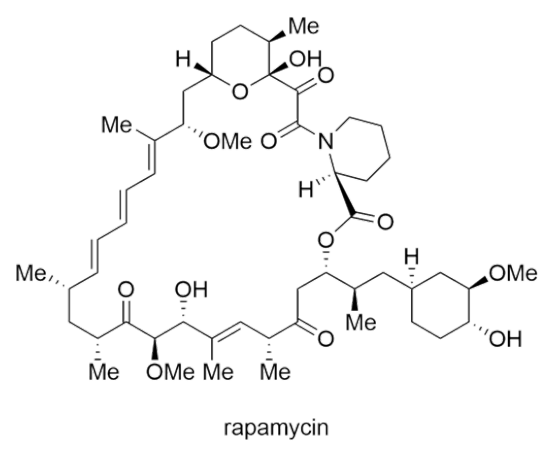
Rapamycin was isolated from the bacterium Streptomyces hygroscopicus in 1972. It acts as a “molecular glue” and inhibits the activity of the protein mTOR through binding to another protein. It has been used to treat various diseases. The stereochemical complexity of rapamycin makes its total synthesis challenging. The figure shows part of the first total synthesis by the Nicolaou group.
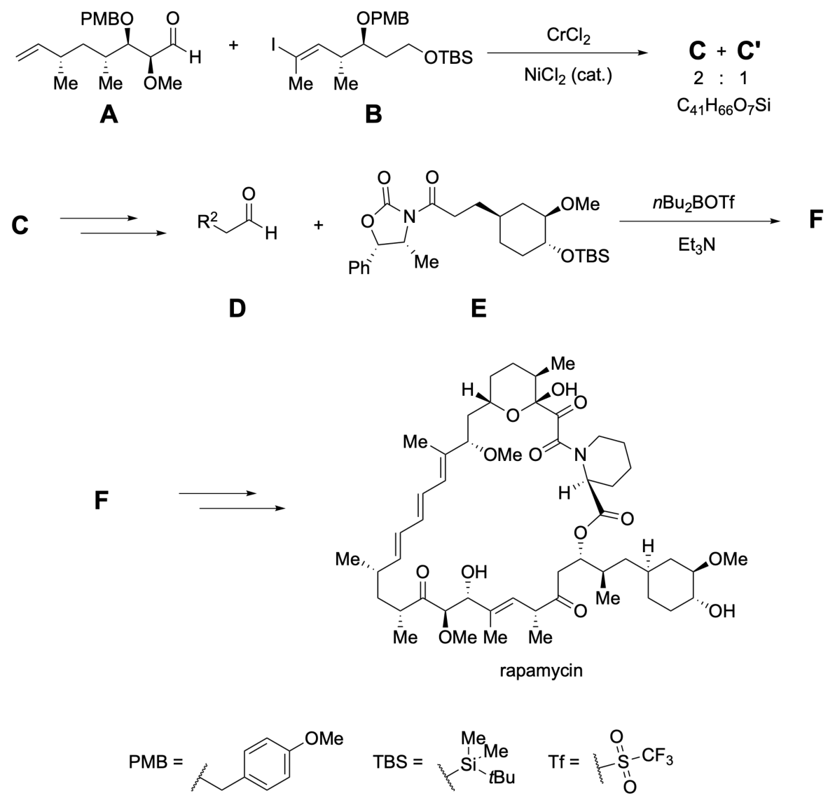
The major stereoisomer (C) is formed from the chromium-mediated coupling reaction of aldehyde A and vinyl iodide B and can be predicted using the Felkin-Anh model without chelation.
2.1a Draw a Newman projection showing the best approach of the nucleophile in this reaction. Use the labels =O, –H, –R\(^{1}\), –OMe, and Nu.
2.1b Draw the full structure of the major diastereomer C showing all stereochemistry.
After a few transformations, C can be converted into an aldehyde D which reacts with E to form F. F can be further transformed into rapamycin.
2.2a Using the Zimmerman-Traxler model, predict the arrangement of the reactants as they come together to form the six-membered ring transition state in the reaction to form F. Draw the substituents at positions labelled with numbers. If a position does not have a substituent, put an “X” in the box. Boxes 1 and 2 have been filled in as an example. You may use the abbreviation R\(^{2}\) as given in the scheme above.
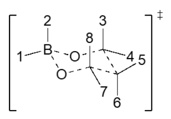
2.2b Draw the structure of F showing all stereochemistry. You may use the abbreviation R\(^{2}\) as given in the scheme above.
Whilst stereochemical control is challenging synthetically, the biological synthesis controls stereochemistry elegantly through an assembly line of various enzymes.
The carbon chain is built up through the addition of monomers. These units are connected via a thioester to Coenzyme A (CoA).
The functions of the various enzymes and proteins are shown in the table.
| Abbreviation | Name | Function |
|---|---|---|
| ACP | Acyl-carrier protein | Carries growing chain between domains |
| CoL | CoA-Ligase | Activates a carboxylic acid substrate to be loaded onto the ACP |
| AT | Acyltransferase | Selects monomer and transfers to ACP |
| KS | Ketosynthase | Accepts growing chain from previous ACP and activates it for Claisen condensation with next monomer-loaded ACP |
| KR | Ketoreductase | Reduces carbonyl of a previous monomer to a hydroxyl |
| DH | Dehydratase | Forms \(\alpha,\beta\)-unsaturated thioester via elimination on the previous monomer |
| ER | Enoylreductase | Reduces \(\alpha,\beta\)-unsaturated thioester to saturated chain on the previous monomer |
The first part of the assembly line is shown with blanks. The enzymes are shown in circles and act in the order of the arrow.
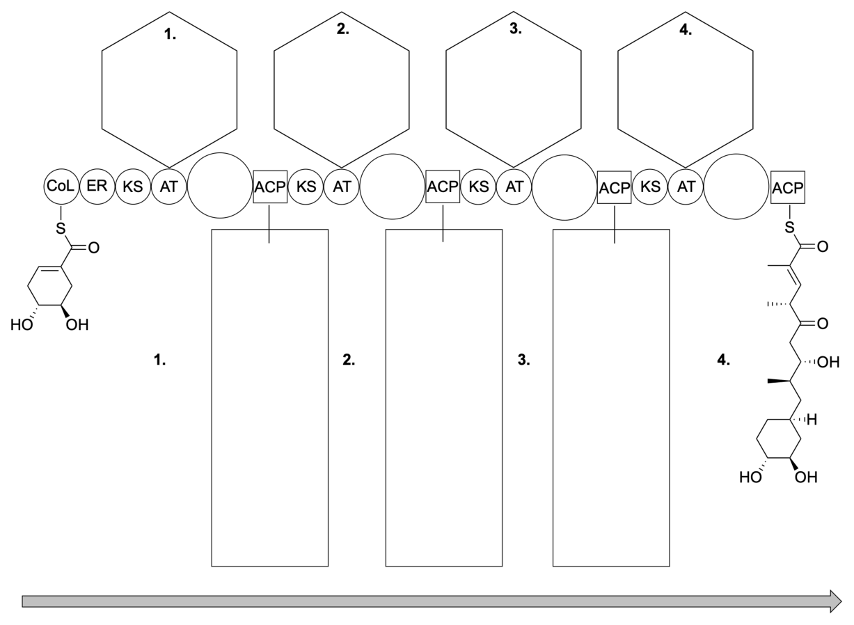
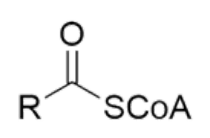
2.3a Draw in the hexagons the structure of the monomer needed by the enzyme AT at each of the points numbered 1-4. Carbon dioxide is released when the monomers are incorporated. The monomers have the following general structure where the R group is anionic:
2.3b Fill in each circle with the abbreviations of enzyme(s) needed. Write “X” if no additional enzyme is needed.
2.3c Complete the rectangles with the structure of the growing molecule attached to the enzyme.
The final steps of the enzymatic synthesis are shown below.
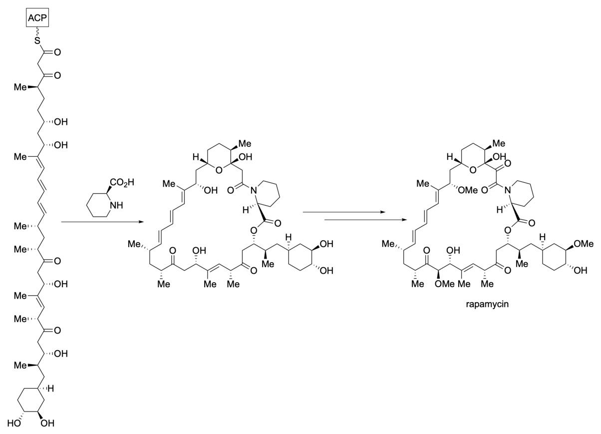
The building blocks involved were determined by feeding the bacterium with three different \(^{13}\)C-labelled starting materials which are incorporated into the monomers (\(^{13}\)C are circled as shown below):

2.4 Circle the carbons in rapamycin that will be \(^{13}\)C-labelled when the bacterium is fed with each of the three starting materials a-c in turn, following the guidelines on the next page. Incorrect circles will be penalised but there is no negative scoring.
Guidelines for circling carbon atoms:
- Each circle should clearly surround one carbon atom
- Do not include multiple carbon atoms in the same circle
- Do not circle any bonds
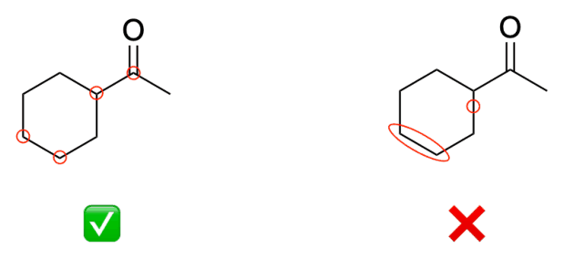
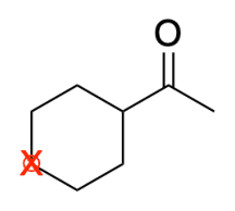
- If you would like to change your answer and cross-out a circle, put an “X” over the circle.
Official English text of the problem.
Answers
In the SMILES below, [*:2] represents the permitted abbreviation \(R^2\). The macromolecular abbreviations are S[*:90] for \(\ce{SCoA}\) and S[*:91] for \(\ce{SACP}\). Other asterisks mark a fragment’s attachment point.
-
2.1a. View from the aldehyde carbon towards its adjacent stereogenic carbon. Put the front \(=\mathrm O\) at 9 o’clock and front H at 3 o’clock. On the rear carbon put OMe at 6 o’clock, \(R^1\) at 10 o’clock, and H at 2 o’clock. Draw Nu approaching the front carbon from above, opposite the rear C–OMe bond, with the usual Bürgi–Dunitz approach.
-
2.1b. The major Felkin–Anh coupling product C has the \(R\) configuration at the newly formed alcohol centre:
C=C[C@@H](C)C[C@@H](C)[C@@H](OCc1ccc(OC)cc1)[C@H](OC)[C@H](O)/C(C)=C/[C@@H](C)[C@@H](OCc1ccc(OC)cc1)CCO[Si](C)(C)C(C)(C)C
- 2.2a. Fill the supplied chair as follows. The auxiliary is attached through its nitrogen; \(R^3\) is the protected cyclohexylmethyl group defined in the solution.
| Position | Entry |
|---|---|
| 1 | \(n\)-Bu |
| 2 | \(n\)-Bu |
| 3 | The \(N\)-attached \((4R,5S)\)-4-methyl-5-phenyloxazolidin-2-one auxiliary |
| 4 | X |
| 5 | H |
| 6 | \(R^3=\ce{CH2}\)–protected cyclohexyl |
| 7 | \(\ce{CH2}R^2\) |
| 8 | H |
- 2.2b. F is the \((2R,3S)\) syn aldol adduct, numbering its acyl chain from the auxiliary-bound carbonyl:
[*:2]C[C@H](O)[C@@H](C[C@H]1C[C@@H](OC)[C@H](O[Si](C)(C)C(C)(C)C)CC1)C(=O)N1[C@H](C)[C@H](c2ccccc2)OC1=O
- 2.3a. The monomers are:
| Hexagon | Monomer | SMILES |
|---|---|---|
| 1 | Methylmalonyl-CoA carboxylate | C[C@@H](C(=O)[O-])C(=O)S[*:90] |
| 2 | Malonyl-CoA carboxylate | O=C([O-])CC(=O)S[*:90] |
| 3 | Methylmalonyl-CoA carboxylate | C[C@@H](C(=O)[O-])C(=O)S[*:90] |
| 4 | Methylmalonyl-CoA carboxylate | C[C@@H](C(=O)[O-])C(=O)S[*:90] |
The methylmalonyl SMILES specifies the conventional \((2S)\) substrate; the problem itself does not give the extender's absolute configuration.
-
2.3b. The circles, in order, contain KR, DH, ER; KR; X; KR, DH.
-
2.3c. The three ACP-bound intermediates are:
Rectangle 1 — first extension product:
[*:91]SC(=O)[C@H](C)C[C@H]1C[C@@H](O)[C@H](O)CC1
Rectangle 2 — second extension product:
[*:91]SC(=O)C[C@H](O)[C@H](C)C[C@H]1C[C@@H](O)[C@H](O)CC1
Rectangle 3 — third extension product:
[*:91]SC(=O)[C@@H](C)C(=O)C[C@H](O)[C@H](C)C[C@H]1C[C@@H](O)[C@H](O)CC1
- 2.4. Define \(P_1\) as the polyketide-derived amide carbonyl bonded to the pipecolate nitrogen. Number consecutively along the carbon backbone to \(P_{29}\), the methylene attached to the cyclohexyl ring. An atom-mapped SMILES specifying this numbering appears below.
| Feed | Carbons to circle individually | Number of circles |
|---|---|---|
| a | \(P_4,P_{10},P_{16},P_{18},P_{22},P_{24},P_{28}\), and the methyl carbon attached to each of these seven atoms | 14 |
| b | \(P_1,P_5,P_7,P_{11},P_{13},P_{19},P_{25}\) | 7 |
| c | \(P_2,P_6,P_8,P_{12},P_{14},P_{20},P_{26}\) | 7 |
Solution
2.1a — Felkin–Anh projection
The protected \(\alpha\)-methoxy aldehyde A is:
C=C[C@@H](C)C[C@@H](C)[C@@H](OCc1ccc(OC)cc1)[C@H](OC)C=O
Its carbon adjacent to the aldehyde has the \(S\) configuration. Here \(R^1\) denotes the rest of the carbon chain attached to that centre.
For the non-chelated, polar Felkin–Anh model, orient the C–OMe bond approximately perpendicular to the carbonyl plane. Nucleophilic attack occurs opposite this bond, along a Bürgi–Dunitz trajectory, with a nucleophile–carbonyl-carbon–oxygen angle of approximately \(107^\circ\).
A precise Newman specification is:
| Component | Position when looking from \(\ce{CHO}\) towards the adjacent carbon |
|---|---|
| Front carbonyl oxygen | 9 o’clock |
| Front aldehydic hydrogen | 3 o’clock |
| Rear OMe | 6 o’clock |
| Rear \(R^1\) | 10 o’clock |
| Rear hydrogen | 2 o’clock |
| Incoming Nu | Approaches from above the front carbon, opposite the rear OMe |
The projection fixes both the substrate’s handedness and the face of attack. Ordinary SMILES cannot specify this approach geometry; the substrate and product SMILES specify the corresponding configurations.
2.1b — Structure of C
The protected vinyl iodide B is:
I/C(C)=C/[C@@H](C)[C@@H](OCc1ccc(OC)cc1)CCO[Si](C)(C)C(C)(C)C
Chromium-mediated activation replaces the vinyl C–I bond by a nucleophilic carbon–chromium bond. That vinyl carbon attacks the aldehyde carbon of A. Protonation of the resulting alkoxide gives C.
The formal, atom- and charge-balanced reductive coupling is
\[ \ce{RCHO + R'I + 2e- + H+ -> RCH(OH)R' + I-}. \]
Here \(\ce{RCHO}\) represents A, and \(\ce{R'I}\) represents B. The proton is [H+], and the iodide ion is [I-]. This equation expresses the net redox balance without assigning particular chromium coordination species.
The resulting connectivity is
\[ \begin{aligned} &\ce{CH2=CH-CH(Me)-CH2-CH(Me)-CH(OPMB)-CH(OMe)}\\ &\qquad\ce{-CH(OH)-C(Me)=CH-CH(Me)-CH(OPMB)-CH2-CH2-OTBS}. \end{aligned} \]
All pre-existing stereogenic centres retain their spatial configurations, and the vinyl geometry is retained. The new alcohol centre is \(R\), as specified by the complete SMILES in Answers.
To draw it with the left-hand portion of A kept in its original zigzag orientation, continue the chain through the former aldehyde carbon and draw its new OH bond hashed and upward. The adjacent OMe bond remains solid-wedged and downward.
The completed structure has molecular formula \(\ce{C41H66O7Si}\), matching the formula supplied in the scheme.
2.2a — Zimmerman–Traxler transition state
The aldehyde D is \(R^2\)-substituted ethanal:
[*:2]CC=O
The chiral \(N\)-acyl oxazolidinone E is:
O=C(N1[C@H](C)[C@H](c2ccccc2)OC1=O)CC[C@H]1C[C@@H](OC)[C@H](O[Si](C)(C)C(C)(C)C)CC1
Define the two substituents used in the chair:
| Symbol or description | Name and attachment-point SMILES |
|---|---|
| Auxiliary at position 3 | \(N\)-attached \((4R,5S)\)-4-methyl-5-phenyloxazolidin-2-one: *N1[C@H](C)[C@H](c2ccccc2)OC1=O |
| \(R^3\), at position 6 | Protected cyclohexylmethyl group: *C[C@H]1C[C@@H](OC)[C@H](O[Si](C)(C)C(C)(C)C)CC1 |
| \(n\)-Bu, at positions 1 and 2 | Butan-1-yl group: *CCCC |
The boron reagent and base produce the \(Z\) boron enolate of E. Its constitution and enolate geometry can be represented as:
CCCCB(CCCC)O/C(N1[C@H](C)[C@H](c2ccccc2)OC1=O)=C\C[C@H]1C[C@@H](OC)[C@H](O[Si](C)(C)C(C)(C)C)CC1
The aldehyde oxygen coordinates to boron, and the new carbon–carbon bond forms through a six-membered chair-like transition state. In the particular perspective printed in the question, the ring atoms run in this order:
\[ \mathrm{B} -\mathrm{O}_{E} -\mathrm{C}_{E} -\mathrm{C}_{\alpha} -\mathrm{C}_{D} -\mathrm{O}_{D} -\mathrm{B}. \]
Consequently:
- The upper-right carbon bearing positions 3/4 is the enolate carbonyl-derived carbon.
- The right-hand carbon bearing 5/6 is the enolate \(\alpha\)-carbon.
- The lower-middle carbon bearing 7/8 is the aldehyde carbon.
The entries therefore are:
| Position | Substituent | Reason |
|---|---|---|
| 1 | \(n\)-Bu | First alkyl ligand on boron |
| 2 | \(n\)-Bu | Second alkyl ligand on boron |
| 3 | \(N\)-attached chiral auxiliary | Substituent on the enolate carbonyl-derived carbon |
| 4 | X | No fourth substituent at that carbon |
| 5 | H | Hydrogen on the enolate \(\alpha\)-carbon |
| 6 | \(R^3\) | Original side chain of E |
| 7 | \(\ce{CH2}R^2\) | Aldehyde substituent, in the favourable equatorial position |
| 8 | H | Aldehydic hydrogen |
This assignment gives the \(2R,3S\) product. The identification of positions 3/4 as the enolate carbon and 7/8 as the aldehyde carbon is essential to preserving the handedness of the supplied chair.
2.2b — Structure of F
The aldol addition gives the connectivity
\[ \ce{R^2CH2-CH(OH)-CH(R^3)-C(=O)-N(Aux)}. \]
No carbon atoms are lost. In generic form, the balanced aldol addition is
\[ \ce{RCHO + R'CH2C(=O)N(Aux) -> RCH(OH)CH(R')C(=O)N(Aux)}. \]
The \(Z\) enolate gives the syn relationship between the new OH group and \(R^3\). The auxiliary selects the face that gives \(2R,3S\), counting the auxiliary-bound carbonyl carbon as C1.
For a drawing, place the chain
\[ \ce{C(=O)-CH(R^3)-CH(OH)-CH2R^2} \]
as a conventional zigzag running left to right. Draw both \(R^3\) and OH with solid wedges; their corresponding hydrogens point behind the plane. Retain the auxiliary’s \((4R,5S)\) configuration and all three cyclohexyl stereocentres. This drawing corresponds to the SMILES for F in Answers.
2.3a — Identification of the monomers
Let \(Q\) denote the cyclohexyl group with the ring stereochemistry shown in the supplied product. Reading the four-module product from the starter end towards ACP gives
\[ \ce{ Q-CH2-CH(Me)-CH(OH)-CH2-C(=O)-CH(Me)-CH=C(Me)-C(=O)-SACP }. \]
Every extension contributes two backbone carbons. A methylmalonyl unit also supplies a methyl branch on its newly introduced \(\alpha\)-carbon.
The successive extensions therefore require:
| Module | Substitution of the newly introduced \(\alpha\)-carbon | Required extender |
|---|---|---|
| 1 | Methyl-substituted | Methylmalonyl-CoA |
| 2 | Unsubstituted | Malonyl-CoA |
| 3 | Methyl-substituted | Methylmalonyl-CoA |
| 4 | Methyl-substituted | Methylmalonyl-CoA |
The generic decarboxylative chain extension, written after loading the extender onto its ACP, is
\[ \ce{ R-C(=O)-SACP + ^-OOC-CH(R')-C(=O)-SACP' + H+ -> R-C(=O)-CH(R')-C(=O)-SACP' + CO2 + HSACP }. \]
Here \(R'=\ce{H}\) for malonyl and \(R'=\ce{Me}\) for methylmalonyl. Carbon dioxide is O=C=O. The released carbon is the extender’s free carboxylate carbon.
As a carbon-count check, the seven-carbon starter plus these four extensions gives
\[ 7+3+2+3+3=18 \]
carbons in the displayed four-module acyl chain, including its cyclohexyl ring and methyl branches.
2.3b — Identification of the enzymes
The processing domains act on the carbonyl that becomes the \(\beta\)-carbonyl after the current extension.
| Module | Fate required for that carbonyl | Enzymes, in order |
|---|---|---|
| 1 | Ketone \(\rightarrow\) alcohol \(\rightarrow\) alkene \(\rightarrow\) methylene | KR, DH, ER |
| 2 | Ketone \(\rightarrow\) alcohol | KR |
| 3 | Carbonyl remains a ketone | X |
| 4 | Ketone \(\rightarrow\) alcohol \(\rightarrow\) alkene | KR, DH |
Their net transformations can be written as balanced reduction half-reactions and dehydration:
\[ \begin{aligned} \text{KR:}\quad& \ce{RCOCH(R')COSACP + 2H+ + 2e- -> RCH(OH)CH(R')COSACP} \\[2mm] \text{DH:}\quad& \ce{RCH(OH)CH(R')COSACP -> RCH=C(R')COSACP + H2O} \\[2mm] \text{ER:}\quad& \ce{RCH=C(R')COSACP + 2H+ + 2e- -> RCH2CH(R')COSACP}. \end{aligned} \]
Water has SMILES O. These equations show the reducing-equivalent balance without introducing cofactors absent from the question.
2.3c — The growing ACP-bound chains
The initially supplied ER saturates the starter’s ring double bond. The completed rectangles must then show:
| Rectangle | Chain, written from the cyclohexyl end towards ACP | Configurations of acyl-chain stereocentres, numbered from the thioester carbonyl |
|---|---|---|
| 1 | \(\ce{Q-CH2-CH(Me)-C(=O)-SACP}\) | \(2R\) |
| 2 | \(\ce{Q-CH2-CH(Me)-CH(OH)-CH2-C(=O)-SACP}\) | \(3S,4R\) |
| 3 | \(\ce{Q-CH2-CH(Me)-CH(OH)-CH2-C(=O)-CH(Me)-C(=O)-SACP}\) | \(2S,5S,6R\) |
In all three, the cyclohexyl group has the \((1S,3R,4R)\) configuration for the substituted ring as numbered in these products. The complete stereochemical SMILES are given in Answers.
For a drawing matching the vertical orientation of the supplied final chain:
- Retain the ring’s dashed H, dashed OH and solid-wedged OH.
- Draw the methyl group introduced by module 1 on a solid wedge.
- Draw the alcohol generated by module 2 with a hashed bond.
- Draw the methyl group introduced by module 3 with a hashed bond.
After module 4, the resulting ACP-bound \(\alpha,\beta\)-unsaturated thioester is:
[*:91]SC(=O)/C(C)=C/[C@@H](C)C(=O)C[C@H](O)[C@H](C)C[C@H]1C[C@@H](O)[C@H](O)CC1
Its \(E\) double bond, retained ketone, alcohol and methyl branches reproduce the product printed at the right of the assembly line.
2.4 — Carbon tracing
Use the following explicit numbering of rapamycin:
- \(P_1\): the amide carbonyl directly bonded to the pipecolate nitrogen.
- \(P_2\): the adjacent ketone carbonyl.
- \(P_3\): the OH-bearing hemiketal carbon.
- Continue along the carbon backbone through the six-membered oxygen-containing ring, the triene and the lower portion of the macrocycle.
- \(P_{27}\): the carbon whose oxygen forms the macrocyclic ester.
- \(P_{28}\): the adjacent methyl-bearing side-chain carbon.
- \(P_{29}\): the methylene directly attached to the cyclohexyl ring.
For complete positional identification, rapamycin has the following atom-mapped SMILES. Maps 1–29 are \(P_1\)–\(P_{29}\); map \(100+i\) denotes the methyl attached to \(P_i\):
[C:1]1(=O)[C:2](=O)[C@@:3]2(O)[C@H:4]([CH3:104])[CH2:5][CH2:6][C@H:7](O2)[CH2:8][C@H:9](O[CH3:209])/[C:10]([CH3:110])=[CH:11]/[CH:12]=[CH:13]/[CH:14]=[CH:15]/[C@@H:16]([CH3:116])[CH2:17][C@@H:18]([CH3:118])[C:19](=O)[C@H:20](O[CH3:220])[C@H:21](O)/[C:22]([CH3:122])=[CH:23]/[C@@H:24]([CH3:124])[C:25](=O)[CH2:26][C@@H:27]([C@H:28]([CH3:128])[CH2:29][C@H:30]2[CH2:31][C@@H:32](O[CH3:232])[C@H:33](O)[CH2:34][CH2:35]2)O[C:41](=O)[C@@H:36]2[CH2:37][CH2:38][CH2:39][CH2:40]N12
The formula of this structure is \(\ce{C51H79NO13}\).
The fourteen extender units supply consecutive carbon pairs. Each pair below is written as (original acyl carbonyl carbon, original \(\alpha\)-carbon):
| Acetate-derived pairs | Propionate-derived pairs |
|---|---|
| \((P_1,P_2)\) | \((P_3,P_4)\) |
| \((P_5,P_6)\) | \((P_9,P_{10})\) |
| \((P_7,P_8)\) | \((P_{15},P_{16})\) |
| \((P_{11},P_{12})\) | \((P_{17},P_{18})\) |
| \((P_{13},P_{14})\) | \((P_{21},P_{22})\) |
| \((P_{19},P_{20})\) | \((P_{23},P_{24})\) |
| \((P_{25},P_{26})\) | \((P_{27},P_{28})\) |
Carboxylation converts the acetate- and propionate-derived acyl units into malonyl and methylmalonyl extenders. The newly added carboxyl carbon is subsequently lost as \(\ce{CO2}\); the original acyl carbonyl carbon remains in the product.
Subsequent reduction, dehydration, cyclization, oxidation and methylation alter functional groups but preserve the identities of the backbone carbons.
The carbon count closes as
\[ \underbrace{7(2)}_{\text{acetate units}} +\underbrace{7(3)}_{\text{propionate units}} +\underbrace{7}_{\text{starter}} +\underbrace{6}_{\text{pipecolate}} +\underbrace{3}_{\text{O-methyl groups}} =51. \]
Feed a: sodium [2,3-\(^{13}\)C\(_2\)]propionate
Starting-material SMILES:
[13CH3][13CH2]C(=O)[O-].[Na+]
The labelled methylene carbon becomes the methyl-bearing backbone carbon, and the labelled terminal methyl remains its methyl branch. Therefore circle
\[ P_4,\ P_{10},\ P_{16},\ P_{18},\ P_{22},\ P_{24},\ P_{28}, \]
and the methyl attached to each one: 14 separate circles.
The corresponding labelled rapamycin is:
C1(=O)C(=O)[C@@]2(O)[13C@H]([13CH3])CC[C@H](O2)C[C@H](OC)/[13C]([13CH3])=C/C=C/C=C/[13C@@H]([13CH3])C[13C@@H]([13CH3])C(=O)[C@H](OC)[C@H](O)/[13C]([13CH3])=C/[13C@@H]([13CH3])C(=O)C[C@@H]([13C@H]([13CH3])C[C@H]2C[C@@H](OC)[C@H](O)CC2)OC(=O)[C@@H]2CCCCN12
Feed b: sodium [1-\(^{13}\)C]acetate
Starting-material SMILES:
C[13C](=O)[O-].[Na+]
The labelled carboxyl carbon becomes the first member of each acetate-derived pair. Therefore circle
\[ \boxed{P_1,\ P_5,\ P_7,\ P_{11},\ P_{13},\ P_{19},\ P_{25}} \]
for 7 separate circles.
The corresponding labelled rapamycin is:
[13C]1(=O)C(=O)[C@@]2(O)[C@H](C)[13CH2]C[13C@H](O2)C[C@H](OC)/C(C)=[13CH]/C=[13CH]/C=C/[C@@H](C)C[C@@H](C)[13C](=O)[C@H](OC)[C@H](O)/C(C)=C/[C@@H](C)[13C](=O)C[C@@H]([C@H](C)C[C@H]2C[C@@H](OC)[C@H](O)CC2)OC(=O)[C@@H]2CCCCN12
Feed c: sodium [2-\(^{13}\)C]acetate
Starting-material SMILES:
[13CH3]C(=O)[O-].[Na+]
The labelled methyl carbon becomes the second member of each acetate-derived pair. Therefore circle
\[ \boxed{P_2,\ P_6,\ P_8,\ P_{12},\ P_{14},\ P_{20},\ P_{26}} \]
for 7 separate circles.
The corresponding labelled rapamycin is:
C1(=O)[13C](=O)[C@@]2(O)[C@H](C)C[13CH2][C@H](O2)[13CH2][C@H](OC)/C(C)=C/[13CH]=C/[13CH]=C/[C@@H](C)C[C@@H](C)C(=O)[13C@H](OC)[C@H](O)/C(C)=C/[C@@H](C)C(=O)[13CH2][C@@H]([C@H](C)C[C@H]2C[C@@H](OC)[C@H](O)CC2)OC(=O)[C@@H]2CCCCN12
Under the building-block incorporation model in the question, the starter-derived carbons, pipecolate carbons and three O-methyl carbons receive none of these labels.
The answers were checked against the official answers published by the organisers.
IChO 2025 · Theorieaufgabe 2
Rapamycin - Ein molekularer Klebstoff
6.5% der Gesamtpunktzahl
| 2.1 | 2.2 | 2.3 | 2.4 | Gesamt |
|---|---|---|---|---|
| 12 | 19 | 18 | 18 | 67 |
Rapamycin wurde 1972 aus dem Bakterium Streptomyces hygroscopicus isoliert. Es wirkt als „molekularer Klebstoff“ und hemmt die Aktivität des Proteins mTOR durch Bindung an ein anderes Protein. Es wird zur Behandlung verschiedener Krankheiten eingesetzt. Die stereochemische Komplexität von Rapamycin macht seine Totalsynthese zu einer Herausforderung. Die Abbildung zeigt einen Teil der ersten Totalsynthese durch die Arbeitsgruppe von Nicolaou.
Das Hauptstereoisomer (C) entsteht bei der chromvermittelten Kupplungsreaktion des Aldehyds A mit dem Vinyliodid B und lässt sich mithilfe des Felkin-Anh-Modells ohne Chelatbildung vorhersagen.
2.1a Zeichnen Sie eine Newman-Projektion, die die günstigste Annäherung des Nukleophils bei dieser Reaktion zeigt. Verwenden Sie die Bezeichnungen =O, –H, –R\(^{1}\), –OMe und Nu.
2.1b Zeichnen Sie die vollständige Struktur des Hauptdiastereomers C unter Angabe der gesamten Stereochemie.
Nach einigen Umwandlungen lässt sich C in einen Aldehyd D überführen, der mit E zu F reagiert. F kann anschließend weiter zu Rapamycin umgesetzt werden.
2.2a Sagen Sie mithilfe des Zimmerman-Traxler-Modells die Anordnung der Reaktanden voraus, wenn sie bei der Reaktion zu F zum sechsgliedrigen Übergangszustand zusammentreten. Zeichnen Sie die Substituenten an den mit Zahlen gekennzeichneten Positionen ein. Wenn eine Position keinen Substituenten trägt, setzen Sie ein „X“ in das Kästchen. Die Kästchen 1 und 2 sind als Beispiel bereits ausgefüllt. Sie dürfen die im obigen Schema angegebene Abkürzung R\(^{2}\) verwenden.
2.2b Zeichnen Sie die Struktur von F unter Angabe der gesamten Stereochemie. Sie dürfen die im obigen Schema angegebene Abkürzung R\(^{2}\) verwenden.
Während die stereochemische Kontrolle bei der chemischen Synthese eine Herausforderung darstellt, steuert die biologische Synthese die Stereochemie auf elegante Weise durch eine Reihe verschiedener Enzyme, die wie ein Fließband zusammenarbeiten.
Die Kohlenstoffkette wird durch die Addition von Monomeren aufgebaut. Diese Einheiten sind über einen Thioester an Coenzym A (CoA) gebunden.
Die Funktionen der verschiedenen Enzyme und Proteine sind in der Tabelle aufgeführt.
| Abkürzung | Name | Funktion |
|---|---|---|
| ACP | Acyl-Carrier-Protein | Transportiert die wachsende Kette zwischen den Domänen |
| CoL | CoA-Ligase | Aktiviert ein Carbonsäuresubstrat für die Beladung des ACP |
| AT | Acyltransferase | Wählt das Monomer aus und überträgt es auf ACP |
| KS | Ketosynthase | Übernimmt die wachsende Kette vom vorherigen ACP und aktiviert sie für die Claisen-Kondensation mit dem nächsten monomerbeladenen ACP |
| KR | Ketoreduktase | Reduziert die Carbonylgruppe eines vorherigen Monomers zu einer Hydroxygruppe |
| DH | Dehydratase | Bildet durch Eliminierung am vorherigen Monomer einen \(\alpha,\beta\)-ungesättigten Thioester |
| ER | Enoylreduktase | Reduziert den \(\alpha,\beta\)-ungesättigten Thioester am vorherigen Monomer zu einer gesättigten Kette |
Der erste Teil des Fließbands ist mit Leerstellen dargestellt. Die Enzyme sind in Kreisen dargestellt und wirken in Pfeilrichtung nacheinander.
2.3a Zeichnen Sie in die Sechsecke die Struktur des Monomers ein, das das Enzym AT an jeder der mit 1-4 nummerierten Stellen benötigt. Beim Einbau der Monomere wird Kohlenstoffdioxid freigesetzt. Die Monomere haben die folgende allgemeine Struktur, in der die R-Gruppe anionisch ist:
2.3b Tragen Sie in jeden Kreis die Abkürzungen der benötigten Enzyme ein. Schreiben Sie „X“, wenn kein zusätzliches Enzym benötigt wird.
2.3c Ergänzen Sie die Rechtecke um die Struktur des an das Enzym gebundenen wachsenden Moleküls.
Die letzten Schritte der enzymatischen Synthese sind unten dargestellt.
Die beteiligten Bausteine wurden bestimmt, indem das Bakterium mit drei verschiedenen \(^{13}\)C-markierten Ausgangsstoffen gefüttert wurde, die in die Monomere eingebaut werden (\(^{13}\)C sind wie unten gezeigt eingekreist):
2.4 Kreisen Sie die Kohlenstoffatome in Rapamycin ein, die \(^{13}\)C-markiert werden, wenn das Bakterium jeweils mit einem der drei Ausgangsstoffe a-c gefüttert wird. Beachten Sie dabei die Hinweise auf der nächsten Seite. Für falsch gesetzte Kreise werden Punkte abgezogen, die Punktzahl kann jedoch nicht negativ werden.
Hinweise zum Einkreisen von Kohlenstoffatomen:
- Jeder Kreis sollte eindeutig ein Kohlenstoffatom umschließen
- Schließen Sie nicht mehrere Kohlenstoffatome in denselben Kreis ein
- Kreisen Sie keine Bindungen ein
- Wenn Sie Ihre Antwort ändern und einen Kreis streichen möchten, setzen Sie ein „X“ über den Kreis.
Aufgabentext aus dem offiziellen englischen Text übersetzt.
Antworten
In den folgenden SMILES steht [*:2] für die zulässige Abkürzung \(R^2\). Die makromolekularen Abkürzungen sind S[*:90] für \(\ce{SCoA}\) und S[*:91] für \(\ce{SACP}\). Andere Sternchen kennzeichnen die Verknüpfungsstelle eines Fragments.
-
2.1a. Blicken Sie vom Aldehydkohlenstoff zum benachbarten stereogenen Kohlenstoffatom. Platzieren Sie das vordere \(=\mathrm O\) auf 9 Uhr und das vordere H auf 3 Uhr. Platzieren Sie am hinteren Kohlenstoffatom OMe auf 6 Uhr, \(R^1\) auf 10 Uhr und H auf 2 Uhr. Zeichnen Sie Nu so ein, dass es sich dem vorderen Kohlenstoffatom von oben, gegenüber der hinteren C–OMe-Bindung, entlang der üblichen Bürgi–Dunitz-Trajektorie nähert.
-
2.1b. Das Hauptprodukt C der Felkin–Anh-Kupplung besitzt am neu gebildeten Alkoholzentrum die \(R\)-Konfiguration:
C=C[C@@H](C)C[C@@H](C)[C@@H](OCc1ccc(OC)cc1)[C@H](OC)[C@H](O)/C(C)=C/[C@@H](C)[C@@H](OCc1ccc(OC)cc1)CCO[Si](C)(C)C(C)(C)C
- 2.2a. Füllen Sie die vorgegebene Sesselform wie folgt aus. Das Auxiliar ist über sein Stickstoffatom gebunden; \(R^3\) ist die in der Lösung definierte geschützte Cyclohexylmethylgruppe.
| Position | Eintrag |
|---|---|
| 1 | \(n\)-Bu |
| 2 | \(n\)-Bu |
| 3 | Das über \(N\) gebundene \((4R,5S)\)-4-Methyl-5-phenyloxazolidin-2-on-Auxiliar |
| 4 | X |
| 5 | H |
| 6 | \(R^3=\ce{CH2}\)–geschütztes Cyclohexyl |
| 7 | \(\ce{CH2}R^2\) |
| 8 | H |
- 2.2b. F ist das \((2R,3S)\)-syn-Aldoladdukt, wobei seine Acylkette ausgehend von der an das Auxiliar gebundenen Carbonylgruppe nummeriert wird:
[*:2]C[C@H](O)[C@@H](C[C@H]1C[C@@H](OC)[C@H](O[Si](C)(C)C(C)(C)C)CC1)C(=O)N1[C@H](C)[C@H](c2ccccc2)OC1=O
- 2.3a. Die Monomere sind:
| Sechseck | Monomer | SMILES |
|---|---|---|
| 1 | Methylmalonyl-CoA-Carboxylat | C[C@@H](C(=O)[O-])C(=O)S[*:90] |
| 2 | Malonyl-CoA-Carboxylat | O=C([O-])CC(=O)S[*:90] |
| 3 | Methylmalonyl-CoA-Carboxylat | C[C@@H](C(=O)[O-])C(=O)S[*:90] |
| 4 | Methylmalonyl-CoA-Carboxylat | C[C@@H](C(=O)[O-])C(=O)S[*:90] |
Der Methylmalonyl-SMILES gibt das übliche \((2S)\)-Substrat an; die Aufgabe selbst gibt die absolute Konfiguration des Kettenverlängerungsbausteins nicht vor.
-
2.3b. Die Kreise enthalten der Reihe nach KR, DH, ER; KR; X; KR, DH.
-
2.3c. Die drei ACP-gebundenen Zwischenprodukte sind:
Rechteck 1 — erstes Kettenverlängerungsprodukt:
[*:91]SC(=O)[C@H](C)C[C@H]1C[C@@H](O)[C@H](O)CC1
Rechteck 2 — zweites Kettenverlängerungsprodukt:
[*:91]SC(=O)C[C@H](O)[C@H](C)C[C@H]1C[C@@H](O)[C@H](O)CC1
Rechteck 3 — drittes Kettenverlängerungsprodukt:
[*:91]SC(=O)[C@@H](C)C(=O)C[C@H](O)[C@H](C)C[C@H]1C[C@@H](O)[C@H](O)CC1
- 2.4. Definieren Sie \(P_1\) als die aus dem Polyketid stammende Amidcarbonylgruppe, die an den Pipecolat-Stickstoff gebunden ist. Nummerieren Sie entlang des Kohlenstoffgerüsts fortlaufend bis \(P_{29}\), der an den Cyclohexylring gebundenen Methylengruppe. Ein SMILES mit Atomzuordnungsnummern, der diese Nummerierung festlegt, ist weiter unten angegeben.
| Zugeführter Ausgangsstoff | Einzeln einzukreisende Kohlenstoffatome | Anzahl der Kreise |
|---|---|---|
| a | \(P_4,P_{10},P_{16},P_{18},P_{22},P_{24},P_{28}\), sowie das an jedes dieser sieben Atome gebundene Methylkohlenstoffatom | 14 |
| b | \(P_1,P_5,P_7,P_{11},P_{13},P_{19},P_{25}\) | 7 |
| c | \(P_2,P_6,P_8,P_{12},P_{14},P_{20},P_{26}\) | 7 |
Lösung
2.1a — Felkin–Anh-Projektion
Der geschützte \(\alpha\)-Methoxyaldehyd A ist:
C=C[C@@H](C)C[C@@H](C)[C@@H](OCc1ccc(OC)cc1)[C@H](OC)C=O
Sein zur Aldehydgruppe benachbartes Kohlenstoffatom besitzt die \(S\)-Konfiguration. Hier bezeichnet \(R^1\) den übrigen Teil der an dieses Zentrum gebundenen Kohlenstoffkette.
Für das polare Felkin–Anh-Modell ohne Chelatbildung wird die C–OMe-Bindung annähernd senkrecht zur Carbonylebene ausgerichtet. Der nukleophile Angriff erfolgt gegenüber dieser Bindung entlang einer Bürgi–Dunitz-Trajektorie mit einem Winkel Nukleophil–Carbonylkohlenstoff–Sauerstoff von ungefähr \(107^\circ\).
Eine präzise Beschreibung der Newman-Projektion lautet:
| Bestandteil | Position beim Blick von \(\ce{CHO}\) zum benachbarten Kohlenstoffatom |
|---|---|
| Vorderer Carbonylsauerstoff | 9 Uhr |
| Vorderer Aldehydwasserstoff | 3 Uhr |
| Hinteres OMe | 6 Uhr |
| Hinteres \(R^1\) | 10 Uhr |
| Hinterer Wasserstoff | 2 Uhr |
| Angreifendes Nu | Nähert sich von oberhalb des vorderen Kohlenstoffatoms, gegenüber dem hinteren OMe |
Die Projektion legt sowohl die Händigkeit des Substrats als auch die Angriffsseite fest. Gewöhnliche SMILES können diese Annäherungsgeometrie nicht angeben; die SMILES von Substrat und Produkt legen die entsprechenden Konfigurationen fest.
2.1b — Struktur von C
Das geschützte Vinyliodid B ist:
I/C(C)=C/[C@@H](C)[C@@H](OCc1ccc(OC)cc1)CCO[Si](C)(C)C(C)(C)C
Die chromvermittelte Aktivierung ersetzt die vinylische C–I-Bindung durch eine nukleophile Kohlenstoff–Chrom-Bindung. Dieses Vinylkohlenstoffatom greift den Aldehydkohlenstoff von A an. Die Protonierung des entstehenden Alkoholats liefert C.
Die formale, atom- und ladungsbilanzierte reduktive Kupplung lautet
\[ \ce{RCHO + R'I + 2e- + H+ -> RCH(OH)R' + I-}. \]
Hier steht \(\ce{RCHO}\) für A und \(\ce{R'I}\) für B. Das Proton ist [H+] und das Iodidion [I-]. Diese Gleichung gibt die Nettoredoxbilanz wieder, ohne bestimmte Chrom-Koordinationsspezies festzulegen.
Die resultierende Verknüpfung der Atome ist
\[ \begin{aligned} &\ce{CH2=CH-CH(Me)-CH2-CH(Me)-CH(OPMB)-CH(OMe)}\\ &\qquad\ce{-CH(OH)-C(Me)=CH-CH(Me)-CH(OPMB)-CH2-CH2-OTBS}. \end{aligned} \]
Alle bereits vorhandenen stereogenen Zentren behalten ihre räumlichen Konfigurationen bei, und die Geometrie der Vinylgruppe bleibt erhalten. Das neue Alkoholzentrum ist \(R\)-konfiguriert, wie durch den vollständigen SMILES unter Antworten angegeben.
Um die Struktur unter Beibehaltung der ursprünglichen Zickzackausrichtung des linken Teils von A zu zeichnen, führen Sie die Kette über den ehemaligen Aldehydkohlenstoff fort und zeichnen Sie dessen neue OH-Bindung gestrichelt und nach oben gerichtet. Die benachbarte OMe-Bindung bleibt als ausgefüllter Keil nach unten gerichtet.
Die vervollständigte Struktur hat die Summenformel \(\ce{C41H66O7Si}\), die mit der im Schema angegebenen Formel übereinstimmt.
2.2a — Zimmerman–Traxler-Übergangszustand
Der Aldehyd D ist \(R^2\)-substituiertes Ethanal:
[*:2]CC=O
Das chirale \(N\)-Acyloxazolidinon E ist:
O=C(N1[C@H](C)[C@H](c2ccccc2)OC1=O)CC[C@H]1C[C@@H](OC)[C@H](O[Si](C)(C)C(C)(C)C)CC1
Die beiden in der Sesselform verwendeten Substituenten werden wie folgt definiert:
| Symbol oder Beschreibung | Name und SMILES mit Verknüpfungsstelle |
|---|---|
| Auxiliar an Position 3 | Über \(N\) gebundenes \((4R,5S)\)-4-Methyl-5-phenyloxazolidin-2-on: *N1[C@H](C)[C@H](c2ccccc2)OC1=O |
| \(R^3\), an Position 6 | Geschützte Cyclohexylmethylgruppe: *C[C@H]1C[C@@H](OC)[C@H](O[Si](C)(C)C(C)(C)C)CC1 |
| \(n\)-Bu, an den Positionen 1 und 2 | Butan-1-ylgruppe: *CCCC |
Das Borreagenz und die Base erzeugen das \(Z\)-Borenolat von E. Seine Konstitution und Enolatgeometrie lassen sich wie folgt darstellen:
CCCCB(CCCC)O/C(N1[C@H](C)[C@H](c2ccccc2)OC1=O)=C\C[C@H]1C[C@@H](OC)[C@H](O[Si](C)(C)C(C)(C)C)CC1
Der Aldehydsauerstoff koordiniert an Bor, und die neue Kohlenstoff–Kohlenstoff-Bindung entsteht über einen sechsgliedrigen, sesselförmigen Übergangszustand. In der in der Aufgabe abgebildeten konkreten Perspektive folgen die Ringatome in dieser Reihenfolge aufeinander:
\[ \mathrm{B} -\mathrm{O}_{E} -\mathrm{C}_{E} -\mathrm{C}_{\alpha} -\mathrm{C}_{D} -\mathrm{O}_{D} -\mathrm{B}. \]
Daraus folgt:
- Das Kohlenstoffatom oben rechts mit den Positionen 3/4 ist das aus dem Carbonylkohlenstoff hervorgegangene Kohlenstoffatom des Enolats.
- Das rechte Kohlenstoffatom mit 5/6 ist das \(\alpha\)-Kohlenstoffatom des Enolats.
- Das Kohlenstoffatom unten in der Mitte mit 7/8 ist der Aldehydkohlenstoff.
Die Einträge lauten daher:
| Position | Substituent | Begründung |
|---|---|---|
| 1 | \(n\)-Bu | Erster Alkylligand am Bor |
| 2 | \(n\)-Bu | Zweiter Alkylligand am Bor |
| 3 | Über \(N\) gebundenes chirales Auxiliar | Substituent am aus dem Carbonylkohlenstoff hervorgegangenen Kohlenstoffatom des Enolats |
| 4 | X | Kein vierter Substituent an diesem Kohlenstoffatom |
| 5 | H | Wasserstoff am \(\alpha\)-Kohlenstoffatom des Enolats |
| 6 | \(R^3\) | Ursprüngliche Seitenkette von E |
| 7 | \(\ce{CH2}R^2\) | Aldehydsubstituent in der günstigen äquatorialen Position |
| 8 | H | Aldehydwasserstoff |
Diese Zuordnung ergibt das \(2R,3S\)-Produkt. Die Identifizierung der Positionen 3/4 als Enolatkohlenstoff und 7/8 als Aldehydkohlenstoff ist entscheidend, um die Händigkeit der vorgegebenen Sesselform zu erhalten.
2.2b — Struktur von F
Die Aldoladdition ergibt die Verknüpfung
\[ \ce{R^2CH2-CH(OH)-CH(R^3)-C(=O)-N(Aux)}. \]
Es gehen keine Kohlenstoffatome verloren. In allgemeiner Form lautet die bilanzierte Aldoladdition
\[ \ce{RCHO + R'CH2C(=O)N(Aux) -> RCH(OH)CH(R')C(=O)N(Aux)}. \]
Das \(Z\)-Enolat ergibt die syn-Beziehung zwischen der neuen OH-Gruppe und \(R^3\). Das Auxiliar bestimmt die Angriffsseite, die zu \(2R,3S\) führt, wobei der an das Auxiliar gebundene Carbonylkohlenstoff als C1 gezählt wird.
Ordnen Sie für eine Zeichnung die Kette
\[ \ce{C(=O)-CH(R^3)-CH(OH)-CH2R^2} \]
als üblichen, von links nach rechts verlaufenden Zickzack an. Zeichnen Sie sowohl \(R^3\) als auch OH mit ausgefüllten Keilen; die zugehörigen Wasserstoffatome weisen hinter die Ebene. Behalten Sie die \((4R,5S)\)-Konfiguration des Auxiliars und alle drei Cyclohexyl-Stereozentren bei. Diese Zeichnung entspricht dem SMILES für F unter Antworten.
2.3a — Identifizierung der Monomere
\(Q\) bezeichne die Cyclohexylgruppe mit der im vorgegebenen Produkt dargestellten Ringstereochemie. Liest man das Produkt der vier Module vom Starterende in Richtung ACP, erhält man
\[ \ce{ Q-CH2-CH(Me)-CH(OH)-CH2-C(=O)-CH(Me)-CH=C(Me)-C(=O)-SACP }. \]
Jede Kettenverlängerung trägt zwei Kohlenstoffatome zum Kettengerüst bei. Eine Methylmalonyleinheit liefert außerdem eine Methylverzweigung an ihrem neu eingeführten \(\alpha\)-Kohlenstoffatom.
Die aufeinanderfolgenden Kettenverlängerungen erfordern daher:
| Modul | Substitution des neu eingeführten \(\alpha\)-Kohlenstoffatoms | Erforderlicher Kettenverlängerungsbaustein |
|---|---|---|
| 1 | Methylsubstituiert | Methylmalonyl-CoA |
| 2 | Unsubstituiert | Malonyl-CoA |
| 3 | Methylsubstituiert | Methylmalonyl-CoA |
| 4 | Methylsubstituiert | Methylmalonyl-CoA |
Die allgemeine decarboxylierende Kettenverlängerung, dargestellt nach der Beladung des ACP mit dem Kettenverlängerungsbaustein, lautet
\[ \ce{ R-C(=O)-SACP + ^-OOC-CH(R')-C(=O)-SACP' + H+ -> R-C(=O)-CH(R')-C(=O)-SACP' + CO2 + HSACP }. \]
Hier gilt \(R'=\ce{H}\) für Malonyl und \(R'=\ce{Me}\) für Methylmalonyl. Kohlenstoffdioxid ist O=C=O. Das freigesetzte Kohlenstoffatom ist der freie Carboxylatkohlenstoff des Kettenverlängerungsbausteins.
Zur Kontrolle der Kohlenstoffzahl ergeben der Starter mit sieben Kohlenstoffatomen und diese vier Kettenverlängerungen
\[ 7+3+2+3+3=18 \]
Kohlenstoffatome in der dargestellten Acylkette aus vier Modulen, einschließlich ihres Cyclohexylrings und ihrer Methylverzweigungen.
2.3b — Identifizierung der Enzyme
Die modifizierenden Domänen wirken auf die Carbonylgruppe, die nach der aktuellen Kettenverlängerung zur \(\beta\)-Carbonylgruppe wird.
| Modul | Erforderliche Umwandlung dieser Carbonylgruppe | Enzyme in ihrer Reihenfolge |
|---|---|---|
| 1 | Keton \(\rightarrow\) Alkohol \(\rightarrow\) Alken \(\rightarrow\) Methylengruppe | KR, DH, ER |
| 2 | Keton \(\rightarrow\) Alkohol | KR |
| 3 | Carbonylgruppe bleibt als Keton erhalten | X |
| 4 | Keton \(\rightarrow\) Alkohol \(\rightarrow\) Alken | KR, DH |
Ihre Nettoumwandlungen lassen sich als bilanzierte Reduktionshalbreaktionen und als Dehydratisierung schreiben:
\[ \begin{aligned} \text{KR:}\quad& \ce{RCOCH(R')COSACP + 2H+ + 2e- -> RCH(OH)CH(R')COSACP} \\[2mm] \text{DH:}\quad& \ce{RCH(OH)CH(R')COSACP -> RCH=C(R')COSACP + H2O} \\[2mm] \text{ER:}\quad& \ce{RCH=C(R')COSACP + 2H+ + 2e- -> RCH2CH(R')COSACP}. \end{aligned} \]
Wasser hat den SMILES O. Diese Gleichungen zeigen die Bilanz der Reduktionsäquivalente, ohne in der Aufgabe nicht genannte Cofaktoren einzuführen.
2.3c — Die wachsenden ACP-gebundenen Ketten
Die zu Beginn vorgegebene ER sättigt die Ringdoppelbindung des Starters. Die vervollständigten Rechtecke müssen dann Folgendes zeigen:
| Rechteck | Kette, vom Cyclohexylende in Richtung ACP geschrieben | Konfigurationen der Stereozentren der Acylkette, ausgehend vom Thioestercarbonyl nummeriert |
|---|---|---|
| 1 | \(\ce{Q-CH2-CH(Me)-C(=O)-SACP}\) | \(2R\) |
| 2 | \(\ce{Q-CH2-CH(Me)-CH(OH)-CH2-C(=O)-SACP}\) | \(3S,4R\) |
| 3 | \(\ce{Q-CH2-CH(Me)-CH(OH)-CH2-C(=O)-CH(Me)-C(=O)-SACP}\) | \(2S,5S,6R\) |
In allen drei Fällen besitzt die Cyclohexylgruppe bei der Nummerierung des substituierten Rings in diesen Produkten die \((1S,3R,4R)\)-Konfiguration. Die vollständigen stereochemischen SMILES sind unter Antworten angegeben.
Für eine Zeichnung, die der vertikalen Ausrichtung der vorgegebenen endgültigen Kette entspricht:
- Behalten Sie am Ring die gestrichelte H-Bindung, die gestrichelte OH-Bindung und die OH-Bindung als ausgefüllten Keil bei.
- Zeichnen Sie die durch Modul 1 eingeführte Methylgruppe an einem ausgefüllten Keil.
- Zeichnen Sie den durch Modul 2 erzeugten Alkohol mit einer gestrichelten Bindung.
- Zeichnen Sie die durch Modul 3 eingeführte Methylgruppe mit einer gestrichelten Bindung.
Nach Modul 4 ist der entstehende ACP-gebundene \(\alpha,\beta\)-ungesättigte Thioester:
[*:91]SC(=O)/C(C)=C/[C@@H](C)C(=O)C[C@H](O)[C@H](C)C[C@H]1C[C@@H](O)[C@H](O)CC1
Seine \(E\)-Doppelbindung, die erhaltene Ketongruppe, die Alkoholgruppe und die Methylverzweigungen geben das rechts neben dem Fließband abgebildete Produkt wieder.
2.4 — Verfolgung der Kohlenstoffatome
Verwenden Sie die folgende eindeutige Nummerierung von Rapamycin:
- \(P_1\): die direkt an den Pipecolat-Stickstoff gebundene Amidcarbonylgruppe.
- \(P_2\): die benachbarte Ketoncarbonylgruppe.
- \(P_3\): das OH-tragende Hemiketalkohlenstoffatom.
- Setzen Sie die Nummerierung entlang des Kohlenstoffgerüsts durch den sechsgliedrigen sauerstoffhaltigen Ring, das Trien und den unteren Teil des Makrozyklus fort.
- \(P_{27}\): das Kohlenstoffatom, dessen Sauerstoff den makrozyklischen Ester bildet.
- \(P_{28}\): das benachbarte methyltragende Kohlenstoffatom der Seitenkette.
- \(P_{29}\): die direkt an den Cyclohexylring gebundene Methylengruppe.
Zur vollständigen Identifizierung der Positionen hat Rapamycin den folgenden SMILES mit Atomzuordnungsnummern. Die Nummern 1–29 entsprechen \(P_1\)–\(P_{29}\); die Nummer \(100+i\) bezeichnet die an \(P_i\) gebundene Methylgruppe:
[C:1]1(=O)[C:2](=O)[C@@:3]2(O)[C@H:4]([CH3:104])[CH2:5][CH2:6][C@H:7](O2)[CH2:8][C@H:9](O[CH3:209])/[C:10]([CH3:110])=[CH:11]/[CH:12]=[CH:13]/[CH:14]=[CH:15]/[C@@H:16]([CH3:116])[CH2:17][C@@H:18]([CH3:118])[C:19](=O)[C@H:20](O[CH3:220])[C@H:21](O)/[C:22]([CH3:122])=[CH:23]/[C@@H:24]([CH3:124])[C:25](=O)[CH2:26][C@@H:27]([C@H:28]([CH3:128])[CH2:29][C@H:30]2[CH2:31][C@@H:32](O[CH3:232])[C@H:33](O)[CH2:34][CH2:35]2)O[C:41](=O)[C@@H:36]2[CH2:37][CH2:38][CH2:39][CH2:40]N12
Die Summenformel dieser Struktur lautet \(\ce{C51H79NO13}\).
Die vierzehn Kettenverlängerungseinheiten liefern aufeinanderfolgende Kohlenstoffpaare. Jedes der folgenden Paare ist als (ursprünglicher Acylcarbonylkohlenstoff, ursprünglicher \(\alpha\)-Kohlenstoff) angegeben:
| Aus Acetat stammende Paare | Aus Propionat stammende Paare |
|---|---|
| \((P_1,P_2)\) | \((P_3,P_4)\) |
| \((P_5,P_6)\) | \((P_9,P_{10})\) |
| \((P_7,P_8)\) | \((P_{15},P_{16})\) |
| \((P_{11},P_{12})\) | \((P_{17},P_{18})\) |
| \((P_{13},P_{14})\) | \((P_{21},P_{22})\) |
| \((P_{19},P_{20})\) | \((P_{23},P_{24})\) |
| \((P_{25},P_{26})\) | \((P_{27},P_{28})\) |
Durch Carboxylierung werden die aus Acetat und Propionat stammenden Acyleinheiten in Malonyl- und Methylmalonyl-Kettenverlängerungsbausteine umgewandelt. Der neu hinzugefügte Carboxylkohlenstoff geht anschließend als \(\ce{CO2}\) verloren; der ursprüngliche Acylcarbonylkohlenstoff bleibt im Produkt erhalten.
Nachfolgende Reduktion, Dehydratisierung, Cyclisierung, Oxidation und Methylierung verändern die funktionellen Gruppen, erhalten jedoch die Identität der Kohlenstoffatome des Gerüsts.
Die Kohlenstoffbilanz geht wie folgt auf:
\[ \underbrace{7(2)}_{\text{Acetateinheiten}} +\underbrace{7(3)}_{\text{Propionateinheiten}} +\underbrace{7}_{\text{Starter}} +\underbrace{6}_{\text{Pipecolat}} +\underbrace{3}_{\text{O-Methylgruppen}} =51. \]
Zugeführter Ausgangsstoff a: Natrium[2,3-\(^{13}\)C\(_2\)]propionat
SMILES des Ausgangsstoffs:
[13CH3][13CH2]C(=O)[O-].[Na+]
Der markierte Methylenkohlenstoff wird zum methyltragenden Kohlenstoffatom des Gerüsts, und die markierte endständige Methylgruppe bleibt als dessen Methylverzweigung erhalten. Kreisen Sie daher
\[ P_4,\ P_{10},\ P_{16},\ P_{18},\ P_{22},\ P_{24},\ P_{28}, \]
und die jeweils daran gebundene Methylgruppe ein: 14 einzelne Kreise.
Das entsprechend markierte Rapamycin ist:
C1(=O)C(=O)[C@@]2(O)[13C@H]([13CH3])CC[C@H](O2)C[C@H](OC)/[13C]([13CH3])=C/C=C/C=C/[13C@@H]([13CH3])C[13C@@H]([13CH3])C(=O)[C@H](OC)[C@H](O)/[13C]([13CH3])=C/[13C@@H]([13CH3])C(=O)C[C@@H]([13C@H]([13CH3])C[C@H]2C[C@@H](OC)[C@H](O)CC2)OC(=O)[C@@H]2CCCCN12
Zugeführter Ausgangsstoff b: Natrium[1-\(^{13}\)C]acetat
SMILES des Ausgangsstoffs:
C[13C](=O)[O-].[Na+]
Der markierte Carboxylkohlenstoff wird zum ersten Atom jedes aus Acetat stammenden Paares. Kreisen Sie daher
\[ \boxed{P_1,\ P_5,\ P_7,\ P_{11},\ P_{13},\ P_{19},\ P_{25}} \]
mit 7 einzelnen Kreisen ein.
Das entsprechend markierte Rapamycin ist:
[13C]1(=O)C(=O)[C@@]2(O)[C@H](C)[13CH2]C[13C@H](O2)C[C@H](OC)/C(C)=[13CH]/C=[13CH]/C=C/[C@@H](C)C[C@@H](C)[13C](=O)[C@H](OC)[C@H](O)/C(C)=C/[C@@H](C)[13C](=O)C[C@@H]([C@H](C)C[C@H]2C[C@@H](OC)[C@H](O)CC2)OC(=O)[C@@H]2CCCCN12
Zugeführter Ausgangsstoff c: Natrium[2-\(^{13}\)C]acetat
SMILES des Ausgangsstoffs:
[13CH3]C(=O)[O-].[Na+]
Der markierte Methylkohlenstoff wird zum zweiten Atom jedes aus Acetat stammenden Paares. Kreisen Sie daher
\[ \boxed{P_2,\ P_6,\ P_8,\ P_{12},\ P_{14},\ P_{20},\ P_{26}} \]
mit 7 einzelnen Kreisen ein.
Das entsprechend markierte Rapamycin ist:
C1(=O)[13C](=O)[C@@]2(O)[C@H](C)C[13CH2][C@H](O2)[13CH2][C@H](OC)/C(C)=C/[13CH]=C/[13CH]=C/[C@@H](C)C[C@@H](C)C(=O)[13C@H](OC)[C@H](O)/C(C)=C/[C@@H](C)C(=O)[13CH2][C@@H]([C@H](C)C[C@H]2C[C@@H](OC)[C@H](O)CC2)OC(=O)[C@@H]2CCCCN12
Nach dem in der Aufgabe verwendeten Modell des Bausteineinbaus erhalten die vom Starter stammenden Kohlenstoffatome, die Pipecolat-Kohlenstoffatome und die drei O-Methylkohlenstoffatome keine dieser Markierungen.
Die Ergebnisse wurden mit den von den Veranstaltern veröffentlichten offiziellen Ergebnissen abgeglichen.
IChO 2025 · Teori sorusu 2
Rapamisin - Bir moleküler yapıştırıcı
Toplam puanın %6.5'i
| 2.1 | 2.2 | 2.3 | 2.4 | Toplam |
|---|---|---|---|---|
| 12 | 19 | 18 | 18 | 67 |
Rapamisin, 1972 yılında Streptomyces hygroscopicus bakterisinden izole edilmiştir. Bir “moleküler yapıştırıcı” gibi davranır ve başka bir proteine bağlanarak mTOR proteininin etkinliğini inhibe eder. Çeşitli hastalıkların tedavisinde kullanılmıştır. Rapamisinin stereokimyasal karmaşıklığı, total sentezini güçleştirir. Şekil, Nicolaou grubunun gerçekleştirdiği ilk total sentezin bir bölümünü göstermektedir.
Başlıca stereoizomer (C), aldehit A ile vinil iyodür B'nin krom aracılı eşleşme tepkimesinden oluşur ve şelatlaşmanın olmadığı Felkin-Anh modeli kullanılarak öngörülebilir.
2.1a Bu tepkimede nükleofilin en uygun yaklaşımını gösteren bir Newman izdüşümü çiziniz. =O, –H, –R\(^{1}\), –OMe ve Nu etiketlerini kullanınız.
2.1b Başlıca diastereomer C'nin tüm stereokimyasını gösteren eksiksiz yapısını çiziniz.
Birkaç dönüşümün ardından C, E ile tepkimeye girerek F'yi oluşturan bir D aldehidine dönüştürülebilir. F, daha ileri dönüşümlerle rapamisine dönüştürülebilir.
2.2a Zimmerman-Traxler modelini kullanarak, F'nin oluştuğu tepkimede altı üyeli halkalı geçiş durumunu oluşturmak üzere bir araya gelen reaktanların düzenlenişini öngörünüz. Numaralarla işaretlenmiş konumlardaki sübstitüentleri çiziniz. Bir konumda sübstitüent yoksa kutuya “X” yazınız. Örnek olarak 1 ve 2 numaralı kutular doldurulmuştur. Yukarıdaki şemada verilen R\(^{2}\) kısaltmasını kullanabilirsiniz.
2.2b F'nin tüm stereokimyasını gösteren yapısını çiziniz. Yukarıdaki şemada verilen R\(^{2}\) kısaltmasını kullanabilirsiniz.
Stereokimyasal kontrol sentetik açıdan güç olsa da biyolojik sentez, çeşitli enzimlerden oluşan bir üretim hattı aracılığıyla stereokimyayı zarif bir biçimde kontrol eder.
Karbon zinciri, monomerlerin eklenmesiyle oluşturulur. Bu birimler, bir tiyoester bağı üzerinden Koenzim A'ya (CoA) bağlanır.
Çeşitli enzim ve proteinlerin işlevleri tabloda gösterilmiştir.
| Kısaltma | Adı | İşlevi |
|---|---|---|
| ACP | Açil taşıyıcı protein | Büyüyen zinciri bölgeler arasında taşır |
| CoL | CoA ligaz | ACP üzerine yüklenmek üzere bir karboksilik asit substratını etkinleştirir |
| AT | Açiltransferaz | Monomeri seçer ve ACP'ye aktarır |
| KS | Ketosentaz | Büyüyen zinciri önceki ACP'den alır ve bir sonraki monomer yüklü ACP ile Claisen kondenzasyonu için etkinleştirir |
| KR | Ketoredüktaz | Önceki bir monomerin karbonilini hidroksile indirger |
| DH | Dehidrataz | Önceki monomerde eliminasyon yoluyla \(\alpha,\beta\)-doymamış tiyoester oluşturur |
| ER | Enoilredüktaz | Önceki monomerdeki \(\alpha,\beta\)-doymamış tiyoesteri doymuş zincire indirger |
Üretim hattının ilk bölümü boşluklar bırakılarak gösterilmiştir. Enzimler daireler içinde gösterilmiş olup ok yönündeki sırayla etki eder.
2.3a 1-4 ile numaralandırılmış noktaların her birinde AT enziminin gereksinim duyduğu monomerin yapısını altıgenlerin içine çiziniz. Monomerler zincire katılırken karbondioksit açığa çıkar. Monomerler, R grubunun anyonik olduğu aşağıdaki genel yapıya sahiptir:
2.3b Her daireyi gerekli enzim veya enzimlerin kısaltmalarıyla doldurunuz. Ek bir enzim gerekmiyorsa “X” yazınız.
2.3c Dikdörtgenleri, enzime bağlı büyüyen molekülün yapısıyla tamamlayınız.
Enzimatik sentezin son basamakları aşağıda gösterilmiştir.
Senteze katılan yapı taşları, bakterinin monomerlere katılan üç farklı \(^{13}\)C ile işaretlenmiş başlangıç maddesiyle beslenmesiyle belirlenmiştir (\(^{13}\)C atomları aşağıda gösterildiği gibi daire içine alınmıştır):
2.4 Bakteri sırayla a-c başlangıç maddelerinin her biriyle beslendiğinde rapamisinde \(^{13}\)C ile işaretlenecek karbonları, sonraki sayfadaki yönergeleri izleyerek daire içine alınız. Yanlış daireler için puan düşürülecek, ancak negatif puan verilmeyecektir.
Karbon atomlarını daire içine alma yönergeleri:
- Her daire, bir karbon atomunu açıkça çevrelemelidir
- Aynı dairenin içine birden fazla karbon atomu almayınız
- Hiçbir bağı daire içine almayınız
- Cevabınızı değiştirmek ve bir daireyi iptal etmek isterseniz dairenin üzerine “X” koyunuz.
Soru metni resmi İngilizce metinden çevrilmiştir.
Cevaplar
Aşağıdaki SMILES gösterimlerinde [*:2], kullanılmasına izin verilen \(R^2\) kısaltmasını temsil eder. Makromoleküler kısaltmalar, \(\ce{SCoA}\) için S[*:90] ve \(\ce{SACP}\) için S[*:91] şeklindedir. Diğer yıldız işaretleri, bir parçanın bağlanma noktasını belirtir.
-
2.1a. Aldehit karbonundan bitişiğindeki stereojenik karbona doğru bakınız. Öndeki \(=\mathrm O\)'yu saat 9, öndeki H'yi saat 3 yönüne yerleştiriniz. Arkadaki karbonda OMe'yi saat 6, \(R^1\)'i saat 10 ve H'yi saat 2 yönüne yerleştiriniz. Nu'yu, arkadaki C–OMe bağının karşı tarafından, olağan Bürgi–Dunitz yaklaşımıyla öndeki karbona yukarıdan yaklaşırken çiziniz.
-
2.1b. Başlıca Felkin–Anh eşleşme ürünü C, yeni oluşan alkol merkezinde \(R\) konfigürasyonuna sahiptir:
C=C[C@@H](C)C[C@@H](C)[C@@H](OCc1ccc(OC)cc1)[C@H](OC)[C@H](O)/C(C)=C/[C@@H](C)[C@@H](OCc1ccc(OC)cc1)CCO[Si](C)(C)C(C)(C)C
- 2.2a. Verilen sandalye yapısını aşağıdaki gibi doldurunuz. Yardımcı grup, azotu üzerinden bağlıdır; \(R^3\), çözümde tanımlanan korunmuş sikloheksilmetil grubudur.
| Konum | Yazılacak grup |
|---|---|
| 1 | \(n\)-Bu |
| 2 | \(n\)-Bu |
| 3 | \(N\) üzerinden bağlı \((4R,5S)\)-4-metil-5-feniloksazolidin-2-on yardımcı grubu |
| 4 | X |
| 5 | H |
| 6 | \(R^3=\ce{CH2}\)–korunmuş sikloheksil |
| 7 | \(\ce{CH2}R^2\) |
| 8 | H |
- 2.2b. Açil zinciri, yardımcı gruba bağlı karbonilden başlanarak numaralandırıldığında F, \((2R,3S)\) syn aldol katılma ürünüdür:
[*:2]C[C@H](O)[C@@H](C[C@H]1C[C@@H](OC)[C@H](O[Si](C)(C)C(C)(C)C)CC1)C(=O)N1[C@H](C)[C@H](c2ccccc2)OC1=O
- 2.3a. Monomerler şunlardır:
| Altıgen | Monomer | SMILES |
|---|---|---|
| 1 | Metilmalonil-CoA karboksilatı | C[C@@H](C(=O)[O-])C(=O)S[*:90] |
| 2 | Malonil-CoA karboksilatı | O=C([O-])CC(=O)S[*:90] |
| 3 | Metilmalonil-CoA karboksilatı | C[C@@H](C(=O)[O-])C(=O)S[*:90] |
| 4 | Metilmalonil-CoA karboksilatı | C[C@@H](C(=O)[O-])C(=O)S[*:90] |
Metilmalonil SMILES gösterimi, geleneksel \((2S)\) substratını belirtir; sorunun kendisinde zincir uzatma biriminin mutlak konfigürasyonu verilmemiştir.
-
2.3b. Dairelerde sırasıyla KR, DH, ER; KR; X; KR, DH bulunur.
-
2.3c. ACP'ye bağlı üç ara ürün şunlardır:
Dikdörtgen 1 — birinci uzama ürünü:
[*:91]SC(=O)[C@H](C)C[C@H]1C[C@@H](O)[C@H](O)CC1
Dikdörtgen 2 — ikinci uzama ürünü:
[*:91]SC(=O)C[C@H](O)[C@H](C)C[C@H]1C[C@@H](O)[C@H](O)CC1
Dikdörtgen 3 — üçüncü uzama ürünü:
[*:91]SC(=O)[C@@H](C)C(=O)C[C@H](O)[C@H](C)C[C@H]1C[C@@H](O)[C@H](O)CC1
- 2.4. \(P_1\)'i, poliketit kökenli pipekolat azotuna bağlı amit karbonili olarak tanımlayınız. Karbon iskeleti boyunca, sikloheksil halkasına bağlı metilen olan \(P_{29}\)'a kadar ardışık olarak numaralandırınız. Bu numaralandırmayı belirten, atomları eşlenmiş bir SMILES gösterimi aşağıda verilmiştir.
| Besleme | Tek tek daire içine alınacak karbonlar | Daire sayısı |
|---|---|---|
| a | \(P_4,P_{10},P_{16},P_{18},P_{22},P_{24},P_{28}\), ve bu yedi atomun her birine bağlı metil karbonu | 14 |
| b | \(P_1,P_5,P_7,P_{11},P_{13},P_{19},P_{25}\) | 7 |
| c | \(P_2,P_6,P_8,P_{12},P_{14},P_{20},P_{26}\) | 7 |
Çözüm
2.1a — Felkin–Anh izdüşümü
Korunmuş \(\alpha\)-metoksi aldehit A şöyledir:
C=C[C@@H](C)C[C@@H](C)[C@@H](OCc1ccc(OC)cc1)[C@H](OC)C=O
Aldehide bitişik karbonu \(S\) konfigürasyonuna sahiptir. Burada \(R^1\), o merkeze bağlı karbon zincirinin geri kalanını gösterir.
Şelatlaşmanın olmadığı polar Felkin–Anh modeli için C–OMe bağını karbonil düzlemine yaklaşık dik olacak şekilde yönlendiriniz. Nükleofilik saldırı, bu bağın karşı tarafından, nükleofil–karbonil karbonu–oksijen açısının yaklaşık \(107^\circ\) olduğu bir Bürgi–Dunitz yörüngesi boyunca gerçekleşir.
Newman izdüşümünün kesin tanımı şöyledir:
| Bileşen | \(\ce{CHO}\)'dan bitişik karbona doğru bakıldığında konum |
|---|---|
| Öndeki karbonil oksijeni | Saat 9 yönü |
| Öndeki aldehit hidrojeni | Saat 3 yönü |
| Arkadaki OMe | Saat 6 yönü |
| Arkadaki \(R^1\) | Saat 10 yönü |
| Arkadaki hidrojen | Saat 2 yönü |
| Gelen Nu | Arkadaki OMe'nin karşı tarafından, öndeki karbona yukarıdan yaklaşır |
Bu izdüşüm, hem substratın kiral yönelimini hem de saldırının gerçekleştiği yüzü belirler. Olağan SMILES gösterimi bu yaklaşım geometrisini belirtemez; substratın ve ürünün SMILES gösterimleri, karşılık gelen konfigürasyonları belirtir.
2.1b — C'nin yapısı
Korunmuş vinil iyodür B şöyledir:
I/C(C)=C/[C@@H](C)[C@@H](OCc1ccc(OC)cc1)CCO[Si](C)(C)C(C)(C)C
Krom aracılı etkinleştirme, vinil C–I bağını nükleofilik bir karbon–krom bağına dönüştürür. Bu vinil karbonu, A'nın aldehit karbonuna saldırır. Oluşan alkoksitin protonlanması C'yi verir.
Atom ve yük bakımından denkleştirilmiş biçimsel indirgeyici eşleşme şöyledir:
\[ \ce{RCHO + R'I + 2e- + H+ -> RCH(OH)R' + I-}. \]
Burada \(\ce{RCHO}\), A'yı; \(\ce{R'I}\) ise B'yi temsil eder. Proton [H+], iyodür iyonu ise [I-] şeklindedir. Bu denklem, belirli krom koordinasyon türleri atamadan net redoks dengesini ifade eder.
Ortaya çıkan bağlanma düzeni şöyledir:
\[ \begin{aligned} &\ce{CH2=CH-CH(Me)-CH2-CH(Me)-CH(OPMB)-CH(OMe)}\\ &\qquad\ce{-CH(OH)-C(Me)=CH-CH(Me)-CH(OPMB)-CH2-CH2-OTBS}. \end{aligned} \]
Önceden var olan tüm stereojenik merkezler uzaysal konfigürasyonlarını korur ve vinil geometrisi korunur. Yeni alkol merkezi, Cevaplar bölümündeki eksiksiz SMILES gösteriminde belirtildiği gibi \(R\)'dir.
Yapıyı, A'nın sol tarafındaki kısmı özgün zikzak yöneliminde tutarak çizmek için zinciri eski aldehit karbonu üzerinden devam ettiriniz ve onun yeni OH bağını kesikli kama ile yukarıya doğru çiziniz. Bitişikteki OMe bağı dolu kama ile aşağıya doğru kalır.
Tamamlanmış yapının molekül formülü \(\ce{C41H66O7Si}\) olup şemada verilen formülle uyumludur.
2.2a — Zimmerman–Traxler geçiş durumu
Aldehit D, \(R^2\) sübstitüentli etanaldir:
[*:2]CC=O
Kiral \(N\)-açil oksazolidinon E şöyledir:
O=C(N1[C@H](C)[C@H](c2ccccc2)OC1=O)CC[C@H]1C[C@@H](OC)[C@H](O[Si](C)(C)C(C)(C)C)CC1
Sandalye yapısında kullanılan iki sübstitüenti tanımlayalım:
| Simge veya açıklama | Adı ve bağlanma noktasını gösteren SMILES |
|---|---|
| 3 konumundaki yardımcı grup | \(N\) üzerinden bağlı \((4R,5S)\)-4-metil-5-feniloksazolidin-2-on: *N1[C@H](C)[C@H](c2ccccc2)OC1=O |
| 6 konumundaki \(R^3\) | Korunmuş sikloheksilmetil grubu: *C[C@H]1C[C@@H](OC)[C@H](O[Si](C)(C)C(C)(C)C)CC1 |
| 1 ve 2 konumlarındaki \(n\)-Bu | Bütan-1-il grubu: *CCCC |
Bor reaktifi ve baz, E'nin \(Z\) bor enolatını oluşturur. Bağlanma düzeni ve enolat geometrisi şöyle gösterilebilir:
CCCCB(CCCC)O/C(N1[C@H](C)[C@H](c2ccccc2)OC1=O)=C\C[C@H]1C[C@@H](OC)[C@H](O[Si](C)(C)C(C)(C)C)CC1
Aldehit oksijeni bora koordine olur ve yeni karbon–karbon bağı, altı üyeli sandalye benzeri bir geçiş durumu üzerinden oluşur. Soruda basılı olan belirli perspektifte, halka atomları şu sırayla ilerler:
\[ \mathrm{B} -\mathrm{O}_{E} -\mathrm{C}_{E} -\mathrm{C}_{\alpha} -\mathrm{C}_{D} -\mathrm{O}_{D} -\mathrm{B}. \]
Dolayısıyla:
- 3/4 konumlarını taşıyan sağ üstteki karbon, enolatın karbonil kökenli karbonudur.
- 5/6 konumlarını taşıyan sağdaki karbon, enolatın \(\alpha\)-karbonudur.
- 7/8 konumlarını taşıyan alt ortadaki karbon, aldehit karbonudur.
Bu nedenle yazılacak gruplar şunlardır:
| Konum | Sübstitüent | Gerekçe |
|---|---|---|
| 1 | \(n\)-Bu | Bor üzerindeki birinci alkil ligandı |
| 2 | \(n\)-Bu | Bor üzerindeki ikinci alkil ligandı |
| 3 | \(N\) üzerinden bağlı kiral yardımcı grup | Enolatın karbonil kökenli karbonu üzerindeki sübstitüent |
| 4 | X | Bu karbonda dördüncü bir sübstitüent yoktur |
| 5 | H | Enolatın \(\alpha\)-karbonu üzerindeki hidrojen |
| 6 | \(R^3\) | E'nin özgün yan zinciri |
| 7 | \(\ce{CH2}R^2\) | Elverişli ekvatoryal konumdaki aldehit sübstitüenti |
| 8 | H | Aldehit hidrojeni |
Bu atama \(2R,3S\) ürününü verir. 3/4 konumlarının enolat karbonuna, 7/8 konumlarının ise aldehit karbonuna ait olduğunun belirlenmesi, verilen sandalye yapısının kiral yönelimini korumak için zorunludur.
2.2b — F'nin yapısı
Aldol katılması şu bağlanma düzenini verir:
\[ \ce{R^2CH2-CH(OH)-CH(R^3)-C(=O)-N(Aux)}. \]
Hiçbir karbon atomu kaybolmaz. Denkleştirilmiş aldol katılması genel biçimiyle şöyledir:
\[ \ce{RCHO + R'CH2C(=O)N(Aux) -> RCH(OH)CH(R')C(=O)N(Aux)}. \]
\(Z\) enolatı, yeni OH grubu ile \(R^3\) arasında syn ilişkisini verir. Yardımcı grup, kendisine bağlı karbonil karbonu C1 olarak sayıldığında \(2R,3S\) konfigürasyonunu veren yüzü seçer.
Çizim için
\[ \ce{C(=O)-CH(R^3)-CH(OH)-CH2R^2} \]
zincirini soldan sağa uzanan geleneksel bir zikzak olarak yerleştiriniz. Hem \(R^3\)'ü hem de OH'yi dolu kamalarla çiziniz; bunlara karşılık gelen hidrojenler düzlemin arkasına yönelir. Yardımcı grubun \((4R,5S)\) konfigürasyonunu ve üç sikloheksil stereomerkezinin tümünü koruyunuz. Bu çizim, Cevaplar bölümünde F için verilen SMILES gösterimine karşılık gelir.
2.3a — Monomerlerin belirlenmesi
\(Q\), verilen üründe gösterilen halka stereokimyasına sahip sikloheksil grubunu göstersin. Dört modülün ürününü başlangıç birimi ucundan ACP'ye doğru okuduğumuzda şu yapı elde edilir:
\[ \ce{ Q-CH2-CH(Me)-CH(OH)-CH2-C(=O)-CH(Me)-CH=C(Me)-C(=O)-SACP }. \]
Her uzama, iskelete iki karbon ekler. Bir metilmalonil birimi ayrıca yeni eklenen \(\alpha\)-karbon üzerinde bir metil dalı sağlar.
Dolayısıyla ardışık uzamalar şunları gerektirir:
| Modül | Yeni eklenen \(\alpha\)-karbonun sübstitüsyonu | Gerekli zincir uzatma birimi |
|---|---|---|
| 1 | Metil sübstitüentli | Metilmalonil-CoA |
| 2 | Sübstitüe olmamış | Malonil-CoA |
| 3 | Metil sübstitüentli | Metilmalonil-CoA |
| 4 | Metil sübstitüentli | Metilmalonil-CoA |
Zincir uzatma birimi kendi ACP'sine yüklendikten sonra yazılan genel dekarboksilatif zincir uzaması şöyledir:
\[ \ce{ R-C(=O)-SACP + ^-OOC-CH(R')-C(=O)-SACP' + H+ -> R-C(=O)-CH(R')-C(=O)-SACP' + CO2 + HSACP }. \]
Burada malonil için \(R'=\ce{H}\), metilmalonil için ise \(R'=\ce{Me}\)'dir. Karbondioksit O=C=O şeklindedir. Açığa çıkan karbon, zincir uzatma biriminin serbest karboksilat karbonudur.
Karbon sayısının kontrolü olarak, yedi karbonlu başlangıç birimi ile bu dört uzama, gösterilen dört modüllü açil zincirinde sikloheksil halkası ve metil dalları da dâhil olmak üzere
\[ 7+3+2+3+3=18 \]
karbon verir.
2.3b — Enzimlerin belirlenmesi
İşleme bölgeleri, mevcut uzamadan sonra \(\beta\)-karbonil hâline gelen karbonil üzerinde etki eder.
| Modül | Bu karbonil için gereken dönüşüm | Enzimler, sırasıyla |
|---|---|---|
| 1 | Keton \(\rightarrow\) alkol \(\rightarrow\) alken \(\rightarrow\) metilen | KR, DH, ER |
| 2 | Keton \(\rightarrow\) alkol | KR |
| 3 | Karbonil keton olarak kalır | X |
| 4 | Keton \(\rightarrow\) alkol \(\rightarrow\) alken | KR, DH |
Net dönüşümleri, denkleştirilmiş indirgenme yarı tepkimeleri ve dehidrasyon olarak yazılabilir:
\[ \begin{aligned} \text{KR:}\quad& \ce{RCOCH(R')COSACP + 2H+ + 2e- -> RCH(OH)CH(R')COSACP} \\[2mm] \text{DH:}\quad& \ce{RCH(OH)CH(R')COSACP -> RCH=C(R')COSACP + H2O} \\[2mm] \text{ER:}\quad& \ce{RCH=C(R')COSACP + 2H+ + 2e- -> RCH2CH(R')COSACP}. \end{aligned} \]
Suyun SMILES gösterimi O'dur. Bu denklemler, soruda yer almayan kofaktörleri eklemeden indirgeme eşdeğerlerinin dengesini gösterir.
2.3c — ACP'ye bağlı büyüyen zincirler
Başlangıçta verilen ER, başlangıç biriminin halkasındaki çift bağı doyurur. Tamamlanan dikdörtgenler daha sonra şunları göstermelidir:
| Dikdörtgen | Sikloheksil ucundan ACP'ye doğru yazılan zincir | Tiyoester karbonilinden başlanarak numaralandırılan açil zinciri stereomerkezlerinin konfigürasyonları |
|---|---|---|
| 1 | \(\ce{Q-CH2-CH(Me)-C(=O)-SACP}\) | \(2R\) |
| 2 | \(\ce{Q-CH2-CH(Me)-CH(OH)-CH2-C(=O)-SACP}\) | \(3S,4R\) |
| 3 | \(\ce{Q-CH2-CH(Me)-CH(OH)-CH2-C(=O)-CH(Me)-C(=O)-SACP}\) | \(2S,5S,6R\) |
Üçünün tümünde sikloheksil grubu, bu ürünlerde numaralandırıldığı şekliyle sübstitüe halka için \((1S,3R,4R)\) konfigürasyonuna sahiptir. Eksiksiz stereokimyasal SMILES gösterimleri Cevaplar bölümünde verilmiştir.
Verilen son zincirin düşey yönelimine uygun bir çizim için:
- Halkanın kesikli bağla gösterilen H'sini, kesikli bağla gösterilen OH'sini ve dolu kamayla gösterilen OH'sini koruyunuz.
- Modül 1 tarafından eklenen metil grubunu dolu bir kama üzerinde çiziniz.
- Modül 2 tarafından oluşturulan alkolü kesikli kama bağıyla çiziniz.
- Modül 3 tarafından eklenen metil grubunu kesikli kama bağıyla çiziniz.
Modül 4'ten sonra oluşan ACP'ye bağlı \(\alpha,\beta\)-doymamış tiyoester şöyledir:
[*:91]SC(=O)/C(C)=C/[C@@H](C)C(=O)C[C@H](O)[C@H](C)C[C@H]1C[C@@H](O)[C@H](O)CC1
Bu yapının \(E\) çift bağı, korunan ketonu, alkolü ve metil dalları, üretim hattının sağında basılı olan ürünü verir.
2.4 — Karbonların izlenmesi
Rapamisin için aşağıdaki açık numaralandırmayı kullanınız:
- \(P_1\): pipekolat azotuna doğrudan bağlı amit karbonili.
- \(P_2\): bitişikteki keton karbonili.
- \(P_3\): OH taşıyan hemiketal karbonu.
- Karbon iskeleti boyunca, altı üyeli oksijen içeren halka, trien ve makrohalkanın alt kısmı üzerinden devam ediniz.
- \(P_{27}\): oksijeni makrohalkalı esteri oluşturan karbon.
- \(P_{28}\): bitişikteki, metil taşıyan yan zincir karbonu.
- \(P_{29}\): sikloheksil halkasına doğrudan bağlı metilen.
Konumların eksiksiz belirlenmesi için rapamisinin atomları eşlenmiş SMILES gösterimi aşağıdadır. 1–29 eşleme numaraları \(P_1\)–\(P_{29}\)'dur; \(100+i\) eşleme numarası, \(P_i\)'ye bağlı metili gösterir:
[C:1]1(=O)[C:2](=O)[C@@:3]2(O)[C@H:4]([CH3:104])[CH2:5][CH2:6][C@H:7](O2)[CH2:8][C@H:9](O[CH3:209])/[C:10]([CH3:110])=[CH:11]/[CH:12]=[CH:13]/[CH:14]=[CH:15]/[C@@H:16]([CH3:116])[CH2:17][C@@H:18]([CH3:118])[C:19](=O)[C@H:20](O[CH3:220])[C@H:21](O)/[C:22]([CH3:122])=[CH:23]/[C@@H:24]([CH3:124])[C:25](=O)[CH2:26][C@@H:27]([C@H:28]([CH3:128])[CH2:29][C@H:30]2[CH2:31][C@@H:32](O[CH3:232])[C@H:33](O)[CH2:34][CH2:35]2)O[C:41](=O)[C@@H:36]2[CH2:37][CH2:38][CH2:39][CH2:40]N12
Bu yapının formülü \(\ce{C51H79NO13}\)'tür.
On dört zincir uzatma birimi, ardışık karbon çiftleri sağlar. Aşağıdaki her çift (özgün açil karbonil karbonu, özgün \(\alpha\)-karbon) biçiminde yazılmıştır:
| Asetat kökenli çiftler | Propiyonat kökenli çiftler |
|---|---|
| \((P_1,P_2)\) | \((P_3,P_4)\) |
| \((P_5,P_6)\) | \((P_9,P_{10})\) |
| \((P_7,P_8)\) | \((P_{15},P_{16})\) |
| \((P_{11},P_{12})\) | \((P_{17},P_{18})\) |
| \((P_{13},P_{14})\) | \((P_{21},P_{22})\) |
| \((P_{19},P_{20})\) | \((P_{23},P_{24})\) |
| \((P_{25},P_{26})\) | \((P_{27},P_{28})\) |
Karboksilasyon, asetat ve propiyonat kökenli açil birimlerini malonil ve metilmalonil zincir uzatma birimlerine dönüştürür. Yeni eklenen karboksil karbonu, daha sonra \(\ce{CO2}\) olarak kaybedilir; özgün açil karbonil karbonu üründe kalır.
Sonraki indirgenme, dehidrasyon, halkalaşma, yükseltgenme ve metillenme işlemleri fonksiyonel grupları değiştirir, ancak iskelet karbonlarının kimliklerini korur.
Karbon sayısı şu şekilde tamamlanır:
\[ \underbrace{7(2)}_{\text{asetat birimleri}} +\underbrace{7(3)}_{\text{propiyonat birimleri}} +\underbrace{7}_{\text{başlangıç birimi}} +\underbrace{6}_{\text{pipekolat}} +\underbrace{3}_{\text{O-metil grupları}} =51. \]
Besleme a: sodyum [2,3-\(^{13}\)C\(_2\)]propiyonat
Başlangıç maddesinin SMILES gösterimi:
[13CH3][13CH2]C(=O)[O-].[Na+]
İşaretli metilen karbonu, metil taşıyan iskelet karbonuna dönüşür; işaretli uç metil ise onun metil dalı olarak kalır. Bu nedenle
\[ P_4,\ P_{10},\ P_{16},\ P_{18},\ P_{22},\ P_{24},\ P_{28}, \]
ve her birine bağlı metili daire içine alınız: 14 ayrı daire.
Buna karşılık gelen işaretli rapamisin şöyledir:
C1(=O)C(=O)[C@@]2(O)[13C@H]([13CH3])CC[C@H](O2)C[C@H](OC)/[13C]([13CH3])=C/C=C/C=C/[13C@@H]([13CH3])C[13C@@H]([13CH3])C(=O)[C@H](OC)[C@H](O)/[13C]([13CH3])=C/[13C@@H]([13CH3])C(=O)C[C@@H]([13C@H]([13CH3])C[C@H]2C[C@@H](OC)[C@H](O)CC2)OC(=O)[C@@H]2CCCCN12
Besleme b: sodyum [1-\(^{13}\)C]asetat
Başlangıç maddesinin SMILES gösterimi:
C[13C](=O)[O-].[Na+]
İşaretli karboksil karbonu, asetat kökenli her çiftin birinci üyesine dönüşür. Bu nedenle
\[ \boxed{P_1,\ P_5,\ P_7,\ P_{11},\ P_{13},\ P_{19},\ P_{25}} \]
karbonlarını daire içine alarak 7 ayrı daire çiziniz.
Buna karşılık gelen işaretli rapamisin şöyledir:
[13C]1(=O)C(=O)[C@@]2(O)[C@H](C)[13CH2]C[13C@H](O2)C[C@H](OC)/C(C)=[13CH]/C=[13CH]/C=C/[C@@H](C)C[C@@H](C)[13C](=O)[C@H](OC)[C@H](O)/C(C)=C/[C@@H](C)[13C](=O)C[C@@H]([C@H](C)C[C@H]2C[C@@H](OC)[C@H](O)CC2)OC(=O)[C@@H]2CCCCN12
Besleme c: sodyum [2-\(^{13}\)C]asetat
Başlangıç maddesinin SMILES gösterimi:
[13CH3]C(=O)[O-].[Na+]
İşaretli metil karbonu, asetat kökenli her çiftin ikinci üyesine dönüşür. Bu nedenle
\[ \boxed{P_2,\ P_6,\ P_8,\ P_{12},\ P_{14},\ P_{20},\ P_{26}} \]
karbonlarını daire içine alarak 7 ayrı daire çiziniz.
Buna karşılık gelen işaretli rapamisin şöyledir:
C1(=O)[13C](=O)[C@@]2(O)[C@H](C)C[13CH2][C@H](O2)[13CH2][C@H](OC)/C(C)=C/[13CH]=C/[13CH]=C/[C@@H](C)C[C@@H](C)C(=O)[13C@H](OC)[C@H](O)/C(C)=C/[C@@H](C)C(=O)[13CH2][C@@H]([C@H](C)C[C@H]2C[C@@H](OC)[C@H](O)CC2)OC(=O)[C@@H]2CCCCN12
Sorudaki yapı taşı katılım modeline göre, başlangıç birimi kökenli karbonlar, pipekolat karbonları ve üç O-metil karbonu bu işaretlerin hiçbirini almaz.
Sonuçlar, düzenleyicilerin yayımladığı resmi sonuçlarla karşılaştırılarak kontrol edildi.
IChO 2025 · Problema teórico 2
Rapamicina - Un pegamento molecular
6.5% del total
| 2.1 | 2.2 | 2.3 | 2.4 | Total |
|---|---|---|---|---|
| 12 | 19 | 18 | 18 | 67 |
La rapamicina se aisló de la bacteria Streptomyces hygroscopicus en 1972. Actúa como un «pegamento molecular» e inhibe la actividad de la proteína mTOR mediante su unión a otra proteína. Se ha utilizado para tratar diversas enfermedades. La complejidad estereoquímica de la rapamicina hace que su síntesis total sea un reto. La figura muestra parte de la primera síntesis total realizada por el grupo de Nicolaou.
El estereoisómero mayoritario (C) se forma en la reacción de acoplamiento mediada por cromo entre el aldehído A y el yoduro vinílico B, y puede predecirse mediante el modelo de Felkin-Anh sin quelación.
2.1a Dibuje una proyección de Newman que muestre la aproximación más favorable del nucleófilo en esta reacción. Utilice las etiquetas =O, –H, –R\(^{1}\), –OMe y Nu.
2.1b Dibuje la estructura completa del diastereómero mayoritario C, mostrando toda su estereoquímica.
Tras unas pocas transformaciones, C puede convertirse en un aldehído D que reacciona con E para formar F. A su vez, F puede transformarse en rapamicina.
2.2a Utilizando el modelo de Zimmerman-Traxler, prediga la disposición de los reactivos cuando se aproximan para formar el estado de transición cíclico de seis miembros en la reacción que produce F. Dibuje los sustituyentes en las posiciones indicadas con números. Si una posición no tiene sustituyente, escriba una «X» en la casilla. Las casillas 1 y 2 se han completado a modo de ejemplo. Puede utilizar la abreviatura R\(^{2}\) tal como se indica en el esquema anterior.
2.2b Dibuje la estructura de F, mostrando toda su estereoquímica. Puede utilizar la abreviatura R\(^{2}\) tal como se indica en el esquema anterior.
Aunque el control estereoquímico es un reto en la síntesis química, la síntesis biológica controla la estereoquímica de forma elegante mediante una línea de ensamblaje de diversas enzimas.
La cadena carbonada se construye mediante la adición de monómeros. Estas unidades están unidas a la coenzima A (CoA) mediante un enlace tioéster.
Las funciones de las distintas enzimas y proteínas se muestran en la tabla.
| Abreviatura | Nombre | Función |
|---|---|---|
| ACP | Proteína transportadora de acilos | Transporta la cadena en crecimiento entre dominios |
| CoL | CoA-ligasa | Activa un sustrato de ácido carboxílico para cargarlo en la ACP |
| AT | Aciltransferasa | Selecciona el monómero y lo transfiere a la ACP |
| KS | Cetosintasa | Acepta la cadena en crecimiento de la ACP anterior y la activa para la condensación de Claisen con la siguiente ACP cargada con un monómero |
| KR | Cetorreductasa | Reduce el carbonilo de un monómero anterior a un hidroxilo |
| DH | Deshidratasa | Forma un tioéster \(\alpha,\beta\)-insaturado mediante una eliminación en el monómero anterior |
| ER | Enoilreductasa | Reduce el tioéster \(\alpha,\beta\)-insaturado a una cadena saturada en el monómero anterior |
Se muestra la primera parte de la línea de ensamblaje con espacios en blanco. Las enzimas se representan en círculos y actúan en el orden indicado por la flecha.
2.3a Dibuje en los hexágonos la estructura del monómero que necesita la enzima AT en cada uno de los puntos numerados 1-4. Al incorporarse los monómeros se libera dióxido de carbono. Los monómeros tienen la siguiente estructura general, en la que el grupo R es aniónico:
2.3b Complete cada círculo con las abreviaturas de las enzimas necesarias. Escriba «X» si no se necesita ninguna enzima adicional.
2.3c Complete los rectángulos con la estructura de la molécula en crecimiento unida a la enzima.
Las etapas finales de la síntesis enzimática se muestran a continuación.
Los bloques de construcción implicados se determinaron alimentando la bacteria con tres materiales de partida diferentes marcados con \(^{13}\)C que se incorporan a los monómeros (los \(^{13}\)C están rodeados con círculos, como se muestra a continuación):
2.4 Rodee con un círculo los carbonos de la rapamicina que estarán marcados con \(^{13}\)C cuando se alimente la bacteria con cada uno de los tres materiales de partida a-c por separado, siguiendo las instrucciones de la página siguiente. Los círculos incorrectos se penalizarán, pero la puntuación no podrá ser negativa.
Instrucciones para rodear los átomos de carbono con círculos:
- Cada círculo debe rodear claramente un átomo de carbono
- No incluya varios átomos de carbono en un mismo círculo
- No rodee ningún enlace con un círculo
- Si desea cambiar su respuesta y tachar un círculo, coloque una «X» sobre él.
Enunciado traducido del texto oficial en inglés.
Respuestas
En los SMILES siguientes, [*:2] representa la abreviatura permitida \(R^2\). Las abreviaturas macromoleculares son S[*:90] para \(\ce{SCoA}\) y S[*:91] para \(\ce{SACP}\). Los demás asteriscos indican el punto de unión de un fragmento.
-
2.1a. Mire desde el carbono del aldehído hacia el carbono estereogénico adyacente. Sitúe el \(=\mathrm O\) delantero a las 9 y el H delantero a las 3. En el carbono trasero, sitúe OMe a las 6, \(R^1\) a las 10 y H a las 2. Dibuje Nu aproximándose al carbono delantero desde arriba, por el lado opuesto al enlace C–OMe trasero, con la aproximación habitual de Bürgi–Dunitz.
-
2.1b. El producto mayoritario del acoplamiento de Felkin–Anh C tiene configuración \(R\) en el centro alcohólico recién formado:
C=C[C@@H](C)C[C@@H](C)[C@@H](OCc1ccc(OC)cc1)[C@H](OC)[C@H](O)/C(C)=C/[C@@H](C)[C@@H](OCc1ccc(OC)cc1)CCO[Si](C)(C)C(C)(C)C
- 2.2a. Complete la silla proporcionada del siguiente modo. El auxiliar está unido a través de su nitrógeno; \(R^3\) es el grupo ciclohexilmetilo protegido definido en la solución.
| Posición | Contenido |
|---|---|
| 1 | \(n\)-Bu |
| 2 | \(n\)-Bu |
| 3 | El auxiliar \((4R,5S)\)-4-metil-5-feniloxazolidin-2-ona unido por el \(N\) |
| 4 | X |
| 5 | H |
| 6 | \(R^3=\ce{CH2}\)–ciclohexilo protegido |
| 7 | \(\ce{CH2}R^2\) |
| 8 | H |
- 2.2b. F es el aducto aldólico syn \((2R,3S)\), numerando su cadena acilo desde el carbonilo unido al auxiliar:
[*:2]C[C@H](O)[C@@H](C[C@H]1C[C@@H](OC)[C@H](O[Si](C)(C)C(C)(C)C)CC1)C(=O)N1[C@H](C)[C@H](c2ccccc2)OC1=O
- 2.3a. Los monómeros son:
| Hexágono | Monómero | SMILES |
|---|---|---|
| 1 | Carboxilato de metilmalonil-CoA | C[C@@H](C(=O)[O-])C(=O)S[*:90] |
| 2 | Carboxilato de malonil-CoA | O=C([O-])CC(=O)S[*:90] |
| 3 | Carboxilato de metilmalonil-CoA | C[C@@H](C(=O)[O-])C(=O)S[*:90] |
| 4 | Carboxilato de metilmalonil-CoA | C[C@@H](C(=O)[O-])C(=O)S[*:90] |
El SMILES del metilmalonilo especifica el sustrato convencional \((2S)\); el propio enunciado no proporciona la configuración absoluta de la unidad de extensión.
-
2.3b. Los círculos contienen, en orden, KR, DH, ER; KR; X; KR, DH.
-
2.3c. Los tres intermedios unidos a la ACP son:
Rectángulo 1 — producto de la primera extensión:
[*:91]SC(=O)[C@H](C)C[C@H]1C[C@@H](O)[C@H](O)CC1
Rectángulo 2 — producto de la segunda extensión:
[*:91]SC(=O)C[C@H](O)[C@H](C)C[C@H]1C[C@@H](O)[C@H](O)CC1
Rectángulo 3 — producto de la tercera extensión:
[*:91]SC(=O)[C@@H](C)C(=O)C[C@H](O)[C@H](C)C[C@H]1C[C@@H](O)[C@H](O)CC1
- 2.4. Defina \(P_1\) como el carbonilo de amida derivado del policétido y unido al nitrógeno del pipecolato. Numere consecutivamente a lo largo del esqueleto carbonado hasta \(P_{29}\), el metileno unido al anillo de ciclohexilo. Más adelante se incluye un SMILES con átomos etiquetados que especifica esta numeración.
| Sustrato suministrado | Carbonos que deben rodearse individualmente con círculos | Número de círculos |
|---|---|---|
| a | \(P_4,P_{10},P_{16},P_{18},P_{22},P_{24},P_{28}\), y el carbono del metilo unido a cada uno de estos siete átomos | 14 |
| b | \(P_1,P_5,P_7,P_{11},P_{13},P_{19},P_{25}\) | 7 |
| c | \(P_2,P_6,P_8,P_{12},P_{14},P_{20},P_{26}\) | 7 |
Solución
2.1a — Proyección de Felkin–Anh
El aldehído \(\alpha\)-metoxi protegido A es:
C=C[C@@H](C)C[C@@H](C)[C@@H](OCc1ccc(OC)cc1)[C@H](OC)C=O
Su carbono adyacente al aldehído tiene configuración \(S\). Aquí, \(R^1\) representa el resto de la cadena carbonada unida a ese centro.
Para el modelo polar de Felkin–Anh sin quelación, oriente el enlace C–OMe aproximadamente perpendicular al plano del carbonilo. El ataque nucleófilo tiene lugar por el lado opuesto a este enlace, siguiendo una trayectoria de Bürgi–Dunitz, con un ángulo nucleófilo–carbono carbonílico–oxígeno de aproximadamente \(107^\circ\).
Una especificación precisa de la proyección de Newman es:
| Componente | Posición al mirar desde \(\ce{CHO}\) hacia el carbono adyacente |
|---|---|
| Oxígeno carbonílico delantero | A las 9 |
| Hidrógeno aldehídico delantero | A las 3 |
| OMe trasero | A las 6 |
| \(R^1\) trasero | A las 10 |
| Hidrógeno trasero | A las 2 |
| Nu entrante | Se aproxima desde arriba del carbono delantero, por el lado opuesto al OMe trasero |
La proyección fija tanto la quiralidad del sustrato como la cara de ataque. Los SMILES ordinarios no pueden especificar esta geometría de aproximación; los SMILES del sustrato y del producto especifican las configuraciones correspondientes.
2.1b — Estructura de C
El yoduro vinílico protegido B es:
I/C(C)=C/[C@@H](C)[C@@H](OCc1ccc(OC)cc1)CCO[Si](C)(C)C(C)(C)C
La activación mediada por cromo sustituye el enlace vinílico C–I por un enlace carbono–cromo nucleófilo. Ese carbono vinílico ataca al carbono del aldehído de A. La protonación del alcóxido resultante da C.
El acoplamiento reductor formal, ajustado en átomos y carga, es
\[ \ce{RCHO + R'I + 2e- + H+ -> RCH(OH)R' + I-}. \]
Aquí, \(\ce{RCHO}\) representa A, y \(\ce{R'I}\) representa B. El protón es [H+], y el ion yoduro es [I-]. Esta ecuación expresa el balance redox neto sin asignar especies de coordinación concretas del cromo.
La conectividad resultante es
\[ \begin{aligned} &\ce{CH2=CH-CH(Me)-CH2-CH(Me)-CH(OPMB)-CH(OMe)}\\ &\qquad\ce{-CH(OH)-C(Me)=CH-CH(Me)-CH(OPMB)-CH2-CH2-OTBS}. \end{aligned} \]
Todos los centros estereogénicos preexistentes conservan sus configuraciones espaciales, y se conserva la geometría vinílica. El nuevo centro alcohólico es \(R\), tal como especifica el SMILES completo de Respuestas.
Para dibujarlo manteniendo la porción izquierda de A en su orientación original en zigzag, prolongue la cadena a través del antiguo carbono del aldehído y dibuje su nuevo enlace OH con una cuña discontinua y hacia arriba. El enlace OMe adyacente permanece con una cuña sólida y hacia abajo.
La estructura completa tiene fórmula molecular \(\ce{C41H66O7Si}\), que coincide con la fórmula proporcionada en el esquema.
2.2a — Estado de transición de Zimmerman–Traxler
El aldehído D es etanal sustituido con \(R^2\):
[*:2]CC=O
La \(N\)-aciloxazolidinona quiral E es:
O=C(N1[C@H](C)[C@H](c2ccccc2)OC1=O)CC[C@H]1C[C@@H](OC)[C@H](O[Si](C)(C)C(C)(C)C)CC1
Definamos los dos sustituyentes utilizados en la silla:
| Símbolo o descripción | Nombre y SMILES con el punto de unión |
|---|---|
| Auxiliar en la posición 3 | \((4R,5S)\)-4-metil-5-feniloxazolidin-2-ona unida por el \(N\): *N1[C@H](C)[C@H](c2ccccc2)OC1=O |
| \(R^3\), en la posición 6 | Grupo ciclohexilmetilo protegido: *C[C@H]1C[C@@H](OC)[C@H](O[Si](C)(C)C(C)(C)C)CC1 |
| \(n\)-Bu, en las posiciones 1 y 2 | Grupo butan-1-ilo: *CCCC |
El reactivo de boro y la base producen el enolato de boro \(Z\) de E. Su constitución y la geometría del enolato pueden representarse así:
CCCCB(CCCC)O/C(N1[C@H](C)[C@H](c2ccccc2)OC1=O)=C\C[C@H]1C[C@@H](OC)[C@H](O[Si](C)(C)C(C)(C)C)CC1
El oxígeno del aldehído se coordina al boro, y el nuevo enlace carbono–carbono se forma a través de un estado de transición de seis miembros con conformación de silla. En la perspectiva concreta impresa en el enunciado, los átomos del anillo se suceden en este orden:
\[ \mathrm{B} -\mathrm{O}_{E} -\mathrm{C}_{E} -\mathrm{C}_{\alpha} -\mathrm{C}_{D} -\mathrm{O}_{D} -\mathrm{B}. \]
Por consiguiente:
- El carbono de la parte superior derecha que lleva las posiciones 3/4 es el carbono del enolato derivado del carbonilo.
- El carbono de la derecha que lleva 5/6 es el carbono \(\alpha\) del enolato.
- El carbono de la parte inferior central que lleva 7/8 es el carbono del aldehído.
Por tanto, las casillas se completan así:
| Posición | Sustituyente | Motivo |
|---|---|---|
| 1 | \(n\)-Bu | Primer ligando alquilo del boro |
| 2 | \(n\)-Bu | Segundo ligando alquilo del boro |
| 3 | Auxiliar quiral unido por el \(N\) | Sustituyente del carbono del enolato derivado del carbonilo |
| 4 | X | No hay un cuarto sustituyente en ese carbono |
| 5 | H | Hidrógeno del carbono \(\alpha\) del enolato |
| 6 | \(R^3\) | Cadena lateral original de E |
| 7 | \(\ce{CH2}R^2\) | Sustituyente del aldehído, en la posición ecuatorial favorable |
| 8 | H | Hidrógeno aldehídico |
Esta asignación da el producto \(2R,3S\). La identificación de las posiciones 3/4 como el carbono del enolato y 7/8 como el carbono del aldehído es esencial para conservar la quiralidad de la silla proporcionada.
2.2b — Estructura de F
La adición aldólica da la conectividad
\[ \ce{R^2CH2-CH(OH)-CH(R^3)-C(=O)-N(Aux)}. \]
No se pierde ningún átomo de carbono. De forma genérica, la adición aldólica ajustada es
\[ \ce{RCHO + R'CH2C(=O)N(Aux) -> RCH(OH)CH(R')C(=O)N(Aux)}. \]
El enolato \(Z\) da la relación syn entre el nuevo grupo OH y \(R^3\). El auxiliar selecciona la cara que da \(2R,3S\), contando el carbono carbonílico unido al auxiliar como C1.
Para dibujarlo, disponga la cadena
\[ \ce{C(=O)-CH(R^3)-CH(OH)-CH2R^2} \]
como un zigzag convencional de izquierda a derecha. Dibuje tanto \(R^3\) como OH con cuñas sólidas; sus hidrógenos correspondientes apuntan hacia detrás del plano. Conserve la configuración \((4R,5S)\) del auxiliar y los tres estereocentros del ciclohexilo. Este dibujo corresponde al SMILES de F en Respuestas.
2.3a — Identificación de los monómeros
Sea \(Q\) el grupo ciclohexilo con la estereoquímica del anillo mostrada en el producto proporcionado. Al leer el producto de los cuatro módulos desde el extremo de la unidad iniciadora hacia la ACP se obtiene
\[ \ce{ Q-CH2-CH(Me)-CH(OH)-CH2-C(=O)-CH(Me)-CH=C(Me)-C(=O)-SACP }. \]
Cada extensión aporta dos carbonos al esqueleto. Una unidad de metilmalonilo también aporta una ramificación metilo en su carbono \(\alpha\) recién introducido.
Por tanto, las extensiones sucesivas requieren:
| Módulo | Sustitución del carbono \(\alpha\) recién introducido | Unidad de extensión necesaria |
|---|---|---|
| 1 | Sustituido con metilo | Metilmalonil-CoA |
| 2 | No sustituido | Malonil-CoA |
| 3 | Sustituido con metilo | Metilmalonil-CoA |
| 4 | Sustituido con metilo | Metilmalonil-CoA |
La extensión descarboxilativa genérica de la cadena, escrita después de cargar la unidad de extensión en su ACP, es
\[ \ce{ R-C(=O)-SACP + ^-OOC-CH(R')-C(=O)-SACP' + H+ -> R-C(=O)-CH(R')-C(=O)-SACP' + CO2 + HSACP }. \]
Aquí, \(R'=\ce{H}\) para el malonilo y \(R'=\ce{Me}\) para el metilmalonilo. El dióxido de carbono es O=C=O. El carbono liberado es el carbono del carboxilato libre de la unidad de extensión.
Como comprobación del recuento de carbonos, la unidad iniciadora de siete carbonos más estas cuatro extensiones da
\[ 7+3+2+3+3=18 \]
carbonos en la cadena acilo de los cuatro módulos representada, incluidos su anillo de ciclohexilo y las ramificaciones metilo.
2.3b — Identificación de las enzimas
Los dominios de procesamiento actúan sobre el carbonilo que pasa a ser el carbonilo \(\beta\) tras la extensión actual.
| Módulo | Transformación necesaria de ese carbonilo | Enzimas, en orden |
|---|---|---|
| 1 | Cetona \(\rightarrow\) alcohol \(\rightarrow\) alqueno \(\rightarrow\) metileno | KR, DH, ER |
| 2 | Cetona \(\rightarrow\) alcohol | KR |
| 3 | El carbonilo permanece como cetona | X |
| 4 | Cetona \(\rightarrow\) alcohol \(\rightarrow\) alqueno | KR, DH |
Sus transformaciones netas pueden escribirse como semirreacciones de reducción ajustadas y una deshidratación:
\[ \begin{aligned} \text{KR:}\quad& \ce{RCOCH(R')COSACP + 2H+ + 2e- -> RCH(OH)CH(R')COSACP} \\[2mm] \text{DH:}\quad& \ce{RCH(OH)CH(R')COSACP -> RCH=C(R')COSACP + H2O} \\[2mm] \text{ER:}\quad& \ce{RCH=C(R')COSACP + 2H+ + 2e- -> RCH2CH(R')COSACP}. \end{aligned} \]
El SMILES del agua es O. Estas ecuaciones muestran el balance de equivalentes reductores sin introducir cofactores ausentes del enunciado.
2.3c — Las cadenas en crecimiento unidas a la ACP
La ER indicada al principio satura el doble enlace del anillo de la unidad iniciadora. Los rectángulos completados deben mostrar entonces:
| Rectángulo | Cadena, escrita desde el extremo del ciclohexilo hacia la ACP | Configuraciones de los estereocentros de la cadena acilo, numerados desde el carbonilo del tioéster |
|---|---|---|
| 1 | \(\ce{Q-CH2-CH(Me)-C(=O)-SACP}\) | \(2R\) |
| 2 | \(\ce{Q-CH2-CH(Me)-CH(OH)-CH2-C(=O)-SACP}\) | \(3S,4R\) |
| 3 | \(\ce{Q-CH2-CH(Me)-CH(OH)-CH2-C(=O)-CH(Me)-C(=O)-SACP}\) | \(2S,5S,6R\) |
En los tres, el grupo ciclohexilo tiene configuración \((1S,3R,4R)\) para el anillo sustituido según la numeración de estos productos. Los SMILES estereoquímicos completos se dan en Respuestas.
Para un dibujo que coincida con la orientación vertical de la cadena final proporcionada:
- Conserve el H y el OH con enlaces discontinuos y el OH con una cuña sólida del anillo.
- Dibuje el grupo metilo introducido por el módulo 1 con una cuña sólida.
- Dibuje el alcohol generado por el módulo 2 con un enlace en cuña discontinua.
- Dibuje el grupo metilo introducido por el módulo 3 con un enlace en cuña discontinua.
Tras el módulo 4, el tioéster \(\alpha,\beta\)-insaturado resultante, unido a la ACP, es:
[*:91]SC(=O)/C(C)=C/[C@@H](C)C(=O)C[C@H](O)[C@H](C)C[C@H]1C[C@@H](O)[C@H](O)CC1
Su doble enlace \(E\), la cetona conservada, el alcohol y las ramificaciones metilo reproducen el producto impreso a la derecha de la línea de ensamblaje.
2.4 — Seguimiento de los carbonos
Utilice la siguiente numeración explícita de la rapamicina:
- \(P_1\): el carbonilo de amida unido directamente al nitrógeno del pipecolato.
- \(P_2\): el carbonilo cetónico adyacente.
- \(P_3\): el carbono hemicetálico que lleva el OH.
- Continúe a lo largo del esqueleto carbonado a través del anillo de seis miembros que contiene oxígeno, el trieno y la porción inferior del macrociclo.
- \(P_{27}\): el carbono cuyo oxígeno forma el éster macrocíclico.
- \(P_{28}\): el carbono adyacente de la cadena lateral que lleva un metilo.
- \(P_{29}\): el metileno unido directamente al anillo de ciclohexilo.
Para identificar completamente las posiciones, la rapamicina tiene el siguiente SMILES con átomos etiquetados. Las etiquetas 1–29 son \(P_1\)–\(P_{29}\); la etiqueta \(100+i\) indica el metilo unido a \(P_i\):
[C:1]1(=O)[C:2](=O)[C@@:3]2(O)[C@H:4]([CH3:104])[CH2:5][CH2:6][C@H:7](O2)[CH2:8][C@H:9](O[CH3:209])/[C:10]([CH3:110])=[CH:11]/[CH:12]=[CH:13]/[CH:14]=[CH:15]/[C@@H:16]([CH3:116])[CH2:17][C@@H:18]([CH3:118])[C:19](=O)[C@H:20](O[CH3:220])[C@H:21](O)/[C:22]([CH3:122])=[CH:23]/[C@@H:24]([CH3:124])[C:25](=O)[CH2:26][C@@H:27]([C@H:28]([CH3:128])[CH2:29][C@H:30]2[CH2:31][C@@H:32](O[CH3:232])[C@H:33](O)[CH2:34][CH2:35]2)O[C:41](=O)[C@@H:36]2[CH2:37][CH2:38][CH2:39][CH2:40]N12
La fórmula de esta estructura es \(\ce{C51H79NO13}\).
Las catorce unidades de extensión aportan pares de carbonos consecutivos. Cada par de la tabla siguiente se escribe como (carbono carbonílico del acilo original, carbono \(\alpha\) original):
| Pares derivados del acetato | Pares derivados del propionato |
|---|---|
| \((P_1,P_2)\) | \((P_3,P_4)\) |
| \((P_5,P_6)\) | \((P_9,P_{10})\) |
| \((P_7,P_8)\) | \((P_{15},P_{16})\) |
| \((P_{11},P_{12})\) | \((P_{17},P_{18})\) |
| \((P_{13},P_{14})\) | \((P_{21},P_{22})\) |
| \((P_{19},P_{20})\) | \((P_{23},P_{24})\) |
| \((P_{25},P_{26})\) | \((P_{27},P_{28})\) |
La carboxilación convierte las unidades acilo derivadas del acetato y del propionato en unidades de extensión de malonilo y metilmalonilo. El carbono carboxílico recién añadido se pierde posteriormente como \(\ce{CO2}\); el carbono carbonílico del acilo original permanece en el producto.
Las posteriores reducción, deshidratación, ciclación, oxidación y metilación alteran los grupos funcionales, pero conservan la identidad de los carbonos del esqueleto.
El balance de carbonos se cierra como
\[ \underbrace{7(2)}_{\text{unidades de acetato}} +\underbrace{7(3)}_{\text{unidades de propionato}} +\underbrace{7}_{\text{unidad iniciadora}} +\underbrace{6}_{\text{pipecolato}} +\underbrace{3}_{\text{grupos O-metilo}} =51. \]
Sustrato suministrado a: [2,3-\(^{13}\)C\(_2\)]propionato de sodio
SMILES del material de partida:
[13CH3][13CH2]C(=O)[O-].[Na+]
El carbono metilénico marcado se convierte en el carbono del esqueleto que lleva un metilo, y el metilo terminal marcado permanece como su ramificación metilo. Por tanto, rodee con círculos
\[ P_4,\ P_{10},\ P_{16},\ P_{18},\ P_{22},\ P_{24},\ P_{28}, \]
y el metilo unido a cada uno de ellos: 14 círculos separados.
La rapamicina marcada correspondiente es:
C1(=O)C(=O)[C@@]2(O)[13C@H]([13CH3])CC[C@H](O2)C[C@H](OC)/[13C]([13CH3])=C/C=C/C=C/[13C@@H]([13CH3])C[13C@@H]([13CH3])C(=O)[C@H](OC)[C@H](O)/[13C]([13CH3])=C/[13C@@H]([13CH3])C(=O)C[C@@H]([13C@H]([13CH3])C[C@H]2C[C@@H](OC)[C@H](O)CC2)OC(=O)[C@@H]2CCCCN12
Sustrato suministrado b: [1-\(^{13}\)C]acetato de sodio
SMILES del material de partida:
C[13C](=O)[O-].[Na+]
El carbono carboxílico marcado se convierte en el primer miembro de cada par derivado del acetato. Por tanto, rodee con círculos
\[ \boxed{P_1,\ P_5,\ P_7,\ P_{11},\ P_{13},\ P_{19},\ P_{25}} \]
para obtener 7 círculos separados.
La rapamicina marcada correspondiente es:
[13C]1(=O)C(=O)[C@@]2(O)[C@H](C)[13CH2]C[13C@H](O2)C[C@H](OC)/C(C)=[13CH]/C=[13CH]/C=C/[C@@H](C)C[C@@H](C)[13C](=O)[C@H](OC)[C@H](O)/C(C)=C/[C@@H](C)[13C](=O)C[C@@H]([C@H](C)C[C@H]2C[C@@H](OC)[C@H](O)CC2)OC(=O)[C@@H]2CCCCN12
Sustrato suministrado c: [2-\(^{13}\)C]acetato de sodio
SMILES del material de partida:
[13CH3]C(=O)[O-].[Na+]
El carbono metílico marcado se convierte en el segundo miembro de cada par derivado del acetato. Por tanto, rodee con círculos
\[ \boxed{P_2,\ P_6,\ P_8,\ P_{12},\ P_{14},\ P_{20},\ P_{26}} \]
para obtener 7 círculos separados.
La rapamicina marcada correspondiente es:
C1(=O)[13C](=O)[C@@]2(O)[C@H](C)C[13CH2][C@H](O2)[13CH2][C@H](OC)/C(C)=C/[13CH]=C/[13CH]=C/[C@@H](C)C[C@@H](C)C(=O)[13C@H](OC)[C@H](O)/C(C)=C/[C@@H](C)C(=O)[13CH2][C@@H]([C@H](C)C[C@H]2C[C@@H](OC)[C@H](O)CC2)OC(=O)[C@@H]2CCCCN12
Según el modelo de incorporación de bloques de construcción del enunciado, los carbonos derivados de la unidad iniciadora, los carbonos del pipecolato y los tres carbonos de los grupos O-metilo no reciben ninguna de estas marcas.
Los resultados se comprobaron con los resultados oficiales publicados por los organizadores.
IChO 2025 · 理论题 2
雷帕霉素——一种分子胶
占总分的 6.5%
| 2.1 | 2.2 | 2.3 | 2.4 | 总计 |
|---|---|---|---|---|
| 12 | 19 | 18 | 18 | 67 |
1972 年,人们从吸水链霉菌(Streptomyces hygroscopicus)中分离出了雷帕霉素。它起到“分子胶”的作用,通过与另一种蛋白质结合来抑制 mTOR 蛋白的活性。它已被用于治疗多种疾病。雷帕霉素复杂的立体化学使其全合成极具挑战性。图中展示了 Nicolaou 课题组首次完成其全合成的部分过程。
醛 A 与烯基碘化物 B 经铬介导的偶联反应生成主要立体异构体(C),可用非螯合 Felkin-Anh 模型预测该产物。
2.1a 画出表示该反应中亲核试剂最佳进攻方式的纽曼投影式。使用标签 =O、–H、–R\(^{1}\)、–OMe 和 Nu。
2.1b 画出主要非对映异构体 C 的完整结构,标明全部立体化学信息。
经过数步转化,C 可转化为醛 D,后者与 E 反应生成 F。F 可进一步转化为雷帕霉素。
2.2a 使用 Zimmerman-Traxler 模型,预测生成 F 的反应中,反应物相互接近并形成六元环过渡态时的排列方式。画出数字标记位置上的取代基。如果某个位置没有取代基,请在方框内填入“X”。方框 1 和 2 已填写作为示例。你可以使用上方反应路线中给出的缩写 R\(^{2}\)。
2.2b 画出 F 的结构,标明全部立体化学信息。你可以使用上方反应路线中给出的缩写 R\(^{2}\)。
尽管在化学合成中控制立体化学具有挑战性,生物合成却能通过由多种酶构成的装配线巧妙地控制立体化学。
碳链通过逐个添加单体而构建。这些单元通过硫酯键与辅酶 A(CoA)相连。
各种酶和蛋白质的功能见下表。
| 缩写 | 名称 | 功能 |
|---|---|---|
| ACP | 酰基载体蛋白 | 携带正在延长的链在各结构域之间转移 |
| CoL | 辅酶 A 连接酶 | 活化羧酸底物,使其装载到 ACP 上 |
| AT | 酰基转移酶 | 选择单体并将其转移至 ACP |
| KS | 酮合酶 | 接受来自前一个 ACP 的延长链,并将其活化,使其与下一个装载了单体的 ACP 发生克莱森缩合 |
| KR | 酮还原酶 | 将前一个单体的羰基还原为羟基 |
| DH | 脱水酶 | 通过前一个单体上的消除反应形成 \(\alpha,\beta\)-不饱和硫酯 |
| ER | 烯酰还原酶 | 将前一个单体上的 \(\alpha,\beta\)-不饱和硫酯还原为饱和链 |
下图给出了装配线的第一部分,其中留有空白。酶以圆圈表示,并按箭头所示顺序发挥作用。
2.3a 在各六边形内画出编号 1-4 处的 AT 酶所需单体的结构。单体并入碳链时会释放二氧化碳。这些单体具有如下通式,其中 R 基团带负电荷:
2.3b 在每个圆圈内填入所需酶的缩写。如果不需要额外的酶,填写“X”。
2.3c 在矩形框内补全连接在酶上、正在延长的分子的结构。
酶促合成的最后几步如下所示。
通过分别向该细菌饲喂三种不同的 \(^{13}\)C 标记起始原料,确定了合成所涉及的构建单元;这些起始原料会并入单体中(\(^{13}\)C 如下图所示以圆圈标出):
2.4 按照下一页的指导,分别以三种起始原料 a-c 饲喂细菌时,圈出雷帕霉素中会被 \(^{13}\)C 标记的碳原子。圈错会扣分,但本题得分不会低于零。
圈出碳原子的指导:
- 每个圆圈应清楚地圈住一个碳原子
- 不要在同一个圆圈内包含多个碳原子
- 不要圈住任何化学键
- 如果想修改答案并取消某个圆圈,请在该圆圈上打“X”。
题目译自官方英文原文。
答案
在下列 SMILES 中,[*:2] 表示允许使用的缩写 \(R^2\)。大分子缩写中,S[*:90] 表示 \(\ce{SCoA}\),S[*:91] 表示 \(\ce{SACP}\)。其他星号表示片段的连接点。
-
2.1a. 从醛基碳朝其相邻的立体中心碳观察。将前方的 \(=\mathrm O\) 放在 9 点钟方向,前方的 H 放在 3 点钟方向。在后方碳上,将 OMe 放在 6 点钟方向,\(R^1\) 放在 10 点钟方向,H 放在 2 点钟方向。画出 Nu 从前方碳的上方、与后方 C–OMe 键相反的方向,沿通常的 Bürgi–Dunitz 进攻轨迹接近。
-
2.1b. 主要 Felkin–Anh 偶联产物 C 新形成的醇立体中心具有 \(R\) 构型:
C=C[C@@H](C)C[C@@H](C)[C@@H](OCc1ccc(OC)cc1)[C@H](OC)[C@H](O)/C(C)=C/[C@@H](C)[C@@H](OCc1ccc(OC)cc1)CCO[Si](C)(C)C(C)(C)C
- 2.2a. 按如下方式填写给定的椅式构象。辅基通过其氮原子连接;\(R^3\) 是解答中定义的受保护的环己基甲基。
| 位置 | 填入内容 |
|---|---|
| 1 | \(n\)-Bu |
| 2 | \(n\)-Bu |
| 3 | 通过 \(N\) 连接的 \((4R,5S)\)-4-甲基-5-苯基噁唑烷-2-酮辅基 |
| 4 | X |
| 5 | H |
| 6 | \(R^3=\ce{CH2}\)–受保护的环己基 |
| 7 | \(\ce{CH2}R^2\) |
| 8 | H |
- 2.2b. F 是 \((2R,3S)\) 同向(syn)羟醛加成产物,其酰基链从与辅基相连的羰基开始编号:
[*:2]C[C@H](O)[C@@H](C[C@H]1C[C@@H](OC)[C@H](O[Si](C)(C)C(C)(C)C)CC1)C(=O)N1[C@H](C)[C@H](c2ccccc2)OC1=O
- 2.3a. 单体如下:
| 六边形 | 单体 | SMILES |
|---|---|---|
| 1 | 甲基丙二酰辅酶 A 的羧酸根形式 | C[C@@H](C(=O)[O-])C(=O)S[*:90] |
| 2 | 丙二酰辅酶 A 的羧酸根形式 | O=C([O-])CC(=O)S[*:90] |
| 3 | 甲基丙二酰辅酶 A 的羧酸根形式 | C[C@@H](C(=O)[O-])C(=O)S[*:90] |
| 4 | 甲基丙二酰辅酶 A 的羧酸根形式 | C[C@@H](C(=O)[O-])C(=O)S[*:90] |
甲基丙二酰基的 SMILES 指定了通常的 \((2S)\) 底物;题目本身并未给出延伸单元的绝对构型。
-
2.3b. 各圆圈依次填入 KR, DH, ER;KR;X;KR, DH。
-
2.3c. 三个与 ACP 结合的中间体如下:
矩形框 1——第一次延伸的产物:
[*:91]SC(=O)[C@H](C)C[C@H]1C[C@@H](O)[C@H](O)CC1
矩形框 2——第二次延伸的产物:
[*:91]SC(=O)C[C@H](O)[C@H](C)C[C@H]1C[C@@H](O)[C@H](O)CC1
矩形框 3——第三次延伸的产物:
[*:91]SC(=O)[C@@H](C)C(=O)C[C@H](O)[C@H](C)C[C@H]1C[C@@H](O)[C@H](O)CC1
- 2.4. 将聚酮来源的、与哌啶-2-羧酸残基的氮原子相连的酰胺羰基定义为 \(P_1\)。沿碳骨架连续编号至 \(P_{29}\),即连接环己基环的亚甲基。下文给出了明确标示该编号的原子映射 SMILES。
| 饲喂原料 | 需要逐个圈出的碳原子 | 圆圈数 |
|---|---|---|
| a | \(P_4,P_{10},P_{16},P_{18},P_{22},P_{24},P_{28}\),以及分别连接在这七个原子上的甲基碳 | 14 |
| b | \(P_1,P_5,P_7,P_{11},P_{13},P_{19},P_{25}\) | 7 |
| c | \(P_2,P_6,P_8,P_{12},P_{14},P_{20},P_{26}\) | 7 |
解答
2.1a——Felkin–Anh 投影式
受保护的 \(\alpha\)-甲氧基醛 A 为:
C=C[C@@H](C)C[C@@H](C)[C@@H](OCc1ccc(OC)cc1)[C@H](OC)C=O
其与醛基相邻的碳具有 \(S\) 构型。这里,\(R^1\) 表示连接在该中心上的碳链其余部分。
对于非螯合的极性 Felkin–Anh 模型,应使 C–OMe 键大致垂直于羰基平面。亲核进攻从该键的反方向沿 Bürgi–Dunitz 轨迹发生,亲核试剂–羰基碳–氧的夹角约为 \(107^\circ\)。
纽曼投影式的精确规定如下:
| 组成部分 | 从 \(\ce{CHO}\) 朝相邻碳观察时的位置 |
|---|---|
| 前方羰基氧 | 9 点钟方向 |
| 前方醛基氢 | 3 点钟方向 |
| 后方 OMe | 6 点钟方向 |
| 后方 \(R^1\) | 10 点钟方向 |
| 后方氢 | 2 点钟方向 |
| 进攻的 Nu | 从前方碳的上方接近,与后方 OMe 方向相反 |
该投影式同时确定了底物的手性和受进攻的面。普通 SMILES 无法表示这种进攻几何关系;底物和产物的 SMILES 则明确给出了相应的构型。
2.1b——C 的结构
受保护的烯基碘化物 B 为:
I/C(C)=C/[C@@H](C)[C@@H](OCc1ccc(OC)cc1)CCO[Si](C)(C)C(C)(C)C
铬介导的活化使烯基 C–I 键被具有亲核性的碳–铬键取代。该烯基碳进攻 A 的醛基碳。所得醇盐经质子化生成 C。
形式上的还原偶联反应经原子和电荷配平后为
\[ \ce{RCHO + R'I + 2e- + H+ -> RCH(OH)R' + I-}. \]
这里,\(\ce{RCHO}\) 代表 A,\(\ce{R'I}\) 代表 B。质子为 [H+],碘离子为 [I-]。该方程式表示净氧化还原计量关系,并未指定具体的铬配位物种。
所得连接关系为
\[ \begin{aligned} &\ce{CH2=CH-CH(Me)-CH2-CH(Me)-CH(OPMB)-CH(OMe)}\\ &\qquad\ce{-CH(OH)-C(Me)=CH-CH(Me)-CH(OPMB)-CH2-CH2-OTBS}. \end{aligned} \]
所有原有立体中心均保持其空间构型,烯基的几何构型也保持不变。新生成的醇立体中心为 \(R\) 构型,如答案中的完整 SMILES 所示。
绘图时,保持 A 左侧部分原有的锯齿形取向,使碳链经过原来的醛基碳继续延伸,并将其新形成的 OH 键画成朝上的虚楔键。相邻的 OMe 键仍为朝下的实楔键。
完整结构的分子式为 \(\ce{C41H66O7Si}\),与反应路线中给出的分子式一致。
2.2a——Zimmerman–Traxler 过渡态
醛 D 是 \(R^2\) 取代的乙醛:
[*:2]CC=O
手性 \(N\)-酰基噁唑烷酮 E 为:
O=C(N1[C@H](C)[C@H](c2ccccc2)OC1=O)CC[C@H]1C[C@@H](OC)[C@H](O[Si](C)(C)C(C)(C)C)CC1
定义椅式构象中使用的两个取代基如下:
| 符号或描述 | 名称及标明连接点的 SMILES |
|---|---|
| 位置 3 的辅基 | 通过 \(N\) 连接的 \((4R,5S)\)-4-甲基-5-苯基噁唑烷-2-酮:*N1[C@H](C)[C@H](c2ccccc2)OC1=O |
| 位置 6 的 \(R^3\) | 受保护的环己基甲基:*C[C@H]1C[C@@H](OC)[C@H](O[Si](C)(C)C(C)(C)C)CC1 |
| 位置 1 和 2 的 \(n\)-Bu | 丁-1-基:*CCCC |
硼试剂与碱使 E 生成 \(Z\) 型硼烯醇盐。其构造及烯醇盐的几何构型可表示为:
CCCCB(CCCC)O/C(N1[C@H](C)[C@H](c2ccccc2)OC1=O)=C\C[C@H]1C[C@@H](OC)[C@H](O[Si](C)(C)C(C)(C)C)CC1
醛基氧与硼配位,新的碳–碳键经六元椅式过渡态形成。在题目所印图示的特定观察角度下,环上原子依次排列为:
\[ \mathrm{B} -\mathrm{O}_{E} -\mathrm{C}_{E} -\mathrm{C}_{\alpha} -\mathrm{C}_{D} -\mathrm{O}_{D} -\mathrm{B}. \]
因此:
- 右上方带有位置 3/4 的碳是烯醇盐中源自羰基的碳。
- 右侧带有位置 5/6 的碳是烯醇盐的 \(\alpha\)-碳。
- 中间偏下、带有位置 7/8 的碳是醛基碳。
因此各位置应填写:
| 位置 | 取代基 | 原因 |
|---|---|---|
| 1 | \(n\)-Bu | 硼上的第一个烷基配体 |
| 2 | \(n\)-Bu | 硼上的第二个烷基配体 |
| 3 | 通过 \(N\) 连接的手性辅基 | 烯醇盐中源自羰基的碳上的取代基 |
| 4 | X | 该碳上没有第四个取代基 |
| 5 | H | 烯醇盐 \(\alpha\)-碳上的氢 |
| 6 | \(R^3\) | E 原有的侧链 |
| 7 | \(\ce{CH2}R^2\) | 醛的取代基,处于有利的平伏键位置 |
| 8 | H | 醛基氢 |
这一安排生成 \(2R,3S\) 产物。明确位置 3/4 对应烯醇盐碳,位置 7/8 对应醛基碳,对于保持给定椅式构象的手性至关重要。
2.2b——F 的结构
羟醛加成得到的连接关系为
\[ \ce{R^2CH2-CH(OH)-CH(R^3)-C(=O)-N(Aux)}. \]
没有碳原子损失。羟醛加成反应经配平后的一般形式为
\[ \ce{RCHO + R'CH2C(=O)N(Aux) -> RCH(OH)CH(R')C(=O)N(Aux)}. \]
\(Z\) 型烯醇盐使新生成的 OH 基团与 \(R^3\) 呈同向(syn)关系。将与辅基相连的羰基碳计为 C1 时,辅基选择使产物具有 \(2R,3S\) 构型的反应面。
绘图时,将碳链
\[ \ce{C(=O)-CH(R^3)-CH(OH)-CH2R^2} \]
画成从左至右延伸的常规锯齿形。将 \(R^3\) 和 OH 均画在实楔键上;各自对应的氢指向平面后方。保留辅基的 \((4R,5S)\) 构型以及环己基上的全部三个立体中心。该图对应于答案中 F 的 SMILES。
2.3a——单体的确定
用 \(Q\) 表示具有给定产物中所示环上立体化学的环己基。从起始单元一端朝 ACP 方向读取四个模块作用后的产物,得到
\[ \ce{ Q-CH2-CH(Me)-CH(OH)-CH2-C(=O)-CH(Me)-CH=C(Me)-C(=O)-SACP }. \]
每次延伸贡献两个骨架碳。甲基丙二酰单元还在其新引入的 \(\alpha\)-碳上提供一个甲基支链。
因此,各次延伸依次需要:
| 模块 | 新引入的 \(\alpha\)-碳的取代情况 | 所需延伸单元 |
|---|---|---|
| 1 | 甲基取代 | 甲基丙二酰辅酶 A |
| 2 | 无取代 | 丙二酰辅酶 A |
| 3 | 甲基取代 | 甲基丙二酰辅酶 A |
| 4 | 甲基取代 | 甲基丙二酰辅酶 A |
将延伸单元装载至其 ACP 后,脱羧链延伸的一般反应式为
\[ \ce{ R-C(=O)-SACP + ^-OOC-CH(R')-C(=O)-SACP' + H+ -> R-C(=O)-CH(R')-C(=O)-SACP' + CO2 + HSACP }. \]
对于丙二酰基,\(R'=\ce{H}\);对于甲基丙二酰基,\(R'=\ce{Me}\)。二氧化碳为 O=C=O。释放出的碳是延伸单元中游离羧酸根的碳。
核对碳原子数时,七碳起始单元加上这四次延伸,得到
\[ 7+3+2+3+3=18 \]
个碳原子,位于图示的四模块酰基链中,包括其环己基环和甲基支链。
2.3b——酶的确定
加工结构域作用于本次延伸后成为 \(\beta\)-羰基的那个羰基。
| 模块 | 该羰基所需经历的转化 | 按作用顺序排列的酶 |
|---|---|---|
| 1 | 酮 \(\rightarrow\) 醇 \(\rightarrow\) 烯烃 \(\rightarrow\) 亚甲基 | KR, DH, ER |
| 2 | 酮 \(\rightarrow\) 醇 | KR |
| 3 | 羰基保持为酮羰基 | X |
| 4 | 酮 \(\rightarrow\) 醇 \(\rightarrow\) 烯烃 | KR, DH |
这些净转化可写成配平的还原半反应和脱水反应:
\[ \begin{aligned} \text{KR:}\quad& \ce{RCOCH(R')COSACP + 2H+ + 2e- -> RCH(OH)CH(R')COSACP} \\[2mm] \text{DH:}\quad& \ce{RCH(OH)CH(R')COSACP -> RCH=C(R')COSACP + H2O} \\[2mm] \text{ER:}\quad& \ce{RCH=C(R')COSACP + 2H+ + 2e- -> RCH2CH(R')COSACP}. \end{aligned} \]
水的 SMILES 为 O。这些方程式展示了还原当量的计量关系,没有引入题目中未提及的辅因子。
2.3c——与 ACP 结合的延长链
最初给出的 ER 使起始单元环上的双键饱和。因此,补全后的矩形框应显示:
| 矩形框 | 从环己基一端朝 ACP 方向书写的碳链 | 从硫酯羰基开始编号的酰基链立体中心构型 |
|---|---|---|
| 1 | \(\ce{Q-CH2-CH(Me)-C(=O)-SACP}\) | \(2R\) |
| 2 | \(\ce{Q-CH2-CH(Me)-CH(OH)-CH2-C(=O)-SACP}\) | \(3S,4R\) |
| 3 | \(\ce{Q-CH2-CH(Me)-CH(OH)-CH2-C(=O)-CH(Me)-C(=O)-SACP}\) | \(2S,5S,6R\) |
在这三个结构中,按各产物中取代环的编号,环己基均具有 \((1S,3R,4R)\) 构型。完整的立体化学 SMILES 见答案。
绘图时,若要与给定最终碳链的竖直取向一致:
- 保留环上用虚线键表示的 H、用虚线键表示的 OH 和用实楔键表示的 OH。
- 将模块 1 引入的甲基画在实楔键上。
- 将模块 2 生成的醇羟基用虚楔键表示。
- 将模块 3 引入的甲基画在虚楔键上。
经过模块 4 后,所得与 ACP 结合的 \(\alpha,\beta\)-不饱和硫酯为:
[*:91]SC(=O)/C(C)=C/[C@@H](C)C(=O)C[C@H](O)[C@H](C)C[C@H]1C[C@@H](O)[C@H](O)CC1
其 \(E\) 型双键、保留的酮基、醇羟基和甲基支链与装配线右侧印出的产物一致。
2.4——碳原子示踪
对雷帕霉素采用以下明确编号:
- \(P_1\):直接与哌啶-2-羧酸残基的氮原子相连的酰胺羰基。
- \(P_2\):相邻的酮羰基。
- \(P_3\):带有 OH 的半缩酮碳。
- 沿碳骨架继续编号,依次经过六元含氧环、三烯以及大环的下部。
- \(P_{27}\):其所连氧原子参与形成大环酯的碳。
- \(P_{28}\):相邻的带甲基侧链碳。
- \(P_{29}\):直接连接环己基环的亚甲基。
为完整确定各位置,雷帕霉素的原子映射 SMILES 如下。映射编号 1–29 对应 \(P_1\)–\(P_{29}\);映射编号 \(100+i\) 表示连接在 \(P_i\) 上的甲基:
[C:1]1(=O)[C:2](=O)[C@@:3]2(O)[C@H:4]([CH3:104])[CH2:5][CH2:6][C@H:7](O2)[CH2:8][C@H:9](O[CH3:209])/[C:10]([CH3:110])=[CH:11]/[CH:12]=[CH:13]/[CH:14]=[CH:15]/[C@@H:16]([CH3:116])[CH2:17][C@@H:18]([CH3:118])[C:19](=O)[C@H:20](O[CH3:220])[C@H:21](O)/[C:22]([CH3:122])=[CH:23]/[C@@H:24]([CH3:124])[C:25](=O)[CH2:26][C@@H:27]([C@H:28]([CH3:128])[CH2:29][C@H:30]2[CH2:31][C@@H:32](O[CH3:232])[C@H:33](O)[CH2:34][CH2:35]2)O[C:41](=O)[C@@H:36]2[CH2:37][CH2:38][CH2:39][CH2:40]N12
该结构的分子式为 \(\ce{C51H79NO13}\)。
十四个延伸单元提供了连续的碳原子对。下表中每一对均按(原酰基羰基碳,原 \(\alpha\)-碳)的顺序书写:
| 乙酸来源的碳原子对 | 丙酸来源的碳原子对 |
|---|---|
| \((P_1,P_2)\) | \((P_3,P_4)\) |
| \((P_5,P_6)\) | \((P_9,P_{10})\) |
| \((P_7,P_8)\) | \((P_{15},P_{16})\) |
| \((P_{11},P_{12})\) | \((P_{17},P_{18})\) |
| \((P_{13},P_{14})\) | \((P_{21},P_{22})\) |
| \((P_{19},P_{20})\) | \((P_{23},P_{24})\) |
| \((P_{25},P_{26})\) | \((P_{27},P_{28})\) |
羧化作用将乙酸和丙酸来源的酰基单元分别转化为丙二酰和甲基丙二酰延伸单元。新加入的羧基碳随后以 \(\ce{CO2}\) 的形式脱去;原有的酰基羰基碳则保留在产物中。
随后的还原、脱水、环化、氧化和甲基化改变了官能团,但保持了骨架碳原子的对应关系。
碳原子总数核算为
\[ \underbrace{7(2)}_{\text{乙酸单元}} +\underbrace{7(3)}_{\text{丙酸单元}} +\underbrace{7}_{\text{起始单元}} +\underbrace{6}_{\text{哌啶-2-羧酸残基}} +\underbrace{3}_{\text{O-甲基}} =51. \]
饲喂原料 a:[2,3-\(^{13}\)C\(_2\)]丙酸钠
起始原料的 SMILES:
[13CH3][13CH2]C(=O)[O-].[Na+]
标记的亚甲基碳成为带甲基的骨架碳,标记的末端甲基仍作为其甲基支链。因此,应圈出
\[ P_4,\ P_{10},\ P_{16},\ P_{18},\ P_{22},\ P_{24},\ P_{28}, \]
以及分别连接在各碳上的甲基:共 14 个独立的圆圈。
相应的标记雷帕霉素为:
C1(=O)C(=O)[C@@]2(O)[13C@H]([13CH3])CC[C@H](O2)C[C@H](OC)/[13C]([13CH3])=C/C=C/C=C/[13C@@H]([13CH3])C[13C@@H]([13CH3])C(=O)[C@H](OC)[C@H](O)/[13C]([13CH3])=C/[13C@@H]([13CH3])C(=O)C[C@@H]([13C@H]([13CH3])C[C@H]2C[C@@H](OC)[C@H](O)CC2)OC(=O)[C@@H]2CCCCN12
饲喂原料 b:[1-\(^{13}\)C]乙酸钠
起始原料的 SMILES:
C[13C](=O)[O-].[Na+]
标记的羧基碳成为每对乙酸来源碳原子中的第一个。因此,应圈出
\[ \boxed{P_1,\ P_5,\ P_7,\ P_{11},\ P_{13},\ P_{19},\ P_{25}} \]
共 7 个独立的圆圈。
相应的标记雷帕霉素为:
[13C]1(=O)C(=O)[C@@]2(O)[C@H](C)[13CH2]C[13C@H](O2)C[C@H](OC)/C(C)=[13CH]/C=[13CH]/C=C/[C@@H](C)C[C@@H](C)[13C](=O)[C@H](OC)[C@H](O)/C(C)=C/[C@@H](C)[13C](=O)C[C@@H]([C@H](C)C[C@H]2C[C@@H](OC)[C@H](O)CC2)OC(=O)[C@@H]2CCCCN12
饲喂原料 c:[2-\(^{13}\)C]乙酸钠
起始原料的 SMILES:
[13CH3]C(=O)[O-].[Na+]
标记的甲基碳成为每对乙酸来源碳原子中的第二个。因此,应圈出
\[ \boxed{P_2,\ P_6,\ P_8,\ P_{12},\ P_{14},\ P_{20},\ P_{26}} \]
共 7 个独立的圆圈。
相应的标记雷帕霉素为:
C1(=O)[13C](=O)[C@@]2(O)[C@H](C)C[13CH2][C@H](O2)[13CH2][C@H](OC)/C(C)=C/[13CH]=C/[13CH]=C/[C@@H](C)C[C@@H](C)C(=O)[13C@H](OC)[C@H](O)/C(C)=C/[C@@H](C)C(=O)[13CH2][C@@H]([C@H](C)C[C@H]2C[C@@H](OC)[C@H](O)CC2)OC(=O)[C@@H]2CCCCN12
按照题目中的构建单元并入模型,起始单元来源的碳、哌啶-2-羧酸残基的碳以及三个 O-甲基碳均不会获得这些标记。
结果已与主办方公布的官方答案核对。
IChO 2025 · Теоретическая задача 2
Рапамицин — молекулярный клей
6.5% от общего числа баллов
| 2.1 | 2.2 | 2.3 | 2.4 | Всего |
|---|---|---|---|---|
| 12 | 19 | 18 | 18 | 67 |
Рапамицин был выделен из бактерии Streptomyces hygroscopicus в 1972 году. Он действует как «молекулярный клей» и подавляет активность белка mTOR, связываясь с другим белком. Его применяют для лечения различных заболеваний. Стереохимическая сложность рапамицина затрудняет его полный синтез. На рисунке показана часть первого полного синтеза, осуществлённого группой Николау.
Основной стереоизомер (C) образуется в реакции сочетания альдегида A и винилиодида B при участии хрома; его строение можно предсказать с помощью модели Фелкина—Ана без хелатирования.
2.1a Нарисуйте проекцию Ньюмена, показывающую наиболее благоприятное направление атаки нуклеофила в этой реакции. Используйте обозначения =O, –H, –R\(^{1}\), –OMe и Nu.
2.1b Нарисуйте полную структуру основного диастереомера C, указав всю стереохимию.
После нескольких превращений C можно превратить в альдегид D, который реагирует с E с образованием F. F затем можно превратить в рапамицин.
2.2a Используя модель Циммермана—Тракслера, предскажите взаимное расположение реагентов при их сближении с образованием шестичленного циклического переходного состояния в реакции образования F. Нарисуйте заместители в положениях, обозначенных цифрами. Если в каком-либо положении нет заместителя, поставьте в ячейке «X». Ячейки 1 и 2 заполнены в качестве примера. Можно использовать сокращённое обозначение R\(^{2}\), приведённое на схеме выше.
2.2b Нарисуйте структуру F, указав всю стереохимию. Можно использовать сокращённое обозначение R\(^{2}\), приведённое на схеме выше.
Хотя стереохимический контроль при химическом синтезе представляет трудности, в биосинтезе стереохимия изящно контролируется благодаря конвейеру из различных ферментов.
Углеродная цепь наращивается путём присоединения мономеров. Эти звенья связаны с коферментом A (CoA) тиоэфирной связью.
Функции различных ферментов и белков приведены в таблице.
| Сокращение | Название | Функция |
|---|---|---|
| ACP | Ацилпереносящий белок | Переносит растущую цепь между доменами |
| CoL | CoA-лигаза | Активирует субстрат — карбоновую кислоту — для присоединения к ACP |
| AT | Ацилтрансфераза | Выбирает мономер и переносит его на ACP |
| KS | Кетосинтаза | Принимает растущую цепь от предыдущего ACP и активирует её для конденсации Кляйзена со следующим ACP, несущим мономер |
| KR | Кеторедуктаза | Восстанавливает карбонильную группу предыдущего мономера до гидроксильной |
| DH | Дегидратаза | Образует \(\alpha,\beta\)-ненасыщенный тиоэфир путём элиминирования в предыдущем мономере |
| ER | Еноилредуктаза | Восстанавливает \(\alpha,\beta\)-ненасыщенный тиоэфир до насыщенной цепи в предыдущем мономере |
Первая часть конвейера показана с пропусками. Ферменты обозначены кружками и действуют в порядке, указанном стрелкой.
2.3a Нарисуйте в шестиугольниках структуру мономера, необходимого ферменту AT в каждой из точек, обозначенных цифрами 1-4. При включении мономеров выделяется углекислый газ. Мономеры имеют следующую общую структуру, в которой группа R представляет собой анион:
2.3b Впишите в каждый кружок сокращённые обозначения необходимых ферментов. Напишите «X», если дополнительный фермент не требуется.
2.3c Заполните прямоугольники структурами растущей молекулы, связанной с ферментом.
Последние стадии ферментативного синтеза показаны ниже.
Используемые строительные блоки были определены путём выращивания бактерии с добавлением трёх различных исходных веществ, меченных \(^{13}\)C, которые включаются в состав мономеров (\(^{13}\)C обведены кружками, как показано ниже):
2.4 Обведите кружками атомы углерода в рапамицине, которые будут мечены \(^{13}\)C при поочерёдном добавлении к культуре бактерии каждого из трёх исходных веществ a-c, следуя указаниям на следующей странице. За неверно обведённые атомы баллы будут сниматься, однако итоговая оценка не может быть отрицательной.
Указания по обведению атомов углерода:
- Каждый кружок должен чётко охватывать один атом углерода
- Не включайте несколько атомов углерода в один кружок
- Не обводите никакие связи
- Если вы хотите изменить ответ и отменить кружок, перечеркните его знаком «X».
Условие переведено с официального английского текста.
Ответы
В приведённых ниже SMILES [*:2] обозначает разрешённое сокращение \(R^2\). Для макромолекулярных фрагментов используются сокращения S[*:90] для \(\ce{SCoA}\) и S[*:91] для \(\ce{SACP}\). Другие звёздочки отмечают точку присоединения фрагмента.
-
2.1a. Смотрите от альдегидного атома углерода в сторону соседнего стереогенного атома углерода. Расположите передний \(=\mathrm O\) в положении 9 часов, а передний H — в положении 3 часов. У заднего атома углерода расположите OMe в положении 6 часов, \(R^1\) — в положении 10 часов, а H — в положении 2 часов. Изобразите Nu, приближающийся к переднему атому углерода сверху, с противоположной стороны от задней связи C–OMe, по обычной траектории Бюрги—Дуница.
-
2.1b. Основной продукт сочетания по Фелкину—Ану C имеет \(R\)-конфигурацию новообразованного спиртового центра:
C=C[C@@H](C)C[C@@H](C)[C@@H](OCc1ccc(OC)cc1)[C@H](OC)[C@H](O)/C(C)=C/[C@@H](C)[C@@H](OCc1ccc(OC)cc1)CCO[Si](C)(C)C(C)(C)C
- 2.2a. Заполните приведённую конформацию кресла следующим образом. Вспомогательная группа присоединена через атом азота; \(R^3\) — защищённая циклогексилметильная группа, определённая в решении.
| Положение | Запись |
|---|---|
| 1 | \(n\)-Bu |
| 2 | \(n\)-Bu |
| 3 | Вспомогательная группа \((4R,5S)\)-4-метил-5-фенилоксазолидин-2-она, присоединённая через \(N\) |
| 4 | X |
| 5 | H |
| 6 | \(R^3=\ce{CH2}\)–защищённый циклогексил |
| 7 | \(\ce{CH2}R^2\) |
| 8 | H |
- 2.2b. F представляет собой \((2R,3S)\)-син-альдольный аддукт, если нумеровать его ацильную цепь от карбонильной группы, связанной со вспомогательной группой:
[*:2]C[C@H](O)[C@@H](C[C@H]1C[C@@H](OC)[C@H](O[Si](C)(C)C(C)(C)C)CC1)C(=O)N1[C@H](C)[C@H](c2ccccc2)OC1=O
- 2.3a. Мономеры:
| Шестиугольник | Мономер | SMILES |
|---|---|---|
| 1 | Карбоксилат метилмалонил-CoA | C[C@@H](C(=O)[O-])C(=O)S[*:90] |
| 2 | Карбоксилат малонил-CoA | O=C([O-])CC(=O)S[*:90] |
| 3 | Карбоксилат метилмалонил-CoA | C[C@@H](C(=O)[O-])C(=O)S[*:90] |
| 4 | Карбоксилат метилмалонил-CoA | C[C@@H](C(=O)[O-])C(=O)S[*:90] |
SMILES метилмалонильного фрагмента задаёт обычный \((2S)\)-субстрат; в самом условии абсолютная конфигурация удлиняющего звена не указана.
-
2.3b. В кружках по порядку: KR, DH, ER; KR; X; KR, DH.
-
2.3c. Три промежуточных соединения, связанных с ACP:
Прямоугольник 1 — продукт первого удлинения:
[*:91]SC(=O)[C@H](C)C[C@H]1C[C@@H](O)[C@H](O)CC1
Прямоугольник 2 — продукт второго удлинения:
[*:91]SC(=O)C[C@H](O)[C@H](C)C[C@H]1C[C@@H](O)[C@H](O)CC1
Прямоугольник 3 — продукт третьего удлинения:
[*:91]SC(=O)[C@@H](C)C(=O)C[C@H](O)[C@H](C)C[C@H]1C[C@@H](O)[C@H](O)CC1
- 2.4. Обозначим через \(P_1\) происходящий из поликетидной цепи амидный карбонильный атом углерода, связанный с атомом азота пипеколатного остатка. Пронумеруем атомы последовательно вдоль углеродного скелета до \(P_{29}\) — метиленовой группы, присоединённой к циклогексильному кольцу. Ниже приведён SMILES с метками атомов, задающий эту нумерацию.
| Добавляемое вещество | Атомы углерода, которые нужно обвести по отдельности | Число кружков |
|---|---|---|
| a | \(P_4,P_{10},P_{16},P_{18},P_{22},P_{24},P_{28}\) и атом углерода метильной группы, присоединённой к каждому из этих семи атомов | 14 |
| b | \(P_1,P_5,P_7,P_{11},P_{13},P_{19},P_{25}\) | 7 |
| c | \(P_2,P_6,P_8,P_{12},P_{14},P_{20},P_{26}\) | 7 |
Решение
2.1a — Проекция Фелкина—Ана
Защищённый \(\alpha\)-метоксиальдегид A:
C=C[C@@H](C)C[C@@H](C)[C@@H](OCc1ccc(OC)cc1)[C@H](OC)C=O
Его атом углерода, соседний с альдегидной группой, имеет \(S\)-конфигурацию. Здесь \(R^1\) обозначает остальную часть углеродной цепи, присоединённую к этому центру.
В полярной модели Фелкина—Ана без хелатирования связь C–OMe располагают приблизительно перпендикулярно плоскости карбонильной группы. Нуклеофильная атака происходит с противоположной стороны от этой связи по траектории Бюрги—Дуница, причём угол нуклеофил–карбонильный атом углерода–кислород составляет приблизительно \(107^\circ\).
Точное описание проекции Ньюмена:
| Компонент | Положение при взгляде от \(\ce{CHO}\) в сторону соседнего атома углерода |
|---|---|
| Передний карбонильный кислород | 9 часов |
| Передний альдегидный водород | 3 часа |
| Задняя OMe | 6 часов |
| Задний \(R^1\) | 10 часов |
| Задний водород | 2 часа |
| Атакующий Nu | Приближается сверху к переднему атому углерода, с противоположной стороны от задней OMe |
Проекция определяет как хиральность субстрата, так и сторону атаки. Обычный SMILES не позволяет задать такую геометрию сближения; SMILES субстрата и продукта задают соответствующие конфигурации.
2.1b — Структура C
Защищённый винилиодид B:
I/C(C)=C/[C@@H](C)[C@@H](OCc1ccc(OC)cc1)CCO[Si](C)(C)C(C)(C)C
Активация при участии хрома заменяет винильную связь C–I нуклеофильной связью углерод–хром. Этот винильный атом углерода атакует альдегидный атом углерода A. Протонирование образовавшегося алкоголята даёт C.
Формальное уравнение восстановительного сочетания, сбалансированное по атомам и заряду:
\[ \ce{RCHO + R'I + 2e- + H+ -> RCH(OH)R' + I-}. \]
Здесь \(\ce{RCHO}\) обозначает A, а \(\ce{R'I}\) — B. Протон записывается как [H+], а иодид-ион — как [I-]. Это уравнение выражает суммарный окислительно-восстановительный баланс без указания конкретных координационных форм хрома.
Получается следующая последовательность связей:
\[ \begin{aligned} &\ce{CH2=CH-CH(Me)-CH2-CH(Me)-CH(OPMB)-CH(OMe)}\\ &\qquad\ce{-CH(OH)-C(Me)=CH-CH(Me)-CH(OPMB)-CH2-CH2-OTBS}. \end{aligned} \]
Все ранее существовавшие стереогенные центры сохраняют свои пространственные конфигурации; геометрия винильного фрагмента также сохраняется. Новый спиртовой центр имеет конфигурацию \(R\), как указано в полном SMILES в разделе Ответы.
Чтобы изобразить его, сохранив исходную зигзагообразную ориентацию левой части A, продолжите цепь через бывший альдегидный атом углерода и нарисуйте его новую связь с OH штриховым клином, направленным вверх. Соседняя связь с OMe остаётся сплошным клином, направленным вниз.
Полная структура имеет молекулярную формулу \(\ce{C41H66O7Si}\), совпадающую с формулой, приведённой на схеме.
2.2a — Переходное состояние Циммермана—Тракслера
Альдегид D представляет собой \(R^2\)-замещённый этаналь:
[*:2]CC=O
Хиральный \(N\)-ацилоксазолидинон E:
O=C(N1[C@H](C)[C@H](c2ccccc2)OC1=O)CC[C@H]1C[C@@H](OC)[C@H](O[Si](C)(C)C(C)(C)C)CC1
Определим два заместителя, используемых в конформации кресла:
| Обозначение или описание | Название и SMILES с точкой присоединения |
|---|---|
| Вспомогательная группа в положении 3 | \((4R,5S)\)-4-метил-5-фенилоксазолидин-2-он, присоединённый через \(N\): *N1[C@H](C)[C@H](c2ccccc2)OC1=O |
| \(R^3\), в положении 6 | Защищённая циклогексилметильная группа: *C[C@H]1C[C@@H](OC)[C@H](O[Si](C)(C)C(C)(C)C)CC1 |
| \(n\)-Bu, в положениях 1 и 2 | Бутан-1-ильная группа: *CCCC |
Борсодержащий реагент и основание образуют \(Z\)-енолят бора из E. Его строение и геометрию енолята можно представить следующим образом:
CCCCB(CCCC)O/C(N1[C@H](C)[C@H](c2ccccc2)OC1=O)=C\C[C@H]1C[C@@H](OC)[C@H](O[Si](C)(C)C(C)(C)C)CC1
Кислород альдегидной группы координируется с бором, и новая связь углерод–углерод образуется через шестичленное переходное состояние в конформации кресла. В конкретной проекции, приведённой в условии, атомы цикла расположены в следующем порядке:
\[ \mathrm{B} -\mathrm{O}_{E} -\mathrm{C}_{E} -\mathrm{C}_{\alpha} -\mathrm{C}_{D} -\mathrm{O}_{D} -\mathrm{B}. \]
Следовательно:
- Верхний правый атом углерода с положениями 3/4 — это атом углерода енолята, происходящий из карбонильной группы.
- Правый атом углерода с положениями 5/6 — это \(\alpha\)-атом углерода енолята.
- Нижний центральный атом углерода с положениями 7/8 — это альдегидный атом углерода.
Таким образом, следует вписать:
| Положение | Заместитель | Обоснование |
|---|---|---|
| 1 | \(n\)-Bu | Первый алкильный лиганд при боре |
| 2 | \(n\)-Bu | Второй алкильный лиганд при боре |
| 3 | Хиральная вспомогательная группа, присоединённая через \(N\) | Заместитель при атоме углерода енолята, происходящем из карбонильной группы |
| 4 | X | У этого атома углерода нет четвёртого заместителя |
| 5 | H | Водород при \(\alpha\)-атоме углерода енолята |
| 6 | \(R^3\) | Исходная боковая цепь E |
| 7 | \(\ce{CH2}R^2\) | Заместитель альдегида в благоприятном экваториальном положении |
| 8 | H | Альдегидный водород |
Такое расположение даёт продукт \(2R,3S\). Отнесение положений 3/4 к атому углерода енолята, а 7/8 — к альдегидному атому углерода необходимо для сохранения хиральности приведённой конформации кресла.
2.2b — Структура F
Альдольное присоединение даёт следующую последовательность связей:
\[ \ce{R^2CH2-CH(OH)-CH(R^3)-C(=O)-N(Aux)}. \]
Ни один атом углерода не теряется. В общем виде сбалансированное уравнение альдольного присоединения имеет вид:
\[ \ce{RCHO + R'CH2C(=O)N(Aux) -> RCH(OH)CH(R')C(=O)N(Aux)}. \]
\(Z\)-Енолят обеспечивает син-расположение новой группы OH и \(R^3\). Вспомогательная группа определяет сторону атаки, дающую \(2R,3S\), если считать карбонильный атом углерода, связанный со вспомогательной группой, атомом C1.
Для изображения расположите цепь
\[ \ce{C(=O)-CH(R^3)-CH(OH)-CH2R^2} \]
в виде обычного зигзага, идущего слева направо. Изобразите связи как с \(R^3\), так и с OH сплошными клиньями; соответствующие атомы водорода направлены за плоскость рисунка. Сохраните конфигурацию \((4R,5S)\) вспомогательной группы и все три стереоцентра циклогексильного фрагмента. Этот рисунок соответствует SMILES для F в разделе Ответы.
2.3a — Определение мономеров
Пусть \(Q\) обозначает циклогексильную группу со стереохимией кольца, показанной в приведённом продукте. Если читать структуру продукта четырёх модулей от стартового конца в направлении ACP, получаем:
\[ \ce{ Q-CH2-CH(Me)-CH(OH)-CH2-C(=O)-CH(Me)-CH=C(Me)-C(=O)-SACP }. \]
Каждое удлинение добавляет два атома углерода в основную цепь. Метилмалонильное звено также вносит метильное ответвление при своём вновь вводимом \(\alpha\)-атоме углерода.
Поэтому для последовательных удлинений требуются:
| Модуль | Замещение у вновь вводимого \(\alpha\)-атома углерода | Необходимое удлиняющее звено |
|---|---|---|
| 1 | Метилзамещённый | Метилмалонил-CoA |
| 2 | Незамещённый | Малонил-CoA |
| 3 | Метилзамещённый | Метилмалонил-CoA |
| 4 | Метилзамещённый | Метилмалонил-CoA |
Общее уравнение декарбоксилирующего удлинения цепи, записанное после переноса удлиняющего звена на его ACP:
\[ \ce{ R-C(=O)-SACP + ^-OOC-CH(R')-C(=O)-SACP' + H+ -> R-C(=O)-CH(R')-C(=O)-SACP' + CO2 + HSACP }. \]
Здесь \(R'=\ce{H}\) для малонила и \(R'=\ce{Me}\) для метилмалонила. Углекислый газ записывается как O=C=O. Удаляется атом углерода свободной карбоксилатной группы удлиняющего звена.
Для проверки числа атомов углерода: семиуглеродное стартовое звено и эти четыре удлинения дают
\[ 7+3+2+3+3=18 \]
атомов углерода в изображённой ацильной цепи, образованной четырьмя модулями, включая её циклогексильное кольцо и метильные ответвления.
2.3b — Определение ферментов
Модифицирующие домены действуют на карбонильную группу, которая становится \(\beta\)-карбонильной после текущего удлинения.
| Модуль | Необходимое превращение этой карбонильной группы | Ферменты по порядку |
|---|---|---|
| 1 | Кетон \(\rightarrow\) спирт \(\rightarrow\) алкен \(\rightarrow\) метилен | KR, DH, ER |
| 2 | Кетон \(\rightarrow\) спирт | KR |
| 3 | Карбонильная группа остаётся кетонной | X |
| 4 | Кетон \(\rightarrow\) спирт \(\rightarrow\) алкен | KR, DH |
Их суммарные превращения можно записать в виде сбалансированных полуреакций восстановления и реакции дегидратации:
\[ \begin{aligned} \text{KR:}\quad& \ce{RCOCH(R')COSACP + 2H+ + 2e- -> RCH(OH)CH(R')COSACP} \\[2mm] \text{DH:}\quad& \ce{RCH(OH)CH(R')COSACP -> RCH=C(R')COSACP + H2O} \\[2mm] \text{ER:}\quad& \ce{RCH=C(R')COSACP + 2H+ + 2e- -> RCH2CH(R')COSACP}. \end{aligned} \]
SMILES воды — O. Эти уравнения показывают баланс восстановительных эквивалентов без введения кофакторов, отсутствующих в условии.
2.3c — Растущие цепи, связанные с ACP
Первоначально указанный ER восстанавливает двойную связь в кольце стартового звена. Затем в заполненных прямоугольниках должны быть показаны:
| Прямоугольник | Цепь, записанная от циклогексильного конца к ACP | Конфигурации стереоцентров ацильной цепи при нумерации от тиоэфирной карбонильной группы |
|---|---|---|
| 1 | \(\ce{Q-CH2-CH(Me)-C(=O)-SACP}\) | \(2R\) |
| 2 | \(\ce{Q-CH2-CH(Me)-CH(OH)-CH2-C(=O)-SACP}\) | \(3S,4R\) |
| 3 | \(\ce{Q-CH2-CH(Me)-CH(OH)-CH2-C(=O)-CH(Me)-C(=O)-SACP}\) | \(2S,5S,6R\) |
Во всех трёх случаях циклогексильная группа имеет конфигурацию \((1S,3R,4R)\) при нумерации замещённого кольца, принятой в этих продуктах. Полные стереохимические SMILES приведены в разделе Ответы.
Для рисунка, соответствующего вертикальной ориентации приведённой конечной цепи:
- Сохраните штриховые связи с H и OH и сплошную клиновидную связь с OH у кольца.
- Изобразите связь с метильной группой, введённой модулем 1, сплошным клином.
- Изобразите спиртовую группу, образованную модулем 2, со штриховой связью.
- Изобразите метильную группу, введённую модулем 3, со штриховой связью.
После модуля 4 образуется следующий связанный с ACP \(\alpha,\beta\)-ненасыщенный тиоэфир:
[*:91]SC(=O)/C(C)=C/[C@@H](C)C(=O)C[C@H](O)[C@H](C)C[C@H]1C[C@@H](O)[C@H](O)CC1
Его двойная связь с \(E\)-конфигурацией, сохранившаяся кетонная группа, спиртовая группа и метильные ответвления воспроизводят продукт, изображённый справа от конвейера.
2.4 — Прослеживание атомов углерода
Используем следующую явную нумерацию рапамицина:
- \(P_1\): амидный карбонильный атом углерода, непосредственно связанный с атомом азота пипеколатного остатка.
- \(P_2\): соседний кетонный карбонильный атом углерода.
- \(P_3\): полукетальный атом углерода, несущий OH.
- Продолжаем вдоль углеродного скелета через шестичленное кислородсодержащее кольцо, триеновый фрагмент и нижнюю часть макроцикла.
- \(P_{27}\): атом углерода, кислород которого образует макроциклическую сложноэфирную связь.
- \(P_{28}\): соседний атом углерода боковой цепи, несущий метильную группу.
- \(P_{29}\): метиленовая группа, непосредственно присоединённая к циклогексильному кольцу.
Для однозначного определения всех положений приведём следующий SMILES рапамицина с метками атомов. Метки 1–29 соответствуют \(P_1\)–\(P_{29}\); метка \(100+i\) обозначает метильную группу, присоединённую к \(P_i\):
[C:1]1(=O)[C:2](=O)[C@@:3]2(O)[C@H:4]([CH3:104])[CH2:5][CH2:6][C@H:7](O2)[CH2:8][C@H:9](O[CH3:209])/[C:10]([CH3:110])=[CH:11]/[CH:12]=[CH:13]/[CH:14]=[CH:15]/[C@@H:16]([CH3:116])[CH2:17][C@@H:18]([CH3:118])[C:19](=O)[C@H:20](O[CH3:220])[C@H:21](O)/[C:22]([CH3:122])=[CH:23]/[C@@H:24]([CH3:124])[C:25](=O)[CH2:26][C@@H:27]([C@H:28]([CH3:128])[CH2:29][C@H:30]2[CH2:31][C@@H:32](O[CH3:232])[C@H:33](O)[CH2:34][CH2:35]2)O[C:41](=O)[C@@H:36]2[CH2:37][CH2:38][CH2:39][CH2:40]N12
Формула этой структуры — \(\ce{C51H79NO13}\).
Четырнадцать удлиняющих звеньев дают последовательные пары атомов углерода. Каждая пара ниже записана в виде (исходный ацильный карбонильный атом углерода, исходный \(\alpha\)-атом углерода):
| Пары, происходящие из ацетата | Пары, происходящие из пропионата |
|---|---|
| \((P_1,P_2)\) | \((P_3,P_4)\) |
| \((P_5,P_6)\) | \((P_9,P_{10})\) |
| \((P_7,P_8)\) | \((P_{15},P_{16})\) |
| \((P_{11},P_{12})\) | \((P_{17},P_{18})\) |
| \((P_{13},P_{14})\) | \((P_{21},P_{22})\) |
| \((P_{19},P_{20})\) | \((P_{23},P_{24})\) |
| \((P_{25},P_{26})\) | \((P_{27},P_{28})\) |
Карбоксилирование превращает ацильные звенья, происходящие из ацетата и пропионата, в малонильные и метилмалонильные удлиняющие звенья. Вновь добавленный карбоксильный атом углерода впоследствии удаляется в виде \(\ce{CO2}\); исходный ацильный карбонильный атом углерода остаётся в продукте.
Последующие восстановление, дегидратация, циклизация, окисление и метилирование изменяют функциональные группы, но сохраняют идентичность атомов углерода основной цепи.
Баланс атомов углерода сходится:
\[ \underbrace{7(2)}_{\text{ацетатные звенья}} +\underbrace{7(3)}_{\text{пропионатные звенья}} +\underbrace{7}_{\text{стартовое звено}} +\underbrace{6}_{\text{пипеколат}} +\underbrace{3}_{\text{O-метильные группы}} =51. \]
Добавляемое вещество a: [2,3-\(^{13}\)C\(_2\)]пропионат натрия
SMILES исходного вещества:
[13CH3][13CH2]C(=O)[O-].[Na+]
Меченый метиленовый атом углерода становится атомом углерода основной цепи, несущим метильную группу, а меченая концевая метильная группа остаётся его метильным ответвлением. Поэтому следует обвести
\[ P_4,\ P_{10},\ P_{16},\ P_{18},\ P_{22},\ P_{24},\ P_{28}, \]
а также метильную группу, присоединённую к каждому из них: 14 отдельных кружков.
Соответствующий меченый рапамицин:
C1(=O)C(=O)[C@@]2(O)[13C@H]([13CH3])CC[C@H](O2)C[C@H](OC)/[13C]([13CH3])=C/C=C/C=C/[13C@@H]([13CH3])C[13C@@H]([13CH3])C(=O)[C@H](OC)[C@H](O)/[13C]([13CH3])=C/[13C@@H]([13CH3])C(=O)C[C@@H]([13C@H]([13CH3])C[C@H]2C[C@@H](OC)[C@H](O)CC2)OC(=O)[C@@H]2CCCCN12
Добавляемое вещество b: [1-\(^{13}\)C]ацетат натрия
SMILES исходного вещества:
C[13C](=O)[O-].[Na+]
Меченый карбоксильный атом углерода становится первым атомом каждой пары, происходящей из ацетата. Поэтому следует обвести
\[ \boxed{P_1,\ P_5,\ P_7,\ P_{11},\ P_{13},\ P_{19},\ P_{25}} \]
— всего 7 отдельных кружков.
Соответствующий меченый рапамицин:
[13C]1(=O)C(=O)[C@@]2(O)[C@H](C)[13CH2]C[13C@H](O2)C[C@H](OC)/C(C)=[13CH]/C=[13CH]/C=C/[C@@H](C)C[C@@H](C)[13C](=O)[C@H](OC)[C@H](O)/C(C)=C/[C@@H](C)[13C](=O)C[C@@H]([C@H](C)C[C@H]2C[C@@H](OC)[C@H](O)CC2)OC(=O)[C@@H]2CCCCN12
Добавляемое вещество c: [2-\(^{13}\)C]ацетат натрия
SMILES исходного вещества:
[13CH3]C(=O)[O-].[Na+]
Меченый метильный атом углерода становится вторым атомом каждой пары, происходящей из ацетата. Поэтому следует обвести
\[ \boxed{P_2,\ P_6,\ P_8,\ P_{12},\ P_{14},\ P_{20},\ P_{26}} \]
— всего 7 отдельных кружков.
Соответствующий меченый рапамицин:
C1(=O)[13C](=O)[C@@]2(O)[C@H](C)C[13CH2][C@H](O2)[13CH2][C@H](OC)/C(C)=C/[13CH]=C/[13CH]=C/[C@@H](C)C[C@@H](C)C(=O)[13C@H](OC)[C@H](O)/C(C)=C/[C@@H](C)C(=O)[13CH2][C@@H]([C@H](C)C[C@H]2C[C@@H](OC)[C@H](O)CC2)OC(=O)[C@@H]2CCCCN12
В рамках приведённой в условии модели включения строительных блоков атомы углерода стартового звена, пипеколатного остатка и трёх O-метильных групп не получают ни одной из этих меток.
Ответы проверены по официальным ответам, опубликованным организаторами.
IChO 2025 · مسألة نظرية 2
الراباميسين - غراء جزيئي
6.5% من المجموع الكلي
| 2.1 | 2.2 | 2.3 | 2.4 | المجموع |
|---|---|---|---|---|
| 12 | 19 | 18 | 18 | 67 |
عُزل الراباميسين من بكتيريا Streptomyces hygroscopicus عام 1972. وهو يعمل بوصفه «غراءً جزيئيًا» ويثبط نشاط البروتين mTOR من خلال الارتباط ببروتين آخر. وقد استُخدم لعلاج أمراض متنوعة. ويجعل التعقيد الكيميائي الفراغي للراباميسين اصطناعه الكلي أمرًا صعبًا. يوضح الشكل جزءًا من أول اصطناع كلي أجرته مجموعة نيكولاو.
يتكون المتماكب الفراغي الرئيسي (C) من تفاعل الاقتران بوساطة الكروم بين الألدهيد A ويوديد الفينيل B، ويمكن التنبؤ به باستخدام نموذج فلكين–آنه دون استخلاب.
2.1a ارسم إسقاط نيومان يوضح أفضل اقتراب للكاشف المحب للنواة في هذا التفاعل. استخدم التسميات =O و–H و–R\(^{1}\) و–OMe وNu.
2.1b ارسم البنية الكاملة للمتماكب الفراغي غير المرآتي الرئيسي C، موضحًا جميع التفاصيل الكيميائية الفراغية.
بعد بضعة تحولات، يمكن تحويل C إلى ألدهيد D يتفاعل مع E لتكوين F. ويمكن إجراء تحولات إضافية على F للحصول على الراباميسين.
2.2a باستخدام نموذج زيمرمان–تراكسلر، تنبأ بترتيب المتفاعلات عند اقترابها بعضها من بعض لتكوين الحالة الانتقالية ذات الحلقة السداسية في التفاعل المؤدي إلى تكوين F. ارسم المستبدلات في المواضع الموسومة بالأرقام. إذا لم يكن في موضع ما مستبدل، فضع «X» في المربع. مُلئ المربعان 1 و2 على سبيل المثال. يمكنك استخدام الاختصار R\(^{2}\) كما هو معرّف في المخطط أعلاه.
2.2b ارسم بنية F موضحًا جميع التفاصيل الكيميائية الفراغية. يمكنك استخدام الاختصار R\(^{2}\) كما هو معرّف في المخطط أعلاه.
على الرغم من صعوبة التحكم في الكيمياء الفراغية أثناء الاصطناع، فإن الاصطناع الحيوي يتحكم فيها ببراعة من خلال خط تجميع يتألف من إنزيمات متنوعة.
تُبنى السلسلة الكربونية بإضافة وحدات أحادية. وترتبط هذه الوحدات بمرافق الإنزيم A (CoA) عبر رابطة ثيوإستر.
ترد وظائف الإنزيمات والبروتينات المختلفة في الجدول.
| الاختصار | الاسم | الوظيفة |
|---|---|---|
| ACP | البروتين الحامل للأسيل | يحمل السلسلة النامية بين النطاقات |
| CoL | ليغاز CoA | ينشّط ركيزة حمض كربوكسيلي لتحميلها على ACP |
| AT | ناقلة الأسيل | تنتقي الوحدة الأحادية وتنقلها إلى ACP |
| KS | سينثاز الكيتو | يستقبل السلسلة النامية من ACP السابق وينشّطها لتكاثف كلايزن مع ACP التالي المحمّل بوحدة أحادية |
| KR | مختزلة الكيتو | تختزل مجموعة الكربونيل في وحدة أحادية سابقة إلى مجموعة هيدروكسيل |
| DH | نازعة الماء | تكوّن ثيوإستر غير مشبع عند \(\alpha,\beta\) عن طريق تفاعل حذف في الوحدة الأحادية السابقة |
| ER | مختزلة الإينويل | تختزل الثيوإستر غير المشبع عند \(\alpha,\beta\) إلى سلسلة مشبعة في الوحدة الأحادية السابقة |
يُعرض الجزء الأول من خط التجميع مع فراغات. تظهر الإنزيمات داخل دوائر وتعمل بالترتيب الذي يشير إليه السهم.
2.3a ارسم داخل الأشكال السداسية بنية الوحدة الأحادية التي يحتاج إليها الإنزيم AT عند كل نقطة من النقاط المرقمة 1-4. ينطلق ثاني أكسيد الكربون عند إدماج الوحدات الأحادية. وللوحدات الأحادية البنية العامة الآتية، حيث تكون المجموعة R أنيونية:
2.3b املأ كل دائرة باختصارات الإنزيم أو الإنزيمات اللازمة. اكتب «X» إذا لم تكن هناك حاجة إلى إنزيم إضافي.
2.3c أكمل المستطيلات ببنية الجزيء النامي المرتبط بالإنزيم.
تُعرض أدناه الخطوات النهائية للاصطناع الإنزيمي.
حُددت وحدات البناء المشاركة بتغذية البكتيريا بثلاث مواد ابتدائية مختلفة موسومة بـ \(^{13}\)C، تُدمج في الوحدات الأحادية (أُحيطت ذرات \(^{13}\)C بدوائر كما هو موضح أدناه):
2.4 أحط بدوائر ذرات الكربون في الراباميسين التي ستُوسم بـ \(^{13}\)C عند تغذية البكتيريا بكل مادة من المواد الابتدائية الثلاث a-c على حدة، متبعًا الإرشادات الواردة في الصفحة التالية. تُخصم درجات مقابل الدوائر غير الصحيحة، لكن الدرجة لا تنخفض إلى قيمة سالبة.
إرشادات إحاطة ذرات الكربون بدوائر:
- ينبغي أن تحيط كل دائرة بوضوح بذرة كربون واحدة
- لا تضع أكثر من ذرة كربون داخل الدائرة نفسها
- لا تحط أي روابط بدوائر
- إذا أردت تغيير إجابتك وإلغاء دائرة، فضع علامة «X» فوقها.
نص المسألة مترجم عن النص الإنجليزي الرسمي.
الإجابات
في صيغ SMILES أدناه، يمثّل [*:2] الاختصار المسموح به \(R^2\). واختصارات الجزيئات الضخمة هي S[*:90] للدلالة على \(\ce{SCoA}\) وS[*:91] للدلالة على \(\ce{SACP}\). وتشير النجوم الأخرى إلى نقطة ارتباط جزء من الجزيء.
-
2.1a. انظر من ذرة كربون الألدهيد نحو ذرة الكربون المجاورة لها المولّدة للتصاوغ الفراغي. ضع \(=\mathrm O\) الأمامية عند موضع الساعة 9، وH الأمامية عند موضع الساعة 3. وعلى ذرة الكربون الخلفية، ضع OMe عند موضع الساعة 6، و\(R^1\) عند موضع الساعة 10، وH عند موضع الساعة 2. ارسم Nu مقتربًا من ذرة الكربون الأمامية من الأعلى، في الجهة المقابلة لرابطة C–OMe الخلفية، وفق اقتراب بورغي–دونيتز المعتاد.
-
2.1b. لناتج اقتران فلكين–آنه الرئيسي C التشكيل \(R\) عند المركز الكحولي المتكوّن حديثًا:
C=C[C@@H](C)C[C@@H](C)[C@@H](OCc1ccc(OC)cc1)[C@H](OC)[C@H](O)/C(C)=C/[C@@H](C)[C@@H](OCc1ccc(OC)cc1)CCO[Si](C)(C)C(C)(C)C
- 2.2a. املأ الشكل الكرسي المعطى كما يأتي. يرتبط العامل المساعد عبر ذرة النيتروجين فيه؛ وتمثّل \(R^3\) مجموعة سيكلوهكسيل ميثيل المحمية المعرّفة في الحل.
| الموضع | ما يُكتب فيه |
|---|---|
| 1 | \(n\)-Bu |
| 2 | \(n\)-Bu |
| 3 | العامل المساعد \((4R,5S)\)-4-ميثيل-5-فينيل أوكسازوليدين-2-ون المرتبط عبر \(N\) |
| 4 | X |
| 5 | H |
| 6 | \(R^3=\ce{CH2}\)–سيكلوهكسيل محمي |
| 7 | \(\ce{CH2}R^2\) |
| 8 | H |
- 2.2b. المركب F هو ناتج إضافة الألدول المتوافق (syn) ذي التشكيل \((2R,3S)\)، عند ترقيم سلسلة الأسيل فيه بدءًا من مجموعة الكربونيل المرتبطة بالعامل المساعد:
[*:2]C[C@H](O)[C@@H](C[C@H]1C[C@@H](OC)[C@H](O[Si](C)(C)C(C)(C)C)CC1)C(=O)N1[C@H](C)[C@H](c2ccccc2)OC1=O
- 2.3a. الوحدات الأحادية هي:
| الشكل السداسي | الوحدة الأحادية | SMILES |
|---|---|---|
| 1 | كربوكسيلات ميثيل مالونيل-CoA | C[C@@H](C(=O)[O-])C(=O)S[*:90] |
| 2 | كربوكسيلات مالونيل-CoA | O=C([O-])CC(=O)S[*:90] |
| 3 | كربوكسيلات ميثيل مالونيل-CoA | C[C@@H](C(=O)[O-])C(=O)S[*:90] |
| 4 | كربوكسيلات ميثيل مالونيل-CoA | C[C@@H](C(=O)[O-])C(=O)S[*:90] |
تحدد صيغة SMILES لميثيل مالونيل الركيزة المعتادة ذات التشكيل \((2S)\)؛ أما المسألة نفسها فلا تعطي التشكيل المطلق لوحدة الاستطالة.
-
2.3b. تحتوي الدوائر، بالترتيب، على KR, DH, ER؛ KR؛ X؛ KR, DH.
-
2.3c. المركبات الوسيطة الثلاثة المرتبطة بـ ACP هي:
المستطيل 1 — ناتج الاستطالة الأولى:
[*:91]SC(=O)[C@H](C)C[C@H]1C[C@@H](O)[C@H](O)CC1
المستطيل 2 — ناتج الاستطالة الثانية:
[*:91]SC(=O)C[C@H](O)[C@H](C)C[C@H]1C[C@@H](O)[C@H](O)CC1
المستطيل 3 — ناتج الاستطالة الثالثة:
[*:91]SC(=O)[C@@H](C)C(=O)C[C@H](O)[C@H](C)C[C@H]1C[C@@H](O)[C@H](O)CC1
- 2.4. عرّف \(P_1\) بأنه كربون كربونيل الأميد المشتق من عديد الكيتيد والمرتبط بنيتروجين البيبيكولات. رقّم بالتتابع على طول الهيكل الكربوني حتى \(P_{29}\)، وهو كربون الميثيلين المرتبط بحلقة السيكلوهكسيل. ترد أدناه صيغة SMILES ذات ذرات موسومة بأرقام تحدد هذا الترقيم.
| مادة التغذية | ذرات الكربون التي تُحاط كل منها بدائرة منفردة | عدد الدوائر |
|---|---|---|
| a | \(P_4,P_{10},P_{16},P_{18},P_{22},P_{24},P_{28}\)، وذرة كربون الميثيل المرتبطة بكل ذرة من هذه الذرات السبع | 14 |
| b | \(P_1,P_5,P_7,P_{11},P_{13},P_{19},P_{25}\) | 7 |
| c | \(P_2,P_6,P_8,P_{12},P_{14},P_{20},P_{26}\) | 7 |
الحل
2.1a — إسقاط فلكين–آنه
الألدهيد A المحمي الحامل لمجموعة ميثوكسي في الموضع \(\alpha\) هو:
C=C[C@@H](C)C[C@@H](C)[C@@H](OCc1ccc(OC)cc1)[C@H](OC)C=O
لذرة الكربون المجاورة لمجموعة الألدهيد التشكيل \(S\). وترمز \(R^1\) هنا إلى بقية السلسلة الكربونية المرتبطة بذلك المركز.
في نموذج فلكين–آنه القطبي غير المستخلَب، وجّه رابطة C–OMe بحيث تكون عمودية تقريبًا على مستوى مجموعة الكربونيل. يحدث الهجوم المحب للنواة من الجهة المقابلة لهذه الرابطة، على طول مسار بورغي–دونيتز، بحيث تبلغ زاوية الكاشف المحب للنواة–كربون الكربونيل–الأكسجين نحو \(107^\circ\).
يمكن تحديد إسقاط نيومان بدقة كما يأتي:
| المكوّن | الموضع عند النظر من \(\ce{CHO}\) نحو ذرة الكربون المجاورة |
|---|---|
| أكسجين الكربونيل الأمامي | موضع الساعة 9 |
| هيدروجين الألدهيد الأمامي | موضع الساعة 3 |
| OMe الخلفية | موضع الساعة 6 |
| \(R^1\) الخلفية | موضع الساعة 10 |
| الهيدروجين الخلفي | موضع الساعة 2 |
| Nu القادم | يقترب من أعلى ذرة الكربون الأمامية، مقابل OMe الخلفية |
يحدد الإسقاط كلًا من اليدوية الفراغية للركيزة ووجه الهجوم. ولا يمكن لصيغة SMILES العادية أن تحدد هندسة هذا الاقتراب؛ وإنما تحدد صيغتا SMILES للركيزة والناتج التشكيلين الموافقين.
2.1b — بنية C
يوديد الفينيل المحمي B هو:
I/C(C)=C/[C@@H](C)[C@@H](OCc1ccc(OC)cc1)CCO[Si](C)(C)C(C)(C)C
يستبدل التنشيط بوساطة الكروم رابطة C–I الفينيلية برابطة كربون–كروم محبة للنواة. وتهاجم ذرة كربون الفينيل تلك ذرة كربون الألدهيد في A. وتؤدي بروتنة الألكوكسيد الناتج إلى إعطاء C.
يُكتب الاقتران الاختزالي الصوري، الموزون من حيث الذرات والشحنة، كما يأتي:
\[ \ce{RCHO + R'I + 2e- + H+ -> RCH(OH)R' + I-}. \]
تمثل \(\ce{RCHO}\) هنا المركب A، وتمثل \(\ce{R'I}\) المركب B. والبروتون هو [H+]، وأيون اليوديد هو [I-]. تعبّر هذه المعادلة عن المحصلة الكلية لاتزان الأكسدة والاختزال دون تعيين أنواع تناسقية محددة للكروم.
يكون تسلسل الترابط الناتج:
\[ \begin{aligned} &\ce{CH2=CH-CH(Me)-CH2-CH(Me)-CH(OPMB)-CH(OMe)}\\ &\qquad\ce{-CH(OH)-C(Me)=CH-CH(Me)-CH(OPMB)-CH2-CH2-OTBS}. \end{aligned} \]
تحتفظ جميع المراكز المولّدة للتصاوغ الفراغي الموجودة مسبقًا بتشكيلاتها الفراغية، كما تُحفظ هندسة مجموعة الفينيل. ويكون تشكيل المركز الكحولي الجديد \(R\)، كما تحدده صيغة SMILES الكاملة في قسم الإجابات.
لرسمه مع إبقاء الجزء الأيسر من A في اتجاهه المتعرج الأصلي، مدّ السلسلة عبر ذرة كربون الألدهيد السابقة وارسم رابطة OH الجديدة عليها بإسفين مخطط متجه إلى الأعلى. وتبقى رابطة OMe المجاورة بإسفين ممتلئ متجه إلى الأسفل.
الصيغة الجزيئية للبنية المكتملة هي \(\ce{C41H66O7Si}\)، وهي تطابق الصيغة المعطاة في المخطط.
2.2a — الحالة الانتقالية وفق زيمرمان–تراكسلر
الألدهيد D هو إيثانال مستبدل بـ \(R^2\):
[*:2]CC=O
المركب E، وهو \(N\)-أسيل أوكسازوليدينون كيرالي، هو:
O=C(N1[C@H](C)[C@H](c2ccccc2)OC1=O)CC[C@H]1C[C@@H](OC)[C@H](O[Si](C)(C)C(C)(C)C)CC1
نعرّف المستبدلين المستخدمين في الشكل الكرسي:
| الرمز أو الوصف | الاسم وصيغة SMILES التي تبين نقطة الارتباط |
|---|---|
| العامل المساعد في الموضع 3 | \((4R,5S)\)-4-ميثيل-5-فينيل أوكسازوليدين-2-ون المرتبط عبر \(N\): *N1[C@H](C)[C@H](c2ccccc2)OC1=O |
| \(R^3\)، في الموضع 6 | مجموعة سيكلوهكسيل ميثيل المحمية: *C[C@H]1C[C@@H](OC)[C@H](O[Si](C)(C)C(C)(C)C)CC1 |
| \(n\)-Bu، في الموضعين 1 و2 | مجموعة بوتان-1-يل: *CCCC |
ينتج كاشف البورون والقاعدة إينولات البورون ذات التشكيل \(Z\) للمركب E. ويمكن تمثيل بنيتها وهندسة الإينولات كما يأتي:
CCCCB(CCCC)O/C(N1[C@H](C)[C@H](c2ccccc2)OC1=O)=C\C[C@H]1C[C@@H](OC)[C@H](O[Si](C)(C)C(C)(C)C)CC1
يتناسق أكسجين الألدهيد مع البورون، وتتكون رابطة الكربون–الكربون الجديدة عبر حالة انتقالية سداسية الحلقة شبيهة بالكرسي. وفي المنظور المحدد المطبوع في السؤال، تتتابع ذرات الحلقة بالترتيب الآتي:
\[ \mathrm{B} -\mathrm{O}_{E} -\mathrm{C}_{E} -\mathrm{C}_{\alpha} -\mathrm{C}_{D} -\mathrm{O}_{D} -\mathrm{B}. \]
وبناءً على ذلك:
- ذرة الكربون في أعلى اليمين التي تحمل الموضعين 3/4 هي ذرة الكربون المشتقة من كربونيل الإينولات.
- ذرة الكربون اليمنى التي تحمل الموضعين 5/6 هي ذرة كربون \(\alpha\) في الإينولات.
- ذرة الكربون في أسفل الوسط التي تحمل الموضعين 7/8 هي ذرة كربون الألدهيد.
وعليه، تكون الإجابات في المواضع كما يأتي:
| الموضع | المستبدل | السبب |
|---|---|---|
| 1 | \(n\)-Bu | ربيطة الألكيل الأولى على البورون |
| 2 | \(n\)-Bu | ربيطة الألكيل الثانية على البورون |
| 3 | عامل مساعد كيرالي مرتبط عبر \(N\) | مستبدل على ذرة الكربون المشتقة من كربونيل الإينولات |
| 4 | X | لا يوجد مستبدل رابع على ذرة الكربون تلك |
| 5 | H | هيدروجين على ذرة كربون \(\alpha\) في الإينولات |
| 6 | \(R^3\) | السلسلة الجانبية الأصلية للمركب E |
| 7 | \(\ce{CH2}R^2\) | مستبدل الألدهيد، في الموضع الاستوائي المفضّل |
| 8 | H | هيدروجين الألدهيد |
يعطي هذا التعيين الناتج ذا التشكيل \(2R,3S\). ومن الضروري تحديد الموضعين 3/4 على أنهما عند كربون الإينولات، والموضعين 7/8 على أنهما عند كربون الألدهيد للحفاظ على اليدوية الفراغية للشكل الكرسي المعطى.
2.2b — بنية F
تعطي إضافة الألدول تسلسل الترابط:
\[ \ce{R^2CH2-CH(OH)-CH(R^3)-C(=O)-N(Aux)}. \]
لا تُفقد أي ذرات كربون. وتكون معادلة إضافة الألدول الموزونة بصيغتها العامة:
\[ \ce{RCHO + R'CH2C(=O)N(Aux) -> RCH(OH)CH(R')C(=O)N(Aux)}. \]
تعطي الإينولات ذات التشكيل \(Z\) العلاقة المتوافقة (syn) بين مجموعة OH الجديدة و\(R^3\). وينتقي العامل المساعد الوجه الذي يعطي التشكيل \(2R,3S\)، عند عدّ ذرة كربون الكربونيل المرتبطة بالعامل المساعد C1.
لإعداد الرسم، ضع السلسلة
\[ \ce{C(=O)-CH(R^3)-CH(OH)-CH2R^2} \]
في شكل متعرج تقليدي يمتد من اليسار إلى اليمين. ارسم كلًا من \(R^3\) وOH باستخدام إسفين ممتلئ؛ وتتجه ذرتا الهيدروجين المقابلتان لهما إلى خلف المستوى. احتفظ بتشكيل العامل المساعد \((4R,5S)\) وبالمراكز الفراغية الثلاثة جميعها في مجموعة السيكلوهكسيل. يوافق هذا الرسم صيغة SMILES للمركب F في قسم الإجابات.
2.3a — تحديد الوحدات الأحادية
لتكن \(Q\) رمزًا لمجموعة السيكلوهكسيل ذات الكيمياء الفراغية للحلقة الموضحة في الناتج المعطى. وتعطي قراءة ناتج الوحدات الأربع من طرف وحدة البدء باتجاه ACP:
\[ \ce{ Q-CH2-CH(Me)-CH(OH)-CH2-C(=O)-CH(Me)-CH=C(Me)-C(=O)-SACP }. \]
تضيف كل استطالة ذرتي كربون إلى الهيكل الأساسي. وتضيف وحدة ميثيل مالونيل أيضًا تفرعًا ميثيليًا على ذرة كربون \(\alpha\) التي تُدخلها حديثًا.
لذلك تتطلب الاستطالات المتتابعة:
| الوحدة | الاستبدال على ذرة كربون \(\alpha\) المُدخلة حديثًا | وحدة الاستطالة المطلوبة |
|---|---|---|
| 1 | مستبدلة بالميثيل | ميثيل مالونيل-CoA |
| 2 | غير مستبدلة | مالونيل-CoA |
| 3 | مستبدلة بالميثيل | ميثيل مالونيل-CoA |
| 4 | مستبدلة بالميثيل | ميثيل مالونيل-CoA |
تُكتب المعادلة العامة لاستطالة السلسلة المصحوبة بنزع الكربوكسيل، بعد تحميل وحدة الاستطالة على ACP الخاص بها، كما يأتي:
\[ \ce{ R-C(=O)-SACP + ^-OOC-CH(R')-C(=O)-SACP' + H+ -> R-C(=O)-CH(R')-C(=O)-SACP' + CO2 + HSACP }. \]
هنا \(R'=\ce{H}\) في المالونيل و\(R'=\ce{Me}\) في ميثيل مالونيل. وثاني أكسيد الكربون هو O=C=O. وذرة الكربون المنطلقة هي ذرة كربون مجموعة الكربوكسيلات الحرة في وحدة الاستطالة.
للتحقق من عدد ذرات الكربون، تعطي وحدة البدء ذات ذرات الكربون السبع مع هذه الاستطالات الأربع
\[ 7+3+2+3+3=18 \]
ذرة كربون في سلسلة الأسيل المعروضة الناتجة من الوحدات الأربع، بما في ذلك حلقة السيكلوهكسيل والتفرعات الميثيلية.
2.3b — تحديد الإنزيمات
تعمل نطاقات المعالجة على مجموعة الكربونيل التي تصبح مجموعة كربونيل \(\beta\) بعد الاستطالة الحالية.
| الوحدة | المصير المطلوب لمجموعة الكربونيل تلك | الإنزيمات، بالترتيب |
|---|---|---|
| 1 | كيتون \(\rightarrow\) كحول \(\rightarrow\) ألكين \(\rightarrow\) ميثيلين | KR, DH, ER |
| 2 | كيتون \(\rightarrow\) كحول | KR |
| 3 | تبقى مجموعة الكربونيل كيتونًا | X |
| 4 | كيتون \(\rightarrow\) كحول \(\rightarrow\) ألكين | KR, DH |
يمكن كتابة محصلة تحولاتها على هيئة أنصاف تفاعلات اختزال موزونة وتفاعل نزع ماء:
\[ \begin{aligned} \text{KR:}\quad& \ce{RCOCH(R')COSACP + 2H+ + 2e- -> RCH(OH)CH(R')COSACP} \\[2mm] \text{DH:}\quad& \ce{RCH(OH)CH(R')COSACP -> RCH=C(R')COSACP + H2O} \\[2mm] \text{ER:}\quad& \ce{RCH=C(R')COSACP + 2H+ + 2e- -> RCH2CH(R')COSACP}. \end{aligned} \]
صيغة SMILES للماء هي O. توضح هذه المعادلات موازنة المكافئات الاختزالية دون إدخال عوامل مرافقة غير واردة في السؤال.
2.3c — السلاسل النامية المرتبطة بـ ACP
يُشبع إنزيم ER المعطى في البداية الرابطة المزدوجة في حلقة وحدة البدء. ويجب أن تُظهر المستطيلات المكتملة عندئذٍ ما يأتي:
| المستطيل | السلسلة، مكتوبة من طرف السيكلوهكسيل باتجاه ACP | تشكيلات المراكز الفراغية في سلسلة الأسيل، مرقمة بدءًا من كربونيل الثيوإستر |
|---|---|---|
| 1 | \(\ce{Q-CH2-CH(Me)-C(=O)-SACP}\) | \(2R\) |
| 2 | \(\ce{Q-CH2-CH(Me)-CH(OH)-CH2-C(=O)-SACP}\) | \(3S,4R\) |
| 3 | \(\ce{Q-CH2-CH(Me)-CH(OH)-CH2-C(=O)-CH(Me)-C(=O)-SACP}\) | \(2S,5S,6R\) |
في الحالات الثلاث جميعها، يكون تشكيل مجموعة السيكلوهكسيل \((1S,3R,4R)\) للحلقة المستبدلة وفق ترقيمها في هذه النواتج. وترد صيغ SMILES الكاملة التي تحدد الكيمياء الفراغية في قسم الإجابات.
لإعداد رسم يطابق الاتجاه الرأسي للسلسلة النهائية المعطاة:
- احتفظ برابطة H المتقطعة ورابطة OH المتقطعة ورابطة OH ذات الإسفين الممتلئ في الحلقة.
- ارسم مجموعة الميثيل التي تُدخلها الوحدة 1 على إسفين ممتلئ.
- ارسم الكحول المتولد بفعل الوحدة 2 برابطة إسفينية مخططة.
- ارسم مجموعة الميثيل التي تُدخلها الوحدة 3 برابطة إسفينية مخططة.
بعد الوحدة 4، يكون الثيوإستر الناتج المرتبط بـ ACP وغير المشبع عند \(\alpha,\beta\):
[*:91]SC(=O)/C(C)=C/[C@@H](C)C(=O)C[C@H](O)[C@H](C)C[C@H]1C[C@@H](O)[C@H](O)CC1
تطابق رابطته المزدوجة ذات التشكيل \(E\)، ومجموعة الكيتون المحتفظ بها، والكحول، والتفرعات الميثيلية الناتج المطبوع إلى يمين خط التجميع.
2.4 — تتبّع الكربون
استخدم الترقيم الصريح الآتي للراباميسين:
- \(P_1\): كربون كربونيل الأميد المرتبط مباشرة بنيتروجين البيبيكولات.
- \(P_2\): كربون كربونيل الكيتون المجاور.
- \(P_3\): ذرة كربون نصف الكيتال الحاملة لمجموعة OH.
- تابع على طول الهيكل الكربوني مرورًا بالحلقة السداسية المحتوية على الأكسجين، والترايين، والجزء السفلي من الحلقة الكبيرة.
- \(P_{27}\): ذرة الكربون التي تشارك ذرة الأكسجين المرتبطة بها في تكوين إستر الحلقة الكبيرة.
- \(P_{28}\): ذرة كربون السلسلة الجانبية المجاورة الحاملة لمجموعة ميثيل.
- \(P_{29}\): كربون الميثيلين المرتبط مباشرة بحلقة السيكلوهكسيل.
لتحديد المواضع تحديدًا كاملًا، تكون صيغة SMILES ذات الذرات الموسومة بأرقام لـ الراباميسين كما يأتي. تقابل أرقام الوسم 1–29 الذرات \(P_1\)–\(P_{29}\)؛ ويدل رقم الوسم \(100+i\) على الميثيل المرتبط بـ \(P_i\):
[C:1]1(=O)[C:2](=O)[C@@:3]2(O)[C@H:4]([CH3:104])[CH2:5][CH2:6][C@H:7](O2)[CH2:8][C@H:9](O[CH3:209])/[C:10]([CH3:110])=[CH:11]/[CH:12]=[CH:13]/[CH:14]=[CH:15]/[C@@H:16]([CH3:116])[CH2:17][C@@H:18]([CH3:118])[C:19](=O)[C@H:20](O[CH3:220])[C@H:21](O)/[C:22]([CH3:122])=[CH:23]/[C@@H:24]([CH3:124])[C:25](=O)[CH2:26][C@@H:27]([C@H:28]([CH3:128])[CH2:29][C@H:30]2[CH2:31][C@@H:32](O[CH3:232])[C@H:33](O)[CH2:34][CH2:35]2)O[C:41](=O)[C@@H:36]2[CH2:37][CH2:38][CH2:39][CH2:40]N12
صيغة هذه البنية هي \(\ce{C51H79NO13}\).
توفر وحدات الاستطالة الأربع عشرة أزواجًا متتالية من ذرات الكربون. ويُكتب كل زوج أدناه على الصورة (ذرة كربون كربونيل الأسيل الأصلية، ذرة كربون \(\alpha\) الأصلية):
| الأزواج المشتقة من الأسيتات | الأزواج المشتقة من البروبيونات |
|---|---|
| \((P_1,P_2)\) | \((P_3,P_4)\) |
| \((P_5,P_6)\) | \((P_9,P_{10})\) |
| \((P_7,P_8)\) | \((P_{15},P_{16})\) |
| \((P_{11},P_{12})\) | \((P_{17},P_{18})\) |
| \((P_{13},P_{14})\) | \((P_{21},P_{22})\) |
| \((P_{19},P_{20})\) | \((P_{23},P_{24})\) |
| \((P_{25},P_{26})\) | \((P_{27},P_{28})\) |
تحوّل الكربكسلة وحدات الأسيل المشتقة من الأسيتات والبروبيونات إلى وحدات استطالة من المالونيل وميثيل مالونيل. وتُفقد ذرة كربون الكربوكسيل المضافة حديثًا لاحقًا على هيئة \(\ce{CO2}\)؛ بينما تبقى ذرة كربون كربونيل الأسيل الأصلية في الناتج.
تغيّر عمليات الاختزال ونزع الماء والتحلّق والأكسدة والمثيلة اللاحقة المجموعات الوظيفية، لكنها تحافظ على هويات ذرات كربون الهيكل الأساسي.
يكتمل حساب عدد ذرات الكربون كما يأتي:
\[ \underbrace{7(2)}_{\text{وحدات الأسيتات}} +\underbrace{7(3)}_{\text{وحدات البروبيونات}} +\underbrace{7}_{\text{وحدة البدء}} +\underbrace{6}_{\text{البيبيكولات}} +\underbrace{3}_{\text{مجموعات O-ميثيل}} =51. \]
مادة التغذية a: بروبيونات الصوديوم [2,3-\(^{13}\)C\(_2\)]
صيغة SMILES للمادة الابتدائية:
[13CH3][13CH2]C(=O)[O-].[Na+]
تصبح ذرة كربون الميثيلين الموسومة ذرة كربون الهيكل الأساسي الحاملة لمجموعة الميثيل، ويبقى الميثيل الطرفي الموسوم تفرعًا ميثيليًا عليها. لذلك، أحط بدوائر
\[ P_4,\ P_{10},\ P_{16},\ P_{18},\ P_{22},\ P_{24},\ P_{28}, \]
والميثيل المرتبط بكل منها: 14 دائرة منفصلة.
الراباميسين الموسوم الموافق هو:
C1(=O)C(=O)[C@@]2(O)[13C@H]([13CH3])CC[C@H](O2)C[C@H](OC)/[13C]([13CH3])=C/C=C/C=C/[13C@@H]([13CH3])C[13C@@H]([13CH3])C(=O)[C@H](OC)[C@H](O)/[13C]([13CH3])=C/[13C@@H]([13CH3])C(=O)C[C@@H]([13C@H]([13CH3])C[C@H]2C[C@@H](OC)[C@H](O)CC2)OC(=O)[C@@H]2CCCCN12
مادة التغذية b: أسيتات الصوديوم [1-\(^{13}\)C]
صيغة SMILES للمادة الابتدائية:
C[13C](=O)[O-].[Na+]
تصبح ذرة كربون الكربوكسيل الموسومة العضو الأول في كل زوج مشتق من الأسيتات. لذلك، أحط بدوائر
\[ \boxed{P_1,\ P_5,\ P_7,\ P_{11},\ P_{13},\ P_{19},\ P_{25}} \]
ليكون المجموع 7 دوائر منفصلة.
الراباميسين الموسوم الموافق هو:
[13C]1(=O)C(=O)[C@@]2(O)[C@H](C)[13CH2]C[13C@H](O2)C[C@H](OC)/C(C)=[13CH]/C=[13CH]/C=C/[C@@H](C)C[C@@H](C)[13C](=O)[C@H](OC)[C@H](O)/C(C)=C/[C@@H](C)[13C](=O)C[C@@H]([C@H](C)C[C@H]2C[C@@H](OC)[C@H](O)CC2)OC(=O)[C@@H]2CCCCN12
مادة التغذية c: أسيتات الصوديوم [2-\(^{13}\)C]
صيغة SMILES للمادة الابتدائية:
[13CH3]C(=O)[O-].[Na+]
تصبح ذرة كربون الميثيل الموسومة العضو الثاني في كل زوج مشتق من الأسيتات. لذلك، أحط بدوائر
\[ \boxed{P_2,\ P_6,\ P_8,\ P_{12},\ P_{14},\ P_{20},\ P_{26}} \]
ليكون المجموع 7 دوائر منفصلة.
الراباميسين الموسوم الموافق هو:
C1(=O)[13C](=O)[C@@]2(O)[C@H](C)C[13CH2][C@H](O2)[13CH2][C@H](OC)/C(C)=C/[13CH]=C/[13CH]=C/[C@@H](C)C[C@@H](C)C(=O)[13C@H](OC)[C@H](O)/C(C)=C/[C@@H](C)C(=O)[13CH2][C@@H]([C@H](C)C[C@H]2C[C@@H](OC)[C@H](O)CC2)OC(=O)[C@@H]2CCCCN12
وفق نموذج إدماج وحدات البناء الوارد في السؤال، لا تتلقى ذرات الكربون المشتقة من وحدة البدء، ولا ذرات كربون البيبيكولات، ولا ذرات الكربون الثلاث لمجموعات O-ميثيل أيًا من هذه الوسوم.
تمت مراجعة النتائج بمقارنتها بالنتائج الرسمية التي نشرها المنظمون.