IChO 2006 · Theory problem 10
Natural Products - Structural Analysis
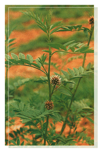
Licorice (Glycyrrhizia. Uralensis)
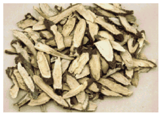
Licorice Root
The flavour extracted from the licorice root is 50 – 150 times sweeter than table sugar. The most important and abundant compound responsible for the sweetness and medicinal effects of licorice is glycyrrhizin (\(\ce{C42H62O16}\)).
Glycyrrhizin requires three equivalents of \(\ce{NaOH}\) to effect neutralization. When glycyrrhizin was subjected to acid hydrolysis, Glycyrrhizinic acid (A (\(\ce{C30H46O4}\))) and B (\(\ce{C6H10O7}\)) were obtainned in a 1:2 molar ratio (Scheme 1).
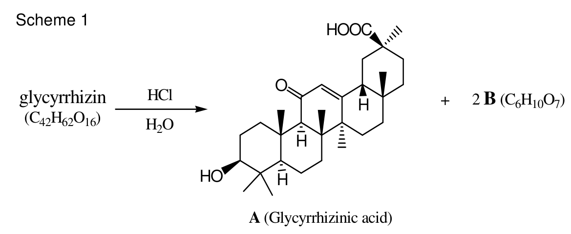
Scheme 1
When glycyrrhizin was methylated with methyl iodide (\(\ce{MeI}\)) at every possible site before hydrolysis, hydrolysis produced A’ (methyl glycyrrhizinate), and C and D (Scheme 2).
B, C and D exist as mixtures of anomers.

Scheme 2
Methylation of C and D with \(\ce{MeI}\) produced the same isomeric mixture of compounds, J (Scheme 3).

Scheme 3
C was reduced with \(\ce{LiAlH4}\) to give K, and L was produced by the reduction of K. Oxidative cleavage of vicinal diol of L with \(\ce{NaIO4}\) produced M and two equivalents of formaldehyde. Reduction of M produced N. The structure and stereochemistry of N was confirmed by the synthesis of N from D-(-)-tartaric acid through methylation followed by reduction (Scheme 4). A \(\ce{^{1}H}\)-NMR spectrum of L showed two distinct peaks for methyl groups. (There is no symmetry in L)
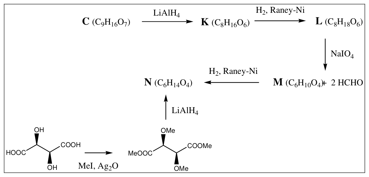
Scheme 4
10.1 Complete structures for L, M, and N in the answer sheet.
10.2 How many structures for C are possible? Complete possible structures for C.
To determine the correct structure of C, following set of reactions were performed.
J was reduced to E, and acid hydrolysis of E produced F. Reduction of F generated G, and G was oxidized with \(\ce{NaIO4}\) to H with formation of one equivalent of formaldehyde. I was obtained from H through reduction. Among all compounds from A to I, only I was optically inactive (Scheme 5).
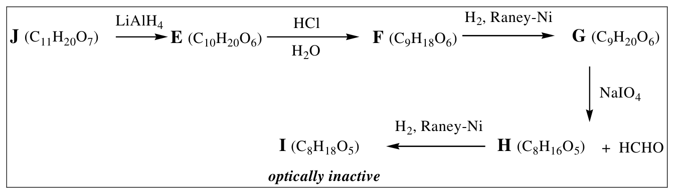
Scheme 5
10.3 Complete structures for G and I.
10.4 Which one is the correct structure for C among ones you have drawn in 10-2?
10.5 Complete structures for B, D, and J.
10.6 Complete the structure for Glycyrrhizin.
Official English text of the problem.
Answers
- 10.1.
L: 3,4-di-O-methyl-D-glucitol, \(\ce{C8H18O6}\).
SMILES: OC[C@H](O)[C@@H](OC)[C@H](OC)[C@H](O)CO
M: (2S,3S)-2,3-dimethoxybutane-1,4-dial, \(\ce{C6H10O4}\).
SMILES: O=C[C@@H](OC)[C@H](OC)C=O
N: (2R,3R)-2,3-dimethoxybutane-1,4-diol, \(\ce{C6H14O4}\).
SMILES: OC[C@@H](OC)[C@H](OC)CO
- 10.2. There are two possible sugar configurations, each occurring as two anomers: thus two anomeric pairs, or four individual cyclic stereoisomers.
Candidate 1: methyl 3,4-di-O-methyl-D-glucopyranuronate.
α: COC(=O)[C@H]1O[C@H](O)[C@H](O)[C@@H](OC)[C@@H]1OC
β: COC(=O)[C@H]1O[C@@H](O)[C@H](O)[C@@H](OC)[C@@H]1OC
Candidate 2: methyl 3,4-di-O-methyl-L-gulopyranuronate.
α: COC(=O)[C@@H]1O[C@@H](O)[C@@H](O)[C@@H](OC)[C@@H]1OC
β: COC(=O)[C@@H]1O[C@H](O)[C@@H](O)[C@@H](OC)[C@@H]1OC
- 10.3.
G: 2,3,4-tri-O-methyl-D-glucitol, \(\ce{C9H20O6}\).
SMILES: OC[C@H](OC)[C@@H](OC)[C@H](OC)[C@H](O)CO
I: meso-2,3,4-tri-O-methylxylitol, \(\ce{C8H18O5}\).
SMILES: OC[C@H](OC)[C@@H](OC)[C@H](OC)CO
-
10.4. Candidate 1 is correct: C is methyl 3,4-di-O-methyl-D-glucopyranuronate, as the α/β pair specified in 10.2.
-
10.5.
B: D-glucopyranuronic acid, \(\ce{C6H10O7}\).
α: O=C(O)[C@H]1O[C@H](O)[C@H](O)[C@@H](O)[C@@H]1O
β: O=C(O)[C@H]1O[C@@H](O)[C@H](O)[C@@H](O)[C@@H]1O
D: methyl 2,3,4-tri-O-methyl-D-glucopyranuronate, \(\ce{C10H18O7}\).
α: COC(=O)[C@H]1O[C@H](O)[C@H](OC)[C@@H](OC)[C@@H]1OC
β: COC(=O)[C@H]1O[C@@H](O)[C@H](OC)[C@@H](OC)[C@@H]1OC
J: methyl 1,2,3,4-tetra-O-methyl-D-glucopyranuronate, \(\ce{C11H20O7}\); equivalently, the methyl ester bearing a methyl glycoside at C1 and methoxy groups at C2, C3 and C4.
α: COC(=O)[C@H]1O[C@H](OC)[C@H](OC)[C@@H](OC)[C@@H]1OC
β: COC(=O)[C@H]1O[C@@H](OC)[C@H](OC)[C@@H](OC)[C@@H]1OC
- 10.6. The established connectivity is A 3-O-[D-glucopyranuronosyl-(1→2)-D-glucopyranuronoside], with all three carboxyl groups free.
The conventional β,β structure of glycyrrhizin, \(\ce{C42H62O16}\), is:
CC1(C)[C@@H](O[C@@H]6O[C@H](C(=O)O)[C@@H](O)[C@H](O)[C@H]6O[C@@H]7O[C@H](C(=O)O)[C@@H](O)[C@H](O)[C@H]7O)CC[C@]2(C)[C@H]3C(=O)C=C4[C@@H]5C[C@@](C)(C(=O)O)CC[C@]5(C)CC[C@@]4(C)[C@]3(C)CC[C@@H]12
Qualification: the supplied degradation experiments determine the connectivity and sugar configurations, but do not independently establish the two original β anomeric configurations.
Solution
In the Fischer projections described below, the carbon chain is vertical, horizontal bonds point toward the viewer, and hydrogen occupies the position opposite the stated oxygen substituent. Sugar positions refer to the original six-carbon chain.
For the reduction balances, \(\ce{[H]}\) denotes a formal reducing hydrogen equivalent. Ester reduction is written as
\[ \ce{RCOOCH3 + 4[H] -> RCH2OH + CH3OH}. \]
This expresses the net organic transformation after work-up.
10.1. Structures of L, M and N
Hydrolysis has the balanced stoichiometry
\[ \ce{C42H62O16 + 2H2O ->[HCl] C30H46O4 + 2C6H10O7}. \]
The aglycone contains one carboxyl group. Since glycyrrhizin consumes three equivalents of base and yields two identical molecules of B, each B supplies one further carboxyl group.
Replacing an O–H hydrogen by methyl adds \(\ce{CH2}\). Relative to B, C contains three such increments:
\[ \ce{C9H16O7}=\ce{C6H10O7}+3\ce{CH2}. \]
One methyl group forms the carboxylic methyl ester; the other two are methyl ethers. Ester reduction removes the ester methyl carbon from the isolated carbohydrate product as methanol, giving K, followed by reduction to the alditol L:
\[ \ce{C9H16O7 + 4[H] ->[LiAlH4] C8H16O6 + CH3OH} \qquad(\mathbf C\rightarrow\mathbf K), \]
\[ \ce{C8H16O6 + H2 ->[Raney-Ni] C8H18O6} \qquad(\mathbf K\rightarrow\mathbf L). \]
The production of two equivalents of formaldehyde requires a terminal vicinal diol at each end of L. Its constitution must therefore be
\[ \ce{HOCH2-CH(OH)-CH(OCH3)-CH(OCH3)-CH(OH)-CH2OH}. \]
Periodate cleaves the C1–C2 and C5–C6 bonds, leaving
\[ \ce{OHC-CH(OCH3)-CH(OCH3)-CHO} \]
as M. Reduction of its two aldehyde groups gives N:
\[ \ce{HOCH2-CH(OCH3)-CH(OCH3)-CH2OH}. \]
The balanced reactions are
\[ \ce{C8H18O6 + 2NaIO4 -> C6H10O4 + 2HCHO + 2NaIO3 + 2H2O}, \]
\[ \ce{C6H10O4 + 2H2 ->[Raney-Ni] C6H14O4}. \]
The tartaric-acid experiment fixes the absolute configurations of the two central carbons.
D-(-)-tartaric acid is (2S,3S)-2,3-dihydroxybutanedioic acid, SMILES
O=C(O)[C@@H](O)[C@H](O)C(=O)O.
Its fully methylated derivative is dimethyl (2S,3S)-2,3-dimethoxybutanedioate, SMILES
COC(=O)[C@@H](OC)[C@H](OC)C(=O)OC.
The balances are
\[ \ce{C4H6O6 + 4CH3I + 2Ag2O -> C8H14O6 + 4AgI + 2H2O}, \]
\[ \ce{C8H14O6 + 8[H] ->[LiAlH4] C6H14O4 + 2CH3OH}. \]
Neither transformation changes the spatial configuration at the two existing stereocentres. The Fischer projections of M and N, with identical terminal groups at the top and bottom, consequently have methoxy left, then methoxy right.
Thus M is \((2S,3S)\), whereas N is \((2R,3R)\). This change of descriptors results from changed CIP priorities upon reduction; it does not indicate inversion.
For L, the central methoxy groups likewise lie left at C3 and right at C4. The remaining possibilities concern the hydroxyl groups at C2 and C5:
| C2 hydroxyl | C5 hydroxyl | Consequence |
|---|---|---|
| Right | Left | End-exchange symmetry makes the methoxy groups equivalent |
| Left | Right | End-exchange symmetry makes the methoxy groups equivalent |
| Right | Right | Methoxy groups are inequivalent |
| Left | Left | Same alditol as the preceding row after rotating the Fischer projection by \(180^\circ\) |
The two distinct methyl resonances therefore select the single asymmetric alditol L given in the Answers.
To draw it, place \(\ce{CH2OH}\) at both ends and arrange the oxygen substituents from C2 through C5 as:
\[ \boxed{\text{OH right;\ OMe left;\ OMe right;\ OH right}.} \]
10.2. Possible structures of C
Reduction to L makes both ends of the original sugar into \(\ce{CH2OH}\). It therefore erases the distinction between the original reducing end and the original carboxyl end.
Assigning those ends in the two possible ways gives the following Fischer configurations. The top is the original C1 aldehyde position and the bottom is the C6 methyl ester.
| Candidate for C | C2–OH | C3–OMe | C4–OMe | C5–OH |
|---|---|---|---|---|
| D-glucuronic configuration | Right | Left | Right | Right |
| L-guluronic configuration | Left | Left | Right | Left |
In the cyclic structures, the C5 oxygen forms the pyranose ring, and C1 is a hemiacetal bearing a free hydroxyl group. Each configuration therefore gives an α and a β anomer.
Hence there are two configurational candidates, represented by four individual anomers.
For a conventional Haworth drawing with the ring oxygen at the upper right:
- D-glucuronic candidate: C5–\(\ce{COOCH3}\) up, C2–OH down, C3–OMe up, C4–OMe down; C1–OH down for α and up for β.
- L-guluronic candidate: C5–\(\ce{COOCH3}\) down, C2–OH up, C3–OMe up, C4–OMe down; C1–OH up for α and down for β.
These are the structures encoded by the four SMILES in 10.2.
10.3. Structures of G and I
Exhaustive methylation gives J with methyl groups at the carboxyl oxygen and at O1, O2, O3 and O4.
Reduction of the ester produces a C6 primary alcohol. Acid hydrolysis then removes the anomeric methyl group, while retaining the ordinary methyl ethers at C2, C3 and C4. Reduction of the reducing sugar consequently gives an alditol of constitution
\[ \ce{HOCH2-CH(OCH3)-CH(OCH3)-CH(OCH3)-CH(OH)-CH2OH}. \]
For the D-glucuronic candidate, this is G, 2,3,4-tri-O-methyl-D-glucitol. Its Fischer oxygen-substituent sequence is
\[ \boxed{\text{OMe right;\ OMe left;\ OMe right;\ OH right}.} \]
The intermediate structures for this candidate are:
| Compound | Identification | Representative β-anomer SMILES |
|---|---|---|
| E | Methyl 2,3,4-tri-O-methyl-D-glucopyranoside | OC[C@H]1O[C@@H](OC)[C@H](OC)[C@@H](OC)[C@@H]1OC |
| F | 2,3,4-Tri-O-methyl-D-glucopyranose | OC[C@H]1O[C@@H](O)[C@H](OC)[C@@H](OC)[C@@H]1OC |
The corresponding α forms differ only at C1. The balanced transformations are
\[ \ce{C11H20O7 + 4[H] ->[LiAlH4] C10H20O6 + CH3OH}, \]
\[ \ce{C10H20O6 + H2O ->[HCl] C9H18O6 + CH3OH}, \]
\[ \ce{C9H18O6 + H2 ->[Raney-Ni] C9H20O6}. \]
Only C5–C6 in G is a free vicinal diol. Its cleavage produces one molecule of formaldehyde and the aldehyde H:
\[ \ce{HOCH2-CH(OCH3)-CH(OCH3)-CH(OCH3)-CHO}. \]
H is open-chain 2,3,4-tri-O-methyl-L-xylose, SMILES
OC[C@H](OC)[C@@H](OC)[C@H](OC)C=O.
Reduction gives I, with \(\ce{CH2OH}\) at both ends and the Fischer methoxy sequence right–left–right:
\[ \ce{HOCH2-CH(OCH3)-CH(OCH3)-CH(OCH3)-CH2OH}. \]
The balanced reactions are
\[ \ce{C9H20O6 + NaIO4 -> C8H16O5 + HCHO + NaIO3 + H2O}, \]
\[ \ce{C8H16O5 + H2 ->[Raney-Ni] C8H18O5}. \]
The two ends of I are identical. Its internal mirror symmetry exchanges C2 with C4, making it a meso compound and therefore optically inactive. This gives the required structure of I.
10.4. Selection of C
The L-guluronic candidate would instead give a pentitol with the Fischer methoxy sequence left–left–right.
That alternative is 2,3,4-tri-O-methyl-D-lyxitol, SMILES
OC[C@@H](OC)[C@@H](OC)[C@H](OC)CO.
It is chiral and would remain optically active. The stated optical inactivity of I therefore selects the D-glucuronic candidate:
\[ \boxed{\mathbf C=\text{methyl 3,4-di-O-methyl-D-glucopyranuronate}.} \]
The terminal aldehyde reduction does not generate a new stereocentre, so the optical inactivity is explained by the meso structure, without invoking racemization.
With this selection, K is 3,4-di-O-methyl-D-glucopyranose; a representative β-anomer SMILES is
OC[C@H]1O[C@@H](O)[C@H](O)[C@@H](OC)[C@@H]1OC.
10.5. Structures of B, D and J
Removing the methyl groups from the identified C gives B, D-glucuronic acid.
The substitution patterns are:
| Compound | C1 substituent | C2 substituent | C3 substituent | C4 substituent | C6 group |
|---|---|---|---|---|---|
| B | \(\ce{OH}\) | \(\ce{OH}\) | \(\ce{OH}\) | \(\ce{OH}\) | \(\ce{COOH}\) |
| C | \(\ce{OH}\) | \(\ce{OH}\) | \(\ce{OCH3}\) | \(\ce{OCH3}\) | \(\ce{COOCH3}\) |
| D | \(\ce{OH}\) | \(\ce{OCH3}\) | \(\ce{OCH3}\) | \(\ce{OCH3}\) | \(\ce{COOCH3}\) |
| J | \(\ce{OCH3}\) | \(\ce{OCH3}\) | \(\ce{OCH3}\) | \(\ce{OCH3}\) | \(\ce{COOCH3}\) |
All retain the D-gluco configuration at C2–C5.
The formula differences confirm these assignments:
\[ \begin{aligned} \mathbf C &: \quad \ce{C6H10O7}+3\ce{CH2}=\ce{C9H16O7},\\ \mathbf D &: \quad \ce{C6H10O7}+4\ce{CH2}=\ce{C10H18O7},\\ \mathbf J &: \quad \ce{C6H10O7}+5\ce{CH2}=\ce{C11H20O7}. \end{aligned} \]
To reach J, C requires methylation of O1 and O2, whereas D requires methylation only of O1:
\[ \ce{C9H16O7 + 2CH3I + Ag2O -> C11H20O7 + 2AgI + H2O}, \]
\[ \ce{2C10H18O7 + 2CH3I + Ag2O -> 2C11H20O7 + 2AgI + H2O}. \]
Thus both give the same pair of methyl-glycoside anomers of J, as required.
10.6. Structure of glycyrrhizin
Methylation analysis identifies the original glycosidic attachment positions: hydroxyl groups exposed only by hydrolysis were unavailable for methylation in intact glycyrrhizin.
Attachment to the aglycone. The depicted aglycone A is commonly called glycyrrhetinic acid, although the problem labels it “Glycyrrhizinic acid.” Its stereochemical SMILES is
CC1(C)[C@@H](O)CC[C@]2(C)[C@H]3C(=O)C=C4[C@@H]5C[C@@](C)(C(=O)O)CC[C@]5(C)CC[C@@]4(C)[C@]3(C)CC[C@@H]12
After methylation and hydrolysis, A’ contains only one added methyl group. It is methyl glycyrrhetinate, SMILES
CC1(C)[C@@H](O)CC[C@]2(C)[C@H]3C(=O)C=C4[C@@H]5C[C@@](C)(C(=O)OC)CC[C@]5(C)CC[C@@]4(C)[C@]3(C)CC[C@@H]12
Its carboxyl group was methylated, but its C3 hydroxyl was protected by a glycosidic linkage during methylation. Hydrolysis restores that hydroxyl. Consequently, the sugar chain is attached through the aglycone’s C3 oxygen.
Attachment between the sugars. The inner sugar gives C, which has an unmethylated hydroxyl at C2 after hydrolysis. Its O2 must therefore have participated in the intersugar glycosidic bond. The terminal sugar gives D, methylated at all three non-anomeric hydroxyl positions.
The connectivity is therefore
\[ \boxed{ \text{terminal D-GlcpA} \xrightarrow{\;1\to2\;} \text{inner D-GlcpA} \xrightarrow{\;1\to O3\;} \mathbf A } \]
where GlcpA denotes a glucopyranuronic-acid residue.
For the drawing, copy the aglycone in Scheme 1 and:
- Replace the hydrogen of its C3 hydroxyl by the anomeric carbon of the inner D-glucopyranuronic-acid residue.
- Attach the terminal residue’s anomeric carbon through the inner residue’s O2.
- Retain the carboxyl group on the aglycone and the C6 carboxyl group of each sugar as free acids.
- For the conventional β,β glycyrrhizin structure, draw both anomeric glycosidic substituents on the same side as the corresponding C5 carboxyl substituent.
The three free carboxyl groups explain the neutralization:
\[ \ce{C42H62O16 + 3NaOH -> C42H59Na3O16 + 3H2O}. \]
The composition also agrees with condensation of A and two molecules of B:
\[ \ce{C30H46O4 + 2C6H10O7 -> C42H62O16 + 2H2O}. \]
There are five free alcoholic hydroxyl groups and three free carboxyl groups in glycyrrhizin, giving eight methylations. The combined balance for exhaustive methylation followed by hydrolysis is
\[ \ce{C42H62O16 + 8CH3I + 4Ag2O -> C31H48O4 + C9H16O7 + C10H18O7 + 8AgI + 2H2O}, \]
whose organic products are A’, C and D, respectively.
For completeness, the small species used in these balances have the following SMILES: formaldehyde C=O; methanol CO; water O; hydrogen [H][H]; methyl iodide CI; hydrogen chloride Cl; lithium aluminium hydride [Li+].[AlH4-]; silver(I) oxide [Ag+].[Ag+].[O-2]; silver iodide [Ag+].[I-]; sodium periodate [Na+].[O-]I(=O)(=O)=O; sodium iodate [Na+].[O-]I(=O)=O; sodium hydroxide [Na+].[OH-]. Raney nickel is a porous solid catalyst, for which [Ni] represents the element but not the catalyst morphology.
The solution was checked against the official solution.
IChO 2006 · Theorieaufgabe 10
Naturstoffe - Strukturanalyse
Süßholz (Glycyrrhizia. Uralensis)
Süßholzwurzel
Der aus der Süßholzwurzel gewonnene Aromastoff ist 50 – 150 Mal süßer als Haushaltszucker. Die wichtigste und am häufigsten vorkommende Verbindung, die für die Süße und die medizinischen Wirkungen des Süßholzes verantwortlich ist, ist Glycyrrhizin (\(\ce{C42H62O16}\)).
Zur Neutralisation von Glycyrrhizin sind drei Äquivalente \(\ce{NaOH}\) erforderlich. Bei der sauren Hydrolyse von Glycyrrhizin wurden Glycyrrhizinsäure (A (\(\ce{C30H46O4}\))) und B (\(\ce{C6H10O7}\)) im Stoffmengenverhältnis 1:2 erhalten (Schema 1).
Schema 1
Wurde Glycyrrhizin vor der Hydrolyse an allen möglichen Stellen mit Methyliodid (\(\ce{MeI}\)) methyliert, so entstanden bei der Hydrolyse A’ (Methylglycyrrhizinat) sowie C und D (Schema 2).
B, C und D liegen als Anomerengemische vor.
Schema 2
Die Methylierung von C und D mit \(\ce{MeI}\) ergab dasselbe Isomerengemisch von Verbindungen, J (Schema 3).
Schema 3
C wurde mit \(\ce{LiAlH4}\) zu K reduziert, und durch Reduktion von K wurde L erhalten. Die oxidative Spaltung der vicinalen Diolgruppen von L mit \(\ce{NaIO4}\) ergab M und zwei Äquivalente Formaldehyd. Die Reduktion von M ergab N. Die Struktur und Stereochemie von N wurden durch die Synthese von N aus D-(-)-Weinsäure durch Methylierung mit anschließender Reduktion bestätigt (Schema 4). Ein \(\ce{^{1}H}\)-NMR-Spektrum von L zeigte zwei getrennte Signale für Methylgruppen. (In L liegt keine Symmetrie vor.)
Schema 4
10.1 Vervollständigen Sie die Strukturen von L, M, und N auf dem Antwortbogen.
10.2 Wie viele Strukturen sind für C möglich? Vervollständigen Sie die möglichen Strukturen von C.
Um die richtige Struktur von C zu bestimmen, wurde die folgende Reaktionsfolge durchgeführt.
J wurde zu E reduziert, und die saure Hydrolyse von E ergab F. Die Reduktion von F lieferte G, und G wurde mit \(\ce{NaIO4}\) unter Bildung eines Äquivalents Formaldehyd zu H oxidiert. I wurde durch Reduktion aus H erhalten. Von allen Verbindungen von A bis I war nur I optisch inaktiv (Schema 5).
Schema 5
10.3 Vervollständigen Sie die Strukturen von G und I.
10.4 Welche der von Ihnen in 10-2 gezeichneten Strukturen ist die richtige Struktur für C?
10.5 Vervollständigen Sie die Strukturen von B, D, und J.
10.6 Vervollständigen Sie die Struktur von Glycyrrhizin.
Aufgabentext aus dem offiziellen englischen Text übersetzt.
Antworten
- 10.1.
L: 3,4-Di-O-methyl-D-glucitol, \(\ce{C8H18O6}\).
SMILES: OC[C@H](O)[C@@H](OC)[C@H](OC)[C@H](O)CO
M: (2S,3S)-2,3-Dimethoxybutan-1,4-dial, \(\ce{C6H10O4}\).
SMILES: O=C[C@@H](OC)[C@H](OC)C=O
N: (2R,3R)-2,3-Dimethoxybutan-1,4-diol, \(\ce{C6H14O4}\).
SMILES: OC[C@@H](OC)[C@H](OC)CO
- 10.2. Es gibt zwei mögliche Zuckerkonfigurationen, die jeweils als zwei Anomere auftreten: somit zwei Anomerenpaare oder vier einzelne cyclische Stereoisomere.
Kandidat 1: Methyl-3,4-di-O-methyl-D-glucopyranuronat.
α: COC(=O)[C@H]1O[C@H](O)[C@H](O)[C@@H](OC)[C@@H]1OC
β: COC(=O)[C@H]1O[C@@H](O)[C@H](O)[C@@H](OC)[C@@H]1OC
Kandidat 2: Methyl-3,4-di-O-methyl-L-gulopyranuronat.
α: COC(=O)[C@@H]1O[C@@H](O)[C@@H](O)[C@@H](OC)[C@@H]1OC
β: COC(=O)[C@@H]1O[C@H](O)[C@@H](O)[C@@H](OC)[C@@H]1OC
- 10.3.
G: 2,3,4-Tri-O-methyl-D-glucitol, \(\ce{C9H20O6}\).
SMILES: OC[C@H](OC)[C@@H](OC)[C@H](OC)[C@H](O)CO
I: meso-2,3,4-Tri-O-methylxylitol, \(\ce{C8H18O5}\).
SMILES: OC[C@H](OC)[C@@H](OC)[C@H](OC)CO
-
10.4. Kandidat 1 ist richtig: C ist Methyl-3,4-di-O-methyl-D-glucopyranuronat in Form des in 10.2 angegebenen α/β-Paares.
-
10.5.
B: D-Glucopyranuronsäure, \(\ce{C6H10O7}\).
α: O=C(O)[C@H]1O[C@H](O)[C@H](O)[C@@H](O)[C@@H]1O
β: O=C(O)[C@H]1O[C@@H](O)[C@H](O)[C@@H](O)[C@@H]1O
D: Methyl-2,3,4-tri-O-methyl-D-glucopyranuronat, \(\ce{C10H18O7}\).
α: COC(=O)[C@H]1O[C@H](O)[C@H](OC)[C@@H](OC)[C@@H]1OC
β: COC(=O)[C@H]1O[C@@H](O)[C@H](OC)[C@@H](OC)[C@@H]1OC
J: Methyl-1,2,3,4-tetra-O-methyl-D-glucopyranuronat, \(\ce{C11H20O7}\); gleichbedeutend damit ist der Methylester mit einer Methylglycosidgruppe an C1 und Methoxygruppen an C2, C3 und C4.
α: COC(=O)[C@H]1O[C@H](OC)[C@H](OC)[C@@H](OC)[C@@H]1OC
β: COC(=O)[C@H]1O[C@@H](OC)[C@H](OC)[C@@H](OC)[C@@H]1OC
- 10.6. Die ermittelte Verknüpfung ist A-3-O-[D-glucopyranuronosyl-(1→2)-D-glucopyranuronosid], wobei alle drei Carboxylgruppen frei sind.
Die übliche β,β-Struktur von Glycyrrhizin, \(\ce{C42H62O16}\), lautet:
CC1(C)[C@@H](O[C@@H]6O[C@H](C(=O)O)[C@@H](O)[C@H](O)[C@H]6O[C@@H]7O[C@H](C(=O)O)[C@@H](O)[C@H](O)[C@H]7O)CC[C@]2(C)[C@H]3C(=O)C=C4[C@@H]5C[C@@](C)(C(=O)O)CC[C@]5(C)CC[C@@]4(C)[C@]3(C)CC[C@@H]12
Einschränkung: Die angegebenen Abbauexperimente bestimmen die Verknüpfung und die Zuckerkonfigurationen, belegen jedoch nicht unabhängig die beiden ursprünglichen β-Konfigurationen an den anomeren Zentren.
Lösung
In den nachfolgend beschriebenen Fischer-Projektionen verläuft die Kohlenstoffkette senkrecht, waagerechte Bindungen zeigen zum Betrachter, und Wasserstoff nimmt die Position gegenüber dem jeweils angegebenen Sauerstoffsubstituenten ein. Die Positionsangaben für die Zucker beziehen sich auf die ursprüngliche Kette aus sechs Kohlenstoffatomen.
In den Reduktionsbilanzen bezeichnet \(\ce{[H]}\) ein formales reduzierendes Wasserstoffäquivalent. Die Esterreduktion wird wie folgt geschrieben:
\[ \ce{RCOOCH3 + 4[H] -> RCH2OH + CH3OH}. \]
Dies beschreibt die Nettoumwandlung der organischen Verbindungen nach der Aufarbeitung.
10.1. Strukturen von L, M und N
Die ausgeglichene stöchiometrische Gleichung der Hydrolyse lautet
\[ \ce{C42H62O16 + 2H2O ->[HCl] C30H46O4 + 2C6H10O7}. \]
Das Aglycon enthält eine Carboxylgruppe. Da Glycyrrhizin drei Äquivalente Base verbraucht und zwei identische Moleküle B liefert, trägt jedes B eine weitere Carboxylgruppe bei.
Wird ein O–H-Wasserstoff durch Methyl ersetzt, kommt \(\ce{CH2}\) hinzu. Gegenüber B enthält C drei solche zusätzlichen Einheiten:
\[ \ce{C9H16O7}=\ce{C6H10O7}+3\ce{CH2}. \]
Eine Methylgruppe bildet den Carbonsäuremethylester; die anderen beiden bilden Methylether. Bei der Esterreduktion wird der Kohlenstoff der Estermethylgruppe als Methanol aus dem isolierten Kohlenhydratprodukt entfernt, wobei K entsteht, gefolgt von der Reduktion zum Alditol L:
\[ \ce{C9H16O7 + 4[H] ->[LiAlH4] C8H16O6 + CH3OH} \qquad(\mathbf C\rightarrow\mathbf K), \]
\[ \ce{C8H16O6 + H2 ->[Raney-Ni] C8H18O6} \qquad(\mathbf K\rightarrow\mathbf L). \]
Die Bildung von zwei Äquivalenten Formaldehyd erfordert an jedem Ende von L eine endständige vicinale Diolgruppe. Seine Konstitution muss daher wie folgt sein:
\[ \ce{HOCH2-CH(OH)-CH(OCH3)-CH(OCH3)-CH(OH)-CH2OH}. \]
Periodat spaltet die Bindungen C1–C2 und C5–C6, wobei
\[ \ce{OHC-CH(OCH3)-CH(OCH3)-CHO} \]
als M zurückbleibt. Die Reduktion seiner beiden Aldehydgruppen ergibt N:
\[ \ce{HOCH2-CH(OCH3)-CH(OCH3)-CH2OH}. \]
Die ausgeglichenen Reaktionsgleichungen lauten
\[ \ce{C8H18O6 + 2NaIO4 -> C6H10O4 + 2HCHO + 2NaIO3 + 2H2O}, \]
\[ \ce{C6H10O4 + 2H2 ->[Raney-Ni] C6H14O4}. \]
Das Experiment mit Weinsäure legt die absoluten Konfigurationen der beiden mittleren Kohlenstoffatome fest.
D-(-)-Weinsäure ist (2S,3S)-2,3-Dihydroxybutandisäure, SMILES
O=C(O)[C@@H](O)[C@H](O)C(=O)O.
Ihr vollständig methyliertes Derivat ist Dimethyl-(2S,3S)-2,3-dimethoxybutandioat, SMILES
COC(=O)[C@@H](OC)[C@H](OC)C(=O)OC.
Die Bilanzen lauten
\[ \ce{C4H6O6 + 4CH3I + 2Ag2O -> C8H14O6 + 4AgI + 2H2O}, \]
\[ \ce{C8H14O6 + 8[H] ->[LiAlH4] C6H14O4 + 2CH3OH}. \]
Keine der beiden Umwandlungen verändert die räumliche Konfiguration an den beiden vorhandenen Stereozentren. In den Fischer-Projektionen von M und N, mit identischen endständigen Gruppen oben und unten, stehen die Methoxygruppen daher zuerst links, dann rechts.
Somit ist M \((2S,3S)\), während N \((2R,3R)\) ist. Diese Änderung der Deskriptoren ergibt sich aus den bei der Reduktion veränderten CIP-Prioritäten; sie bedeutet keine Inversion.
Bei L stehen die mittleren Methoxygruppen ebenfalls an C3 links und an C4 rechts. Die verbleibenden Möglichkeiten betreffen die Hydroxylgruppen an C2 und C5:
| Hydroxylgruppe an C2 | Hydroxylgruppe an C5 | Konsequenz |
|---|---|---|
| Rechts | Links | Die Symmetrie beim Vertauschen der Enden macht die Methoxygruppen äquivalent |
| Links | Rechts | Die Symmetrie beim Vertauschen der Enden macht die Methoxygruppen äquivalent |
| Rechts | Rechts | Die Methoxygruppen sind nicht äquivalent |
| Links | Links | Dasselbe Alditol wie in der vorhergehenden Zeile nach Drehung der Fischer-Projektion um \(180^\circ\) |
Die beiden unterschiedlichen Methylresonanzen legen daher das einzige asymmetrische Alditol L fest, das unter Antworten angegeben ist.
Um es zu zeichnen, setzen Sie \(\ce{CH2OH}\) an beide Enden und ordnen Sie die Sauerstoffsubstituenten von C2 bis C5 wie folgt an:
\[ \boxed{\text{OH rechts;\ OMe links;\ OMe rechts;\ OH rechts}.} \]
10.2. Mögliche Strukturen von C
Durch die Reduktion zu L werden beide Enden des ursprünglichen Zuckers zu \(\ce{CH2OH}\). Dadurch geht die Unterscheidung zwischen dem ursprünglichen reduzierenden Ende und dem ursprünglichen Carboxylende verloren.
Die Zuordnung dieser Enden auf die beiden möglichen Arten ergibt die folgenden Fischer-Konfigurationen. Oben liegt die ursprüngliche C1-Aldehydposition und unten der C6-Methylester.
| Kandidat für C | C2–OH | C3–OMe | C4–OMe | C5–OH |
|---|---|---|---|---|
| D-Glucuronsäure-Konfiguration | Rechts | Links | Rechts | Rechts |
| L-Guluronsäure-Konfiguration | Links | Links | Rechts | Links |
In den cyclischen Strukturen bildet der Sauerstoff an C5 den Pyranosering, und C1 ist ein Halbacetal mit einer freien Hydroxylgruppe. Jede Konfiguration ergibt daher ein α- und ein β-Anomer.
Es gibt folglich zwei Konfigurationskandidaten, die durch vier einzelne Anomere repräsentiert werden.
Für eine übliche Haworth-Darstellung mit dem Ringsauerstoff oben rechts gilt:
- D-Glucuronsäure-Kandidat: C5–\(\ce{COOCH3}\) oben, C2–OH unten, C3–OMe oben, C4–OMe unten; C1–OH bei α unten und bei β oben.
- L-Guluronsäure-Kandidat: C5–\(\ce{COOCH3}\) unten, C2–OH oben, C3–OMe oben, C4–OMe unten; C1–OH bei α oben und bei β unten.
Dies sind die Strukturen, die durch die vier SMILES in 10.2 codiert werden.
10.3. Strukturen von G und I
Vollständige Methylierung ergibt J mit Methylgruppen am Carboxylsauerstoff sowie an O1, O2, O3 und O4.
Die Reduktion des Esters erzeugt einen primären Alkohol an C6. Die anschließende saure Hydrolyse entfernt die anomere Methylgruppe, während die gewöhnlichen Methylether an C2, C3 und C4 erhalten bleiben. Die Reduktion des reduzierenden Zuckers ergibt daher ein Alditol mit der Konstitution
\[ \ce{HOCH2-CH(OCH3)-CH(OCH3)-CH(OCH3)-CH(OH)-CH2OH}. \]
Für den D-Glucuronsäure-Kandidaten ist dies G, 2,3,4-Tri-O-methyl-D-glucitol. Die Abfolge seiner Sauerstoffsubstituenten in der Fischer-Projektion lautet
\[ \boxed{\text{OMe rechts;\ OMe links;\ OMe rechts;\ OH rechts}.} \]
Die Zwischenprodukte für diesen Kandidaten haben folgende Strukturen:
| Verbindung | Identifizierung | Repräsentativer SMILES des β-Anomers |
|---|---|---|
| E | Methyl-2,3,4-tri-O-methyl-D-glucopyranosid | OC[C@H]1O[C@@H](OC)[C@H](OC)[C@@H](OC)[C@@H]1OC |
| F | 2,3,4-Tri-O-methyl-D-glucopyranose | OC[C@H]1O[C@@H](O)[C@H](OC)[C@@H](OC)[C@@H]1OC |
Die entsprechenden α-Formen unterscheiden sich nur an C1. Die ausgeglichenen Reaktionsgleichungen der Umwandlungen lauten
\[ \ce{C11H20O7 + 4[H] ->[LiAlH4] C10H20O6 + CH3OH}, \]
\[ \ce{C10H20O6 + H2O ->[HCl] C9H18O6 + CH3OH}, \]
\[ \ce{C9H18O6 + H2 ->[Raney-Ni] C9H20O6}. \]
Nur C5–C6 bildet in G eine freie vicinale Diolgruppe. Ihre Spaltung erzeugt ein Molekül Formaldehyd und den Aldehyd H:
\[ \ce{HOCH2-CH(OCH3)-CH(OCH3)-CH(OCH3)-CHO}. \]
H ist offenkettige 2,3,4-Tri-O-methyl-L-xylose, SMILES
OC[C@H](OC)[C@@H](OC)[C@H](OC)C=O.
Die Reduktion ergibt I, mit \(\ce{CH2OH}\) an beiden Enden und der Methoxyabfolge rechts–links–rechts in der Fischer-Projektion:
\[ \ce{HOCH2-CH(OCH3)-CH(OCH3)-CH(OCH3)-CH2OH}. \]
Die ausgeglichenen Reaktionsgleichungen lauten
\[ \ce{C9H20O6 + NaIO4 -> C8H16O5 + HCHO + NaIO3 + H2O}, \]
\[ \ce{C8H16O5 + H2 ->[Raney-Ni] C8H18O5}. \]
Die beiden Enden von I sind identisch. Seine innere Spiegelsymmetrie vertauscht C2 und C4, wodurch es eine meso-Verbindung und daher optisch inaktiv ist. Damit ergibt sich die gesuchte Struktur von I.
10.4. Auswahl von C
Der L-Guluronsäure-Kandidat würde stattdessen ein Pentitol mit der Methoxyabfolge links–links–rechts in der Fischer-Projektion ergeben.
Diese Alternative ist 2,3,4-Tri-O-methyl-D-lyxitol, SMILES
OC[C@@H](OC)[C@@H](OC)[C@H](OC)CO.
Sie ist chiral und würde optisch aktiv bleiben. Die angegebene optische Inaktivität von I legt daher den D-Glucuronsäure-Kandidaten fest:
\[ \boxed{\mathbf C=\text{Methyl-3,4-di-O-methyl-D-glucopyranuronat}.} \]
Die Reduktion der endständigen Aldehydgruppe erzeugt kein neues Stereozentrum, sodass die optische Inaktivität durch die meso-Struktur erklärt wird, ohne eine Racemisierung anzunehmen.
Mit dieser Auswahl ist K 3,4-Di-O-methyl-D-glucopyranose; ein repräsentativer SMILES des β-Anomers lautet
OC[C@H]1O[C@@H](O)[C@H](O)[C@@H](OC)[C@@H]1OC.
10.5. Strukturen von B, D und J
Das Entfernen der Methylgruppen aus dem identifizierten C ergibt B, D-Glucuronsäure.
Die Substitutionsmuster lauten:
| Verbindung | C1-Substituent | C2-Substituent | C3-Substituent | C4-Substituent | C6-Gruppe |
|---|---|---|---|---|---|
| B | \(\ce{OH}\) | \(\ce{OH}\) | \(\ce{OH}\) | \(\ce{OH}\) | \(\ce{COOH}\) |
| C | \(\ce{OH}\) | \(\ce{OH}\) | \(\ce{OCH3}\) | \(\ce{OCH3}\) | \(\ce{COOCH3}\) |
| D | \(\ce{OH}\) | \(\ce{OCH3}\) | \(\ce{OCH3}\) | \(\ce{OCH3}\) | \(\ce{COOCH3}\) |
| J | \(\ce{OCH3}\) | \(\ce{OCH3}\) | \(\ce{OCH3}\) | \(\ce{OCH3}\) | \(\ce{COOCH3}\) |
Alle behalten die D-gluco-Konfiguration an C2–C5 bei.
Die Unterschiede der Summenformeln bestätigen diese Zuordnungen:
\[ \begin{aligned} \mathbf C &: \quad \ce{C6H10O7}+3\ce{CH2}=\ce{C9H16O7},\\ \mathbf D &: \quad \ce{C6H10O7}+4\ce{CH2}=\ce{C10H18O7},\\ \mathbf J &: \quad \ce{C6H10O7}+5\ce{CH2}=\ce{C11H20O7}. \end{aligned} \]
Um J zu erhalten, müssen bei C O1 und O2 methyliert werden, bei D dagegen nur O1:
\[ \ce{C9H16O7 + 2CH3I + Ag2O -> C11H20O7 + 2AgI + H2O}, \]
\[ \ce{2C10H18O7 + 2CH3I + Ag2O -> 2C11H20O7 + 2AgI + H2O}. \]
Somit liefern beide wie gefordert dasselbe Paar von Methylglycosid-Anomeren von J.
10.6. Struktur von Glycyrrhizin
Die Methylierungsanalyse identifiziert die ursprünglichen glycosidischen Verknüpfungsstellen: Hydroxylgruppen, die erst durch Hydrolyse freigelegt wurden, standen im intakten Glycyrrhizin nicht für die Methylierung zur Verfügung.
Verknüpfung mit dem Aglycon. Das dargestellte Aglycon A wird gewöhnlich als Glycyrrhetinsäure bezeichnet, obwohl es in der Aufgabe „Glycyrrhizinsäure“ genannt wird. Sein stereochemischer SMILES lautet
CC1(C)[C@@H](O)CC[C@]2(C)[C@H]3C(=O)C=C4[C@@H]5C[C@@](C)(C(=O)O)CC[C@]5(C)CC[C@@]4(C)[C@]3(C)CC[C@@H]12
Nach Methylierung und Hydrolyse enthält A’ nur eine zusätzlich eingeführte Methylgruppe. Es handelt sich um Methylglycyrrhetinat, SMILES
CC1(C)[C@@H](O)CC[C@]2(C)[C@H]3C(=O)C=C4[C@@H]5C[C@@](C)(C(=O)OC)CC[C@]5(C)CC[C@@]4(C)[C@]3(C)CC[C@@H]12
Seine Carboxylgruppe wurde methyliert, während seine C3-Hydroxylgruppe während der Methylierung durch eine glycosidische Bindung geschützt war. Die Hydrolyse stellt diese Hydroxylgruppe wieder her. Folglich ist die Zuckerkette über den C3-Sauerstoff des Aglycons gebunden.
Verknüpfung zwischen den Zuckern. Der innere Zucker ergibt C, das nach der Hydrolyse an C2 eine unmethylierte Hydroxylgruppe aufweist. Sein O2 muss daher an der glycosidischen Bindung zwischen den Zuckern beteiligt gewesen sein. Der endständige Zucker ergibt D, das an allen drei nichtanomeren Hydroxylpositionen methyliert ist.
Die Verknüpfung ist daher
\[ \boxed{ \text{endständiger D-GlcpA} \xrightarrow{\;1\to2\;} \text{innerer D-GlcpA} \xrightarrow{\;1\to O3\;} \mathbf A } \]
wobei GlcpA einen Glucopyranuronsäurerest bezeichnet.
Übernehmen Sie für die Zeichnung das Aglycon aus Schema 1 und gehen Sie wie folgt vor:
- Ersetzen Sie den Wasserstoff seiner C3-Hydroxylgruppe durch das anomere Kohlenstoffatom des inneren D-Glucopyranuronsäurerests.
- Binden Sie das anomere Kohlenstoffatom des endständigen Rests über O2 des inneren Rests an.
- Belassen Sie die Carboxylgruppe am Aglycon und die C6-Carboxylgruppe jedes Zuckers als freie Säuren.
- Zeichnen Sie für die übliche β,β-Struktur von Glycyrrhizin beide anomeren glycosidischen Substituenten auf derselben Seite wie den jeweils zugehörigen C5-Carboxylsubstituenten.
Die drei freien Carboxylgruppen erklären die Neutralisation:
\[ \ce{C42H62O16 + 3NaOH -> C42H59Na3O16 + 3H2O}. \]
Die Zusammensetzung stimmt auch mit der Kondensation von A und zwei Molekülen B überein:
\[ \ce{C30H46O4 + 2C6H10O7 -> C42H62O16 + 2H2O}. \]
Glycyrrhizin enthält fünf freie alkoholische Hydroxylgruppen und drei freie Carboxylgruppen, sodass acht Methylierungen erfolgen. Die Gesamtbilanz für die vollständige Methylierung mit anschließender Hydrolyse lautet
\[ \ce{C42H62O16 + 8CH3I + 4Ag2O -> C31H48O4 + C9H16O7 + C10H18O7 + 8AgI + 2H2O}, \]
wobei die organischen Produkte der Reihe nach A’, C und D sind.
Der Vollständigkeit halber haben die in diesen Bilanzen verwendeten kleinen Spezies folgende SMILES: Formaldehyd C=O; Methanol CO; Wasser O; Wasserstoff [H][H]; Methyliodid CI; Chlorwasserstoff Cl; Lithiumaluminiumhydrid [Li+].[AlH4-]; Silber(I)-oxid [Ag+].[Ag+].[O-2]; Silberiodid [Ag+].[I-]; Natriumperiodat [Na+].[O-]I(=O)(=O)=O; Natriumiodat [Na+].[O-]I(=O)=O; Natriumhydroxid [Na+].[OH-]. Raney-Nickel ist ein poröser Feststoffkatalysator, für den [Ni] das Element, jedoch nicht die Morphologie des Katalysators darstellt.
Die Lösung wurde mit der offiziellen Lösung abgeglichen.
IChO 2006 · Teori sorusu 10
Doğal Ürünler - Yapı Analizi
Meyan (Glycyrrhizia. Uralensis)
Meyan Kökü
Meyan kökünden elde edilen tatlandırıcı, sofra şekerinden 50 – 150 kat daha tatlıdır. Meyanın tatlılığından ve tıbbi etkilerinden sorumlu en önemli ve en bol bulunan bileşik glisirizindir (\(\ce{C42H62O16}\)).
Glisirizinin nötralleşmesi için üç eşdeğer \(\ce{NaOH}\) gerekir. Glisirizin asit hidrolizine tabi tutulduğunda, 1:2 mol oranında Glisirizinik asit (A (\(\ce{C30H46O4}\))) ve B (\(\ce{C6H10O7}\)) elde edilmiştir (Şema 1).
Şema 1
Glisirizin, hidrolizden önce metil iyodür (\(\ce{MeI}\)) ile mümkün olan her konumda metillendiğinde, hidroliz sonucunda A’ (metil glisirizinat), C ve D oluşmuştur (Şema 2).
B, C ve D, anomer karışımları hâlinde bulunur.
Şema 2
C ve D'nin \(\ce{MeI}\) ile metillenmesi, aynı izomerik bileşik karışımını, J'yi vermiştir (Şema 3).
Şema 3
C, \(\ce{LiAlH4}\) ile indirgenerek K elde edilmiş, K'nin indirgenmesiyle de L oluşmuştur. L'nin komşu diol gruplarının \(\ce{NaIO4}\) ile yükseltgenerek parçalanması, M ve iki eşdeğer formaldehit vermiştir. M'nin indirgenmesiyle N oluşmuştur. N'nin yapısı ve stereokimyası, D-(-)-tartarik asidin önce metillenmesi, ardından indirgenmesi yoluyla N'nin sentezlenmesiyle doğrulanmıştır (Şema 4). L'nin \(\ce{^{1}H}\)-NMR spektrumunda metil gruplarına ait iki farklı pik görülmüştür. (L'de simetri yoktur.)
Şema 4
10.1 Cevap kâğıdında L, M ve N'nin yapılarını tamamlayınız.
10.2 C için kaç yapı mümkündür? C'nin olası yapılarını tamamlayınız.
C'nin doğru yapısını belirlemek için aşağıdaki tepkime dizisi gerçekleştirilmiştir.
J indirgenerek E elde edilmiş, E'nin asit hidroliziyle F oluşmuştur. F'nin indirgenmesiyle G elde edilmiş ve G, \(\ce{NaIO4}\) ile yükseltgenerek bir eşdeğer formaldehit oluşumuyla H'ye dönüştürülmüştür. H'nin indirgenmesiyle I elde edilmiştir. A'dan I'ye kadar olan tüm bileşikler arasında yalnızca I optikçe inaktiftir (Şema 5).
Şema 5
10.3 G ve I'nın yapılarını tamamlayınız.
10.4 10-2'de çizdiğiniz yapılar arasında C için doğru yapı hangisidir?
10.5 B, D ve J'nin yapılarını tamamlayınız.
10.6 Glisirizinin yapısını tamamlayınız.
Soru metni resmi İngilizce metinden çevrilmiştir.
Cevaplar
- 10.1.
L: 3,4-di-O-metil-D-glusitol, \(\ce{C8H18O6}\).
SMILES: OC[C@H](O)[C@@H](OC)[C@H](OC)[C@H](O)CO
M: (2S,3S)-2,3-dimetoksibütan-1,4-dial, \(\ce{C6H10O4}\).
SMILES: O=C[C@@H](OC)[C@H](OC)C=O
N: (2R,3R)-2,3-dimetoksibütan-1,4-diol, \(\ce{C6H14O4}\).
SMILES: OC[C@@H](OC)[C@H](OC)CO
- 10.2. Her biri iki anomer hâlinde bulunan iki olası şeker konfigürasyonu vardır: dolayısıyla iki anomer çifti veya dört ayrı halkalı stereoizomer söz konusudur.
Aday 1: metil 3,4-di-O-metil-D-glukopiranuronat.
α: COC(=O)[C@H]1O[C@H](O)[C@H](O)[C@@H](OC)[C@@H]1OC
β: COC(=O)[C@H]1O[C@@H](O)[C@H](O)[C@@H](OC)[C@@H]1OC
Aday 2: metil 3,4-di-O-metil-L-gulopiranuronat.
α: COC(=O)[C@@H]1O[C@@H](O)[C@@H](O)[C@@H](OC)[C@@H]1OC
β: COC(=O)[C@@H]1O[C@H](O)[C@@H](O)[C@@H](OC)[C@@H]1OC
- 10.3.
G: 2,3,4-tri-O-metil-D-glusitol, \(\ce{C9H20O6}\).
SMILES: OC[C@H](OC)[C@@H](OC)[C@H](OC)[C@H](O)CO
I: mezo-2,3,4-tri-O-metilksilitol, \(\ce{C8H18O5}\).
SMILES: OC[C@H](OC)[C@@H](OC)[C@H](OC)CO
-
10.4. Aday 1 doğrudur: C, 10.2'de belirtilen α/β çifti hâlindeki metil 3,4-di-O-metil-D-glukopiranuronattır.
-
10.5.
B: D-glukopiranuronik asit, \(\ce{C6H10O7}\).
α: O=C(O)[C@H]1O[C@H](O)[C@H](O)[C@@H](O)[C@@H]1O
β: O=C(O)[C@H]1O[C@@H](O)[C@H](O)[C@@H](O)[C@@H]1O
D: metil 2,3,4-tri-O-metil-D-glukopiranuronat, \(\ce{C10H18O7}\).
α: COC(=O)[C@H]1O[C@H](O)[C@H](OC)[C@@H](OC)[C@@H]1OC
β: COC(=O)[C@H]1O[C@@H](O)[C@H](OC)[C@@H](OC)[C@@H]1OC
J: metil 1,2,3,4-tetra-O-metil-D-glukopiranuronat, \(\ce{C11H20O7}\); eşdeğer bir ifadeyle, C1'de bir metil glikozit ve C2, C3 ile C4'te metoksi grupları taşıyan metil ester.
α: COC(=O)[C@H]1O[C@H](OC)[C@H](OC)[C@@H](OC)[C@@H]1OC
β: COC(=O)[C@H]1O[C@@H](OC)[C@H](OC)[C@@H](OC)[C@@H]1OC
- 10.6. Belirlenen bağlanma düzeni, üç karboksil grubunun da serbest olduğu A 3-O-[D-glukopiranuronozil-(1→2)-D-glukopiranuronozit] biçimindedir.
\(\ce{C42H62O16}\) formüllü glisirizinin geleneksel β,β yapısı şöyledir:
CC1(C)[C@@H](O[C@@H]6O[C@H](C(=O)O)[C@@H](O)[C@H](O)[C@H]6O[C@@H]7O[C@H](C(=O)O)[C@@H](O)[C@H](O)[C@H]7O)CC[C@]2(C)[C@H]3C(=O)C=C4[C@@H]5C[C@@](C)(C(=O)O)CC[C@]5(C)CC[C@@]4(C)[C@]3(C)CC[C@@H]12
Sınırlama: verilen parçalanma deneyleri bağlanma düzenini ve şeker konfigürasyonlarını belirler, ancak başlangıçtaki iki β anomerik konfigürasyonu bağımsız olarak ortaya koymaz.
Çözüm
Aşağıda açıklanan Fischer izdüşümlerinde karbon zinciri düşeydir, yatay bağlar gözlemciye doğru yönelir ve hidrojen, belirtilen oksijenli sübstitüentin karşısındaki konumda bulunur. Şeker konumları, başlangıçtaki altı karbonlu zincire göre belirtilmiştir.
İndirgenme denklemlerinde \(\ce{[H]}\), biçimsel bir indirgeme hidrojeni eşdeğerini gösterir. Ester indirgenmesi şöyle yazılır:
\[ \ce{RCOOCH3 + 4[H] -> RCH2OH + CH3OH}. \]
Bu denklem, tepkime sonrası işlemlerden sonraki net organik dönüşümü ifade eder.
10.1. L, M ve N'nin yapıları
Hidrolizin denkleştirilmiş stokiyometrisi şöyledir:
\[ \ce{C42H62O16 + 2H2O ->[HCl] C30H46O4 + 2C6H10O7}. \]
Aglikon bir karboksil grubu içerir. Glisirizin üç eşdeğer baz tükettiğine ve birbirinin aynı iki B molekülü verdiğine göre, her B bir ek karboksil grubu sağlar.
Bir O–H hidrojeninin metille değiştirilmesi, yapıya \(\ce{CH2}\) ekler. B'ye kıyasla C, bu tür üç artış içerir:
\[ \ce{C9H16O7}=\ce{C6H10O7}+3\ce{CH2}. \]
Bir metil grubu karboksilik asidin metil esterini oluşturur; diğer ikisi metil eterleridir. Ester indirgenmesi, esterin metil karbonunu izole edilen karbonhidrat ürününden metanol olarak uzaklaştırır ve K'yi verir; bunu alditol L'ye indirgenme izler:
\[ \ce{C9H16O7 + 4[H] ->[LiAlH4] C8H16O6 + CH3OH} \qquad(\mathbf C\rightarrow\mathbf K), \]
\[ \ce{C8H16O6 + H2 ->[Raney-Ni] C8H18O6} \qquad(\mathbf K\rightarrow\mathbf L). \]
İki eşdeğer formaldehit oluşması, L'nin her iki ucunda birer uç komşu diol grubu bulunmasını gerektirir. Dolayısıyla yapısı şöyle olmalıdır:
\[ \ce{HOCH2-CH(OH)-CH(OCH3)-CH(OCH3)-CH(OH)-CH2OH}. \]
Periyodat, C1–C2 ve C5–C6 bağlarını kopararak M olarak
\[ \ce{OHC-CH(OCH3)-CH(OCH3)-CHO} \]
yapısını bırakır. İki aldehit grubunun indirgenmesi N'yi verir:
\[ \ce{HOCH2-CH(OCH3)-CH(OCH3)-CH2OH}. \]
Denkleştirilmiş tepkimeler şöyledir:
\[ \ce{C8H18O6 + 2NaIO4 -> C6H10O4 + 2HCHO + 2NaIO3 + 2H2O}, \]
\[ \ce{C6H10O4 + 2H2 ->[Raney-Ni] C6H14O4}. \]
Tartarik asit deneyi, merkezdeki iki karbonun mutlak konfigürasyonlarını belirler.
D-(-)-tartarik asit, (2S,3S)-2,3-dihidroksibütandioik asittir; SMILES gösterimi şöyledir:
O=C(O)[C@@H](O)[C@H](O)C(=O)O.
Tamamen metillenmiş türevi dimetil (2S,3S)-2,3-dimetoksibütandioattır; SMILES gösterimi şöyledir:
COC(=O)[C@@H](OC)[C@H](OC)C(=O)OC.
Denkleştirilmiş tepkimeler şöyledir:
\[ \ce{C4H6O6 + 4CH3I + 2Ag2O -> C8H14O6 + 4AgI + 2H2O}, \]
\[ \ce{C8H14O6 + 8[H] ->[LiAlH4] C6H14O4 + 2CH3OH}. \]
Bu dönüşümlerin hiçbiri, mevcut iki stereomerkezin uzaysal konfigürasyonunu değiştirmez. Dolayısıyla üstte ve altta aynı uç grupları bulunan M ve N'nin Fischer izdüşümlerinde önce metoksi solda, sonra metoksi sağdadır.
Böylece M, \((2S,3S)\), N ise \((2R,3R)\) konfigürasyonundadır. Tanımlayıcılardaki bu değişiklik, indirgenme sonucunda CIP önceliklerinin değişmesinden kaynaklanır; bir inversiyona işaret etmez.
L için de merkezdeki metoksi grupları C3'te solda ve C4'te sağda bulunur. Geriye kalan olasılıklar C2 ve C5'teki hidroksil gruplarıyla ilgilidir:
| C2 hidroksili | C5 hidroksili | Sonuç |
|---|---|---|
| Sağda | Solda | Uçların yer değiştirmesine ilişkin simetri, metoksi gruplarını eşdeğer kılar |
| Solda | Sağda | Uçların yer değiştirmesine ilişkin simetri, metoksi gruplarını eşdeğer kılar |
| Sağda | Sağda | Metoksi grupları eşdeğer değildir |
| Solda | Solda | Fischer izdüşümü \(180^\circ\) döndürüldüğünde önceki satırdakiyle aynı alditol elde edilir |
Dolayısıyla iki farklı metil rezonansı, Cevaplar bölümünde verilen tek asimetrik alditol L'yi seçer.
Bu yapıyı çizmek için her iki uca \(\ce{CH2OH}\) yerleştiriniz ve C2'den C5'e kadar oksijenli sübstitüentleri şöyle düzenleyiniz:
\[ \boxed{\text{OH sağda;\ OMe solda;\ OMe sağda;\ OH sağda}.} \]
10.2. C'nin olası yapıları
L'ye indirgenme, başlangıçtaki şekerin her iki ucunu da \(\ce{CH2OH}\) grubuna dönüştürür. Böylece başlangıçtaki indirgen uç ile karboksil ucu arasındaki ayrımı ortadan kaldırır.
Bu uçların iki olası biçimde atanması, aşağıdaki Fischer konfigürasyonlarını verir. Üstte başlangıçtaki C1 aldehit konumu, altta ise C6 metil esteri bulunur.
| C için aday | C2–OH | C3–OMe | C4–OMe | C5–OH |
|---|---|---|---|---|
| D-glukuronik konfigürasyon | Sağda | Solda | Sağda | Sağda |
| L-guluronik konfigürasyon | Solda | Solda | Sağda | Solda |
Halkalı yapılarda C5 oksijeni piranoz halkasını oluşturur ve C1, serbest bir hidroksil grubu taşıyan bir hemiasetaldir. Dolayısıyla her konfigürasyon bir α ve bir β anomeri verir.
Bu nedenle dört ayrı anomerle temsil edilen iki konfigürasyon adayı vardır.
Halka oksijeninin sağ üstte bulunduğu geleneksel bir Haworth çizimi için:
- D-glukuronik aday: C5–\(\ce{COOCH3}\) yukarıda, C2–OH aşağıda, C3–OMe yukarıda, C4–OMe aşağıda; C1–OH, α için aşağıda, β için yukarıdadır.
- L-guluronik aday: C5–\(\ce{COOCH3}\) aşağıda, C2–OH yukarıda, C3–OMe yukarıda, C4–OMe aşağıda; C1–OH, α için yukarıda, β için aşağıdadır.
Bunlar, 10.2'deki dört SMILES gösteriminin kodladığı yapılardır.
10.3. G ve I'nın yapıları
Tam metilleme, karboksil oksijeninde ve O1, O2, O3 ile O4'te metil grupları bulunan J'yi verir.
Esterin indirgenmesi, C6'da bir birincil alkol oluşturur. Ardından asit hidrolizi, C2, C3 ve C4'teki sıradan metil eterlerini korurken anomerik metil grubunu uzaklaştırır. Böylece indirgen şekerin indirgenmesi, aşağıdaki yapıya sahip bir alditol verir:
\[ \ce{HOCH2-CH(OCH3)-CH(OCH3)-CH(OCH3)-CH(OH)-CH2OH}. \]
D-glukuronik aday için bu, G, yani 2,3,4-tri-O-metil-D-glusitoldür. Fischer izdüşümündeki oksijenli sübstitüent sırası şöyledir:
\[ \boxed{\text{OMe sağda;\ OMe solda;\ OMe sağda;\ OH sağda}.} \]
Bu aday için ara ürünlerin yapıları şöyledir:
| Bileşik | Tanımı | Temsilî β-anomerin SMILES gösterimi |
|---|---|---|
| E | Metil 2,3,4-tri-O-metil-D-glukopiranozit | OC[C@H]1O[C@@H](OC)[C@H](OC)[C@@H](OC)[C@@H]1OC |
| F | 2,3,4-Tri-O-metil-D-glukopiranoz | OC[C@H]1O[C@@H](O)[C@H](OC)[C@@H](OC)[C@@H]1OC |
Karşılık gelen α biçimleri yalnızca C1'de farklılık gösterir. Denkleştirilmiş dönüşümler şöyledir:
\[ \ce{C11H20O7 + 4[H] ->[LiAlH4] C10H20O6 + CH3OH}, \]
\[ \ce{C10H20O6 + H2O ->[HCl] C9H18O6 + CH3OH}, \]
\[ \ce{C9H18O6 + H2 ->[Raney-Ni] C9H20O6}. \]
G'de yalnızca C5–C6 serbest bir komşu diol oluşturur. Bunun parçalanması bir formaldehit molekülü ve aldehit H'yi verir:
\[ \ce{HOCH2-CH(OCH3)-CH(OCH3)-CH(OCH3)-CHO}. \]
H, açık zincirli 2,3,4-tri-O-metil-L-ksilozdur; SMILES gösterimi şöyledir:
OC[C@H](OC)[C@@H](OC)[C@H](OC)C=O.
İndirgenme, her iki ucunda \(\ce{CH2OH}\) bulunan ve Fischer izdüşümündeki metoksi sırası sağ–sol–sağ olan I'yı verir:
\[ \ce{HOCH2-CH(OCH3)-CH(OCH3)-CH(OCH3)-CH2OH}. \]
Denkleştirilmiş tepkimeler şöyledir:
\[ \ce{C9H20O6 + NaIO4 -> C8H16O5 + HCHO + NaIO3 + H2O}, \]
\[ \ce{C8H16O5 + H2 ->[Raney-Ni] C8H18O5}. \]
I'nın iki ucu aynıdır. İç ayna simetrisi C2 ile C4'ün yerini değiştirerek onu bir mezo bileşik, dolayısıyla optikçe inaktif bir bileşik yapar. Bu, I'nın istenen yapısını verir.
10.4. C'nin seçilmesi
L-guluronik aday ise Fischer izdüşümündeki metoksi sırası sol–sol–sağ olan bir pentitol verirdi.
Bu alternatif, 2,3,4-tri-O-metil-D-liksitoldür; SMILES gösterimi şöyledir:
OC[C@@H](OC)[C@@H](OC)[C@H](OC)CO.
Bu bileşik kiraldir ve optikçe aktif kalırdı. Dolayısıyla I'nın belirtilen optik inaktifliği, D-glukuronik adayı seçer:
\[ \boxed{\mathbf C=\text{metil 3,4-di-O-metil-D-glukopiranuronat}.} \]
Uçtaki aldehidin indirgenmesi yeni bir stereomerkez oluşturmaz; bu nedenle optik inaktiflik, rasemleşmeye başvurmadan mezo yapıyla açıklanır.
Bu seçimle K, 3,4-di-O-metil-D-glukopiranoz olur; temsilî bir β-anomerin SMILES gösterimi şöyledir:
OC[C@H]1O[C@@H](O)[C@H](O)[C@@H](OC)[C@@H]1OC.
10.5. B, D ve J'nin yapıları
Yapısı belirlenen C'den metil gruplarının uzaklaştırılması, B'yi, yani D-glukuronik asidi verir.
Sübstitüsyon düzenleri şöyledir:
| Bileşik | C1 sübstitüenti | C2 sübstitüenti | C3 sübstitüenti | C4 sübstitüenti | C6 grubu |
|---|---|---|---|---|---|
| B | \(\ce{OH}\) | \(\ce{OH}\) | \(\ce{OH}\) | \(\ce{OH}\) | \(\ce{COOH}\) |
| C | \(\ce{OH}\) | \(\ce{OH}\) | \(\ce{OCH3}\) | \(\ce{OCH3}\) | \(\ce{COOCH3}\) |
| D | \(\ce{OH}\) | \(\ce{OCH3}\) | \(\ce{OCH3}\) | \(\ce{OCH3}\) | \(\ce{COOCH3}\) |
| J | \(\ce{OCH3}\) | \(\ce{OCH3}\) | \(\ce{OCH3}\) | \(\ce{OCH3}\) | \(\ce{COOCH3}\) |
Hepsi C2–C5'te D-gluko konfigürasyonunu korur.
Formüller arasındaki farklar bu atamaları doğrular:
\[ \begin{aligned} \mathbf C &: \quad \ce{C6H10O7}+3\ce{CH2}=\ce{C9H16O7},\\ \mathbf D &: \quad \ce{C6H10O7}+4\ce{CH2}=\ce{C10H18O7},\\ \mathbf J &: \quad \ce{C6H10O7}+5\ce{CH2}=\ce{C11H20O7}. \end{aligned} \]
J'ye ulaşmak için C'de O1 ve O2'nin metillenmesi gerekirken, D'de yalnızca O1'in metillenmesi gerekir:
\[ \ce{C9H16O7 + 2CH3I + Ag2O -> C11H20O7 + 2AgI + H2O}, \]
\[ \ce{2C10H18O7 + 2CH3I + Ag2O -> 2C11H20O7 + 2AgI + H2O}. \]
Böylece her ikisi de beklendiği gibi J'nin aynı metil glikozit anomer çiftini verir.
10.6. Glisirizinin yapısı
Metilleme analizi, başlangıçtaki glikozidik bağlanma konumlarını belirler: yalnızca hidrolizle açığa çıkan hidroksil grupları, parçalanmamış glisirizinde metillenmeye uygun değildi.
Aglikona bağlanma. Gösterilen aglikon A, soruda “Glisirizinik asit” olarak adlandırılsa da yaygın olarak glisirretinik asit adıyla bilinir. Stereokimyasal SMILES gösterimi şöyledir:
CC1(C)[C@@H](O)CC[C@]2(C)[C@H]3C(=O)C=C4[C@@H]5C[C@@](C)(C(=O)O)CC[C@]5(C)CC[C@@]4(C)[C@]3(C)CC[C@@H]12
Metilleme ve hidrolizden sonra A’, yalnızca bir ek metil grubu içerir. Bu bileşik metil glisirretinattır; SMILES gösterimi şöyledir:
CC1(C)[C@@H](O)CC[C@]2(C)[C@H]3C(=O)C=C4[C@@H]5C[C@@](C)(C(=O)OC)CC[C@]5(C)CC[C@@]4(C)[C@]3(C)CC[C@@H]12
Karboksil grubu metillenmiştir, ancak C3 hidroksili metilleme sırasında bir glikozidik bağla korunmuştur. Hidroliz bu hidroksili yeniden oluşturur. Dolayısıyla şeker zinciri, aglikonun C3 oksijeni üzerinden bağlanmıştır.
Şekerler arasındaki bağlanma. İçteki şeker, hidrolizden sonra C2'de metillenmemiş bir hidroksil grubu bulunan C'yi verir. Dolayısıyla O2'si, şekerler arasındaki glikozidik bağa katılmış olmalıdır. Uçtaki şeker, anomerik olmayan üç hidroksil konumunun tümünde metillenmiş olan D'yi verir.
Dolayısıyla bağlanma düzeni şöyledir:
\[ \boxed{ \text{uçtaki D-GlcpA} \xrightarrow{\;1\to2\;} \text{içteki D-GlcpA} \xrightarrow{\;1\to O3\;} \mathbf A } \]
Burada GlcpA, bir glukopiranuronik asit kalıntısını gösterir.
Çizim için Şema 1'deki aglikonu kopyalayınız ve:
- C3 hidroksilinin hidrojenini, içteki D-glukopiranuronik asit kalıntısının anomerik karbonuyla değiştiriniz.
- Uçtaki kalıntının anomerik karbonunu, içteki kalıntının O2'si üzerinden bağlayınız.
- Aglikondaki karboksil grubunu ve her şekerin C6 karboksil grubunu serbest asit olarak koruyunuz.
- Geleneksel β,β glisirizin yapısı için her iki anomerik glikozidik sübstitüenti, karşılık gelen C5 karboksil sübstitüentiyle aynı tarafta çiziniz.
Üç serbest karboksil grubu, nötralleşmeyi açıklar:
\[ \ce{C42H62O16 + 3NaOH -> C42H59Na3O16 + 3H2O}. \]
Bileşim, A ile iki B molekülünün kondenzasyonuyla da uyumludur:
\[ \ce{C30H46O4 + 2C6H10O7 -> C42H62O16 + 2H2O}. \]
Glisirizinde beş serbest alkolik hidroksil grubu ve üç serbest karboksil grubu bulunur; bunlar sekiz metillemeye yol açar. Tam metilleme ve ardından hidroliz için birleşik denkleştirilmiş tepkime şöyledir:
\[ \ce{C42H62O16 + 8CH3I + 4Ag2O -> C31H48O4 + C9H16O7 + C10H18O7 + 8AgI + 2H2O}, \]
Bu tepkimenin organik ürünleri sırasıyla A’, C ve D'dir.
Eksiksiz olması için, bu denklemlerde kullanılan küçük türlerin SMILES gösterimleri aşağıda verilmiştir: formaldehit C=O; metanol CO; su O; hidrojen [H][H]; metil iyodür CI; hidrojen klorür Cl; lityum alüminyum hidrür [Li+].[AlH4-]; gümüş(I) oksit [Ag+].[Ag+].[O-2]; gümüş iyodür [Ag+].[I-]; sodyum periyodat [Na+].[O-]I(=O)(=O)=O; sodyum iyodat [Na+].[O-]I(=O)=O; sodyum hidroksit [Na+].[OH-]. Raney nikeli gözenekli bir katı katalizördür; [Ni], elementi temsil eder, ancak katalizörün morfolojisini temsil etmez.
Çözüm, resmi çözümle karşılaştırılarak kontrol edildi.
IChO 2006 · Problema teórico 10
Productos naturales - Análisis estructural
Regaliz (Glycyrrhizia. Uralensis)
Raíz de regaliz
El saborizante extraído de la raíz de regaliz es entre 50 y 150 veces más dulce que el azúcar de mesa. El compuesto más importante y abundante responsable del dulzor y de los efectos medicinales del regaliz es la glicirricina (\(\ce{C42H62O16}\)).
La glicirricina requiere tres equivalentes de \(\ce{NaOH}\) para su neutralización. Cuando la glicirricina se sometió a hidrólisis ácida, se obtuvieron ácido glicirrícico (A (\(\ce{C30H46O4}\))) y B (\(\ce{C6H10O7}\)) en una proporción molar de 1:2 (Esquema 1).
Esquema 1
Cuando la glicirricina se metiló con yoduro de metilo (\(\ce{MeI}\)) en todas las posiciones posibles antes de la hidrólisis, esta produjo A’ (glicirricinato de metilo), y C y D (Esquema 2).
B, C y D existen como mezclas de anómeros.
Esquema 2
La metilación de C y D con \(\ce{MeI}\) produjo la misma mezcla de compuestos isómeros, J (Esquema 3).
Esquema 3
C se redujo con \(\ce{LiAlH4}\) para dar K, y L se produjo por reducción de K. La escisión oxidativa de los dioles vecinales de L con \(\ce{NaIO4}\) produjo M y dos equivalentes de formaldehído. La reducción de M produjo N. La estructura y la estereoquímica de N se confirmaron mediante la síntesis de N a partir de ácido D-(-)-tartárico por metilación seguida de reducción (Esquema 4). Un espectro de RMN de \(\ce{^{1}H}\) de L mostró dos señales distintas para los grupos metilo. (No hay simetría en L)
Esquema 4
10.1 Complete las estructuras de L, M, y N en la hoja de respuestas.
10.2 ¿Cuántas estructuras son posibles para C? Complete las posibles estructuras de C.
Para determinar la estructura correcta de C, se llevó a cabo la siguiente serie de reacciones.
J se redujo a E, y la hidrólisis ácida de E produjo F. La reducción de F generó G, y G se oxidó con \(\ce{NaIO4}\) a H, con formación de un equivalente de formaldehído. I se obtuvo a partir de H por reducción. De todos los compuestos de A a I, solo I era ópticamente inactivo (Esquema 5).
Esquema 5
10.3 Complete las estructuras de G e I.
10.4 ¿Cuál es la estructura correcta de C entre las que ha dibujado en 10-2?
10.5 Complete las estructuras de B, D, y J.
10.6 Complete la estructura de la glicirricina.
Enunciado traducido del texto oficial en inglés.
Respuestas
- 10.1.
L: 3,4-di-O-metil-D-glucitol, \(\ce{C8H18O6}\).
SMILES: OC[C@H](O)[C@@H](OC)[C@H](OC)[C@H](O)CO
M: (2S,3S)-2,3-dimetoxibutano-1,4-dial, \(\ce{C6H10O4}\).
SMILES: O=C[C@@H](OC)[C@H](OC)C=O
N: (2R,3R)-2,3-dimetoxibutano-1,4-diol, \(\ce{C6H14O4}\).
SMILES: OC[C@@H](OC)[C@H](OC)CO
- 10.2. Hay dos configuraciones posibles del azúcar, cada una presente como dos anómeros: por tanto, dos pares de anómeros, o cuatro estereoisómeros cíclicos individuales.
Candidato 1: 3,4-di-O-metil-D-glucopiranuronato de metilo.
α: COC(=O)[C@H]1O[C@H](O)[C@H](O)[C@@H](OC)[C@@H]1OC
β: COC(=O)[C@H]1O[C@@H](O)[C@H](O)[C@@H](OC)[C@@H]1OC
Candidato 2: 3,4-di-O-metil-L-gulopiranuronato de metilo.
α: COC(=O)[C@@H]1O[C@@H](O)[C@@H](O)[C@@H](OC)[C@@H]1OC
β: COC(=O)[C@@H]1O[C@H](O)[C@@H](O)[C@@H](OC)[C@@H]1OC
- 10.3.
G: 2,3,4-tri-O-metil-D-glucitol, \(\ce{C9H20O6}\).
SMILES: OC[C@H](OC)[C@@H](OC)[C@H](OC)[C@H](O)CO
I: meso-2,3,4-tri-O-metilxilitol, \(\ce{C8H18O5}\).
SMILES: OC[C@H](OC)[C@@H](OC)[C@H](OC)CO
-
10.4. El candidato 1 es el correcto: C es 3,4-di-O-metil-D-glucopiranuronato de metilo, como el par α/β especificado en 10.2.
-
10.5.
B: ácido D-glucopiranurónico, \(\ce{C6H10O7}\).
α: O=C(O)[C@H]1O[C@H](O)[C@H](O)[C@@H](O)[C@@H]1O
β: O=C(O)[C@H]1O[C@@H](O)[C@H](O)[C@@H](O)[C@@H]1O
D: 2,3,4-tri-O-metil-D-glucopiranuronato de metilo, \(\ce{C10H18O7}\).
α: COC(=O)[C@H]1O[C@H](O)[C@H](OC)[C@@H](OC)[C@@H]1OC
β: COC(=O)[C@H]1O[C@@H](O)[C@H](OC)[C@@H](OC)[C@@H]1OC
J: 1,2,3,4-tetra-O-metil-D-glucopiranuronato de metilo, \(\ce{C11H20O7}\); de forma equivalente, el éster metílico con un glicósido de metilo en C1 y grupos metoxi en C2, C3 y C4.
α: COC(=O)[C@H]1O[C@H](OC)[C@H](OC)[C@@H](OC)[C@@H]1OC
β: COC(=O)[C@H]1O[C@@H](OC)[C@H](OC)[C@@H](OC)[C@@H]1OC
- 10.6. La conectividad establecida es 3-O-[D-glucopiranuronosil-(1→2)-D-glucopiranuronósido] de A, con los tres grupos carboxilo libres.
La estructura β,β convencional de la glicirricina, \(\ce{C42H62O16}\), es:
CC1(C)[C@@H](O[C@@H]6O[C@H](C(=O)O)[C@@H](O)[C@H](O)[C@H]6O[C@@H]7O[C@H](C(=O)O)[C@@H](O)[C@H](O)[C@H]7O)CC[C@]2(C)[C@H]3C(=O)C=C4[C@@H]5C[C@@](C)(C(=O)O)CC[C@]5(C)CC[C@@]4(C)[C@]3(C)CC[C@@H]12
Salvedad: los experimentos de degradación proporcionados determinan la conectividad y las configuraciones de los azúcares, pero no establecen de manera independiente las dos configuraciones anoméricas β originales.
Solución
En las proyecciones de Fischer descritas a continuación, la cadena carbonada es vertical, los enlaces horizontales apuntan hacia el observador y el hidrógeno ocupa la posición opuesta al sustituyente oxigenado indicado. Las posiciones del azúcar se refieren a la cadena original de seis carbonos.
En los balances de reducción, \(\ce{[H]}\) representa un equivalente formal de hidrógeno reductor. La reducción de un éster se escribe como
\[ \ce{RCOOCH3 + 4[H] -> RCH2OH + CH3OH}. \]
Esto expresa la transformación orgánica neta tras el tratamiento final de la mezcla de reacción.
10.1. Estructuras de L, M y N
La hidrólisis tiene la siguiente estequiometría ajustada:
\[ \ce{C42H62O16 + 2H2O ->[HCl] C30H46O4 + 2C6H10O7}. \]
La aglicona contiene un grupo carboxilo. Puesto que la glicirricina consume tres equivalentes de base y produce dos moléculas idénticas de B, cada B aporta un grupo carboxilo adicional.
Sustituir un hidrógeno de O–H por un metilo añade \(\ce{CH2}\). En comparación con B, C contiene tres incrementos de este tipo:
\[ \ce{C9H16O7}=\ce{C6H10O7}+3\ce{CH2}. \]
Un grupo metilo forma el éster metílico del carboxilo; los otros dos son éteres metílicos. La reducción del éster elimina el carbono del metilo del éster del producto glucídico aislado en forma de metanol, dando K, seguido de la reducción al alditol L:
\[ \ce{C9H16O7 + 4[H] ->[LiAlH4] C8H16O6 + CH3OH} \qquad(\mathbf C\rightarrow\mathbf K), \]
\[ \ce{C8H16O6 + H2 ->[Raney-Ni] C8H18O6} \qquad(\mathbf K\rightarrow\mathbf L). \]
La producción de dos equivalentes de formaldehído requiere un diol vecinal terminal en cada extremo de L. Por tanto, su constitución debe ser
\[ \ce{HOCH2-CH(OH)-CH(OCH3)-CH(OCH3)-CH(OH)-CH2OH}. \]
El peryodato escinde los enlaces C1–C2 y C5–C6, dejando
\[ \ce{OHC-CH(OCH3)-CH(OCH3)-CHO} \]
como M. La reducción de sus dos grupos aldehído da N:
\[ \ce{HOCH2-CH(OCH3)-CH(OCH3)-CH2OH}. \]
Las reacciones ajustadas son
\[ \ce{C8H18O6 + 2NaIO4 -> C6H10O4 + 2HCHO + 2NaIO3 + 2H2O}, \]
\[ \ce{C6H10O4 + 2H2 ->[Raney-Ni] C6H14O4}. \]
El experimento con ácido tartárico determina las configuraciones absolutas de los dos carbonos centrales.
El ácido D-(-)-tartárico es el ácido (2S,3S)-2,3-dihidroxibutanodioico, SMILES
O=C(O)[C@@H](O)[C@H](O)C(=O)O.
Su derivado completamente metilado es el (2S,3S)-2,3-dimetoxibutanodioato de dimetilo, SMILES
COC(=O)[C@@H](OC)[C@H](OC)C(=O)OC.
Los balances son
\[ \ce{C4H6O6 + 4CH3I + 2Ag2O -> C8H14O6 + 4AgI + 2H2O}, \]
\[ \ce{C8H14O6 + 8[H] ->[LiAlH4] C6H14O4 + 2CH3OH}. \]
Ninguna de las dos transformaciones cambia la configuración espacial de los dos estereocentros existentes. Por consiguiente, las proyecciones de Fischer de M y N, con grupos terminales idénticos arriba y abajo, tienen un metoxi a la izquierda y, a continuación, un metoxi a la derecha.
Así, M es \((2S,3S)\), mientras que N es \((2R,3R)\). Este cambio de descriptores se debe al cambio de las prioridades CIP al producirse la reducción; no indica una inversión.
En L, los grupos metoxi centrales también se sitúan a la izquierda en C3 y a la derecha en C4. Las posibilidades restantes corresponden a los grupos hidroxilo de C2 y C5:
| Hidroxilo de C2 | Hidroxilo de C5 | Consecuencia |
|---|---|---|
| Derecha | Izquierda | La simetría de intercambio de los extremos hace equivalentes los grupos metoxi |
| Izquierda | Derecha | La simetría de intercambio de los extremos hace equivalentes los grupos metoxi |
| Derecha | Derecha | Los grupos metoxi no son equivalentes |
| Izquierda | Izquierda | Es el mismo alditol que el de la fila anterior tras girar la proyección de Fischer \(180^\circ\) |
Por tanto, las dos resonancias distintas de los metilos seleccionan el único alditol asimétrico L indicado en las Respuestas.
Para dibujarlo, coloque \(\ce{CH2OH}\) en ambos extremos y disponga los sustituyentes oxigenados desde C2 hasta C5 así:
\[ \boxed{\text{OH derecha;\ OMe izquierda;\ OMe derecha;\ OH derecha}.} \]
10.2. Posibles estructuras de C
La reducción a L convierte ambos extremos del azúcar original en \(\ce{CH2OH}\). Por tanto, elimina la distinción entre el extremo reductor original y el extremo carboxílico original.
La asignación de esos extremos de las dos maneras posibles da las siguientes configuraciones de Fischer. Arriba está la posición del aldehído C1 original y abajo está el éster metílico de C6.
| Candidato para C | C2–OH | C3–OMe | C4–OMe | C5–OH |
|---|---|---|---|---|
| Configuración D-glucurónica | Derecha | Izquierda | Derecha | Derecha |
| Configuración L-gulurónica | Izquierda | Izquierda | Derecha | Izquierda |
En las estructuras cíclicas, el oxígeno de C5 forma el anillo de piranosa y C1 es un hemiacetal con un grupo hidroxilo libre. Por tanto, cada configuración da un anómero α y uno β.
En consecuencia, hay dos candidatos configuracionales, representados por cuatro anómeros individuales.
Para una representación convencional de Haworth con el oxígeno del anillo en la parte superior derecha:
- Candidato D-glucurónico: C5–\(\ce{COOCH3}\) arriba, C2–OH abajo, C3–OMe arriba, C4–OMe abajo; C1–OH abajo para α y arriba para β.
- Candidato L-gulurónico: C5–\(\ce{COOCH3}\) abajo, C2–OH arriba, C3–OMe arriba, C4–OMe abajo; C1–OH arriba para α y abajo para β.
Estas son las estructuras codificadas por los cuatro SMILES de 10.2.
10.3. Estructuras de G e I
La metilación exhaustiva da J, con grupos metilo en el oxígeno del carboxilo y en O1, O2, O3 y O4.
La reducción del éster produce un alcohol primario en C6. A continuación, la hidrólisis ácida elimina el grupo metilo anomérico, conservando los éteres metílicos ordinarios de C2, C3 y C4. Por consiguiente, la reducción del azúcar reductor da un alditol de constitución
\[ \ce{HOCH2-CH(OCH3)-CH(OCH3)-CH(OCH3)-CH(OH)-CH2OH}. \]
Para el candidato D-glucurónico, este es G, 2,3,4-tri-O-metil-D-glucitol. Su secuencia de sustituyentes oxigenados en la proyección de Fischer es
\[ \boxed{\text{OMe derecha;\ OMe izquierda;\ OMe derecha;\ OH derecha}.} \]
Las estructuras intermedias para este candidato son:
| Compuesto | Identificación | SMILES representativo del anómero β |
|---|---|---|
| E | 2,3,4-tri-O-metil-D-glucopiranósido de metilo | OC[C@H]1O[C@@H](OC)[C@H](OC)[C@@H](OC)[C@@H]1OC |
| F | 2,3,4-Tri-O-metil-D-glucopiranosa | OC[C@H]1O[C@@H](O)[C@H](OC)[C@@H](OC)[C@@H]1OC |
Las formas α correspondientes difieren únicamente en C1. Las transformaciones ajustadas son
\[ \ce{C11H20O7 + 4[H] ->[LiAlH4] C10H20O6 + CH3OH}, \]
\[ \ce{C10H20O6 + H2O ->[HCl] C9H18O6 + CH3OH}, \]
\[ \ce{C9H18O6 + H2 ->[Raney-Ni] C9H20O6}. \]
Solo C5–C6 en G constituye un diol vecinal libre. Su escisión produce una molécula de formaldehído y el aldehído H:
\[ \ce{HOCH2-CH(OCH3)-CH(OCH3)-CH(OCH3)-CHO}. \]
H es 2,3,4-tri-O-metil-L-xilosa de cadena abierta, SMILES
OC[C@H](OC)[C@@H](OC)[C@H](OC)C=O.
La reducción da I, con \(\ce{CH2OH}\) en ambos extremos y la secuencia de metoxi en la proyección de Fischer derecha–izquierda–derecha:
\[ \ce{HOCH2-CH(OCH3)-CH(OCH3)-CH(OCH3)-CH2OH}. \]
Las reacciones ajustadas son
\[ \ce{C9H20O6 + NaIO4 -> C8H16O5 + HCHO + NaIO3 + H2O}, \]
\[ \ce{C8H16O5 + H2 ->[Raney-Ni] C8H18O5}. \]
Los dos extremos de I son idénticos. Su simetría especular interna intercambia C2 con C4, lo que lo convierte en un compuesto meso y, por tanto, ópticamente inactivo. Esto da la estructura requerida de I.
10.4. Selección de C
El candidato L-gulurónico daría, en cambio, un pentitol con la secuencia de metoxi en la proyección de Fischer izquierda–izquierda–derecha.
Esa alternativa es 2,3,4-tri-O-metil-D-lixitol, SMILES
OC[C@@H](OC)[C@@H](OC)[C@H](OC)CO.
Es quiral y seguiría siendo ópticamente activo. Por tanto, la inactividad óptica indicada para I selecciona el candidato D-glucurónico:
\[ \boxed{\mathbf C=\text{3,4-di-O-metil-D-glucopiranuronato de metilo}.} \]
La reducción del aldehído terminal no genera un nuevo estereocentro, de modo que la inactividad óptica se explica por la estructura meso, sin recurrir a la racemización.
Con esta selección, K es 3,4-di-O-metil-D-glucopiranosa; un SMILES representativo del anómero β es
OC[C@H]1O[C@@H](O)[C@H](O)[C@@H](OC)[C@@H]1OC.
10.5. Estructuras de B, D y J
La eliminación de los grupos metilo del C identificado da B, ácido D-glucurónico.
Los patrones de sustitución son:
| Compuesto | Sustituyente de C1 | Sustituyente de C2 | Sustituyente de C3 | Sustituyente de C4 | Grupo de C6 |
|---|---|---|---|---|---|
| B | \(\ce{OH}\) | \(\ce{OH}\) | \(\ce{OH}\) | \(\ce{OH}\) | \(\ce{COOH}\) |
| C | \(\ce{OH}\) | \(\ce{OH}\) | \(\ce{OCH3}\) | \(\ce{OCH3}\) | \(\ce{COOCH3}\) |
| D | \(\ce{OH}\) | \(\ce{OCH3}\) | \(\ce{OCH3}\) | \(\ce{OCH3}\) | \(\ce{COOCH3}\) |
| J | \(\ce{OCH3}\) | \(\ce{OCH3}\) | \(\ce{OCH3}\) | \(\ce{OCH3}\) | \(\ce{COOCH3}\) |
Todos conservan la configuración D-gluco en C2–C5.
Las diferencias entre las fórmulas confirman estas asignaciones:
\[ \begin{aligned} \mathbf C &: \quad \ce{C6H10O7}+3\ce{CH2}=\ce{C9H16O7},\\ \mathbf D &: \quad \ce{C6H10O7}+4\ce{CH2}=\ce{C10H18O7},\\ \mathbf J &: \quad \ce{C6H10O7}+5\ce{CH2}=\ce{C11H20O7}. \end{aligned} \]
Para llegar a J, C requiere la metilación de O1 y O2, mientras que D requiere la metilación únicamente de O1:
\[ \ce{C9H16O7 + 2CH3I + Ag2O -> C11H20O7 + 2AgI + H2O}, \]
\[ \ce{2C10H18O7 + 2CH3I + Ag2O -> 2C11H20O7 + 2AgI + H2O}. \]
Así, ambos dan el mismo par de anómeros del glicósido de metilo J, como se requiere.
10.6. Estructura de la glicirricina
El análisis por metilación identifica las posiciones originales de unión glicosídica: los grupos hidroxilo que quedan expuestos únicamente por hidrólisis no estaban disponibles para la metilación en la glicirricina intacta.
Unión a la aglicona. La aglicona A representada se denomina habitualmente ácido glicirretínico, aunque el problema la denomina «ácido glicirrícico». Su SMILES estereoquímico es
CC1(C)[C@@H](O)CC[C@]2(C)[C@H]3C(=O)C=C4[C@@H]5C[C@@](C)(C(=O)O)CC[C@]5(C)CC[C@@]4(C)[C@]3(C)CC[C@@H]12
Tras la metilación y la hidrólisis, A’ contiene un solo grupo metilo añadido. Es glicirretinato de metilo, SMILES
CC1(C)[C@@H](O)CC[C@]2(C)[C@H]3C(=O)C=C4[C@@H]5C[C@@](C)(C(=O)OC)CC[C@]5(C)CC[C@@]4(C)[C@]3(C)CC[C@@H]12
Su grupo carboxilo se metiló, pero su hidroxilo de C3 estaba protegido por un enlace glicosídico durante la metilación. La hidrólisis restablece ese hidroxilo. Por consiguiente, la cadena de azúcares está unida a través del oxígeno de C3 de la aglicona.
Unión entre los azúcares. El azúcar interno da C, que tiene un hidroxilo sin metilar en C2 tras la hidrólisis. Por tanto, su O2 debe haber participado en el enlace glicosídico entre los azúcares. El azúcar terminal da D, metilado en las tres posiciones de hidroxilos no anoméricos.
La conectividad es, por tanto,
\[ \boxed{ \text{D-GlcpA terminal} \xrightarrow{\;1\to2\;} \text{D-GlcpA interno} \xrightarrow{\;1\to O3\;} \mathbf A } \]
donde GlcpA denota un residuo de ácido glucopiranurónico.
Para el dibujo, copie la aglicona del Esquema 1 y:
- Sustituya el hidrógeno de su hidroxilo de C3 por el carbono anomérico del residuo interno de ácido D-glucopiranurónico.
- Una el carbono anomérico del residuo terminal a través del O2 del residuo interno.
- Mantenga el grupo carboxilo de la aglicona y el grupo carboxilo de C6 de cada azúcar como ácidos libres.
- Para la estructura β,β convencional de la glicirricina, dibuje ambos sustituyentes glicosídicos anoméricos en el mismo lado que el sustituyente carboxilo de C5 correspondiente.
Los tres grupos carboxilo libres explican la neutralización:
\[ \ce{C42H62O16 + 3NaOH -> C42H59Na3O16 + 3H2O}. \]
La composición también concuerda con la condensación de A y dos moléculas de B:
\[ \ce{C30H46O4 + 2C6H10O7 -> C42H62O16 + 2H2O}. \]
En la glicirricina hay cinco grupos hidroxilo alcohólicos libres y tres grupos carboxilo libres, lo que da lugar a ocho metilaciones. El balance combinado de la metilación exhaustiva seguida de hidrólisis es
\[ \ce{C42H62O16 + 8CH3I + 4Ag2O -> C31H48O4 + C9H16O7 + C10H18O7 + 8AgI + 2H2O}, \]
cuyos productos orgánicos son A’, C y D, respectivamente.
Para completar la información, las especies pequeñas utilizadas en estos balances tienen los siguientes SMILES: formaldehído C=O; metanol CO; agua O; hidrógeno [H][H]; yoduro de metilo CI; cloruro de hidrógeno Cl; hidruro de litio y aluminio [Li+].[AlH4-]; óxido de plata(I) [Ag+].[Ag+].[O-2]; yoduro de plata [Ag+].[I-]; peryodato de sodio [Na+].[O-]I(=O)(=O)=O; yodato de sodio [Na+].[O-]I(=O)=O; hidróxido de sodio [Na+].[OH-]. El níquel Raney es un catalizador sólido poroso, para el cual [Ni] representa el elemento, pero no la morfología del catalizador.
La solución se comprobó con la solución oficial.
IChO 2006 · 理论题 10
天然产物 - 结构分析
甘草(Glycyrrhizia. Uralensis)
甘草根
从甘草根中提取的风味物质,其甜度是蔗糖的 50 – 150 倍。赋予甘草甜味和药效的最重要且含量最丰富的化合物是甘草酸(\(\ce{C42H62O16}\))。
甘草酸需要三个当量的 \(\ce{NaOH}\) 才能完全中和。将甘草酸进行酸水解,得到的甘草次酸(A(\(\ce{C30H46O4}\)))和 B(\(\ce{C6H10O7}\))的摩尔比为 1:2(反应示意图 1)。
反应示意图 1
若先用碘甲烷(\(\ce{MeI}\))将甘草酸所有可能的位点甲基化,再进行水解,则水解产物为 A’(甘草次酸甲酯)、C 和 D(反应示意图 2)。
B、C 和 D 均以异头体混合物的形式存在。
反应示意图 2
用 \(\ce{MeI}\) 将 C 和 D 甲基化,得到相同的化合物异构体混合物 J(反应示意图 3)。
反应示意图 3
用 \(\ce{LiAlH4}\) 还原 C 得到 K,再还原 K 得到 L。用 \(\ce{NaIO4}\) 对 L 中的邻二醇进行氧化断裂,得到 M 和两个当量的甲醛。还原 M 得到 N。由 D-(-)-酒石酸先经甲基化、再经还原合成 N,从而确认了 N 的结构和立体化学(反应示意图 4)。L 的 \(\ce{^{1}H}\)-NMR 谱中出现两个不同的甲基峰。(L 不具有对称性)
反应示意图 4
10.1 在答题纸上补全 L、M 和 N 的结构。
10.2 C 有多少种可能的结构?补全 C 的所有可能结构。
为确定 C 的正确结构,进行了以下一系列反应。
将 J 还原为 E,再将 E 酸水解得到 F。还原 F 得到 G,随后用 \(\ce{NaIO4}\) 将 G 氧化为 H,同时生成一个当量的甲醛。还原 H 得到 I。在 A 至 I 的所有化合物中,只有 I 无旋光性(反应示意图 5)。
反应示意图 5
10.3 补全 G 和 I 的结构。
10.4 在你于 10-2 中画出的结构中,哪一个是 C 的正确结构?
10.5 补全 B、D 和 J 的结构。
10.6 补全甘草酸的结构。
题目译自官方英文原文。
答案
- 10.1.
L:3,4-二-O-甲基-D-葡萄糖醇,\(\ce{C8H18O6}\)。
SMILES:OC[C@H](O)[C@@H](OC)[C@H](OC)[C@H](O)CO
M:(2S,3S)-2,3-二甲氧基丁烷-1,4-二醛,\(\ce{C6H10O4}\)。
SMILES:O=C[C@@H](OC)[C@H](OC)C=O
N:(2R,3R)-2,3-二甲氧基丁烷-1,4-二醇,\(\ce{C6H14O4}\)。
SMILES:OC[C@@H](OC)[C@H](OC)CO
- 10.2. 有两种可能的糖构型,每种均有两个异头体:因此共有两对异头体,即四种独立的环状立体异构体。
候选结构 1:3,4-二-O-甲基-D-吡喃葡萄糖醛酸甲酯。
α:COC(=O)[C@H]1O[C@H](O)[C@H](O)[C@@H](OC)[C@@H]1OC
β:COC(=O)[C@H]1O[C@@H](O)[C@H](O)[C@@H](OC)[C@@H]1OC
候选结构 2:3,4-二-O-甲基-L-吡喃古洛糖醛酸甲酯。
α:COC(=O)[C@@H]1O[C@@H](O)[C@@H](O)[C@@H](OC)[C@@H]1OC
β:COC(=O)[C@@H]1O[C@H](O)[C@@H](O)[C@@H](OC)[C@@H]1OC
- 10.3.
G:2,3,4-三-O-甲基-D-葡萄糖醇,\(\ce{C9H20O6}\)。
SMILES:OC[C@H](OC)[C@@H](OC)[C@H](OC)[C@H](O)CO
I:内消旋-2,3,4-三-O-甲基木糖醇,\(\ce{C8H18O5}\)。
SMILES:OC[C@H](OC)[C@@H](OC)[C@H](OC)CO
-
10.4. 候选结构 1 正确: C 是 3,4-二-O-甲基-D-吡喃葡萄糖醛酸甲酯,以 10.2 中所列的 α/β 异头体对的形式存在。
-
10.5.
B:D-吡喃葡萄糖醛酸,\(\ce{C6H10O7}\)。
α:O=C(O)[C@H]1O[C@H](O)[C@H](O)[C@@H](O)[C@@H]1O
β:O=C(O)[C@H]1O[C@@H](O)[C@H](O)[C@@H](O)[C@@H]1O
D:2,3,4-三-O-甲基-D-吡喃葡萄糖醛酸甲酯,\(\ce{C10H18O7}\)。
α:COC(=O)[C@H]1O[C@H](O)[C@H](OC)[C@@H](OC)[C@@H]1OC
β:COC(=O)[C@H]1O[C@@H](O)[C@H](OC)[C@@H](OC)[C@@H]1OC
J:1,2,3,4-四-O-甲基-D-吡喃葡萄糖醛酸甲酯,\(\ce{C11H20O7}\);也就是说,它是 C1 处形成甲基糖苷、C2、C3 和 C4 处带有甲氧基的甲酯。
α:COC(=O)[C@H]1O[C@H](OC)[C@H](OC)[C@@H](OC)[C@@H]1OC
β:COC(=O)[C@H]1O[C@@H](OC)[C@H](OC)[C@@H](OC)[C@@H]1OC
- 10.6. 确定的连接方式为 A 的 3-O-[D-吡喃葡萄糖醛酸基-(1→2)-D-吡喃葡萄糖醛酸苷],三个羧基均为游离羧基。
通常所指的甘草酸的 β,β 结构,\(\ce{C42H62O16}\),为:
CC1(C)[C@@H](O[C@@H]6O[C@H](C(=O)O)[C@@H](O)[C@H](O)[C@H]6O[C@@H]7O[C@H](C(=O)O)[C@@H](O)[C@H](O)[C@H]7O)CC[C@]2(C)[C@H]3C(=O)C=C4[C@@H]5C[C@@](C)(C(=O)O)CC[C@]5(C)CC[C@@]4(C)[C@]3(C)CC[C@@H]12
限定说明:所给的降解实验确定了连接方式和糖的构型,但不能独立确定原有的两个异头构型均为 β。
解答
下文所述的 Fischer 投影式中,碳链竖直排列,水平键指向观察者,氢位于所述含氧取代基的相反位置。糖的位次均指原始六碳链中的位置。
在还原反应的配平方程式中,\(\ce{[H]}\) 表示一个形式上的还原性氢当量。酯的还原写为
\[ \ce{RCOOCH3 + 4[H] -> RCH2OH + CH3OH}. \]
该式表示后处理完成后的有机物净转化。
10.1. L、M 和 N 的结构
水解反应的配平化学计量方程式为
\[ \ce{C42H62O16 + 2H2O ->[HCl] C30H46O4 + 2C6H10O7}. \]
苷元含有一个羧基。由于甘草酸消耗三个当量的碱,并生成两个相同的 B 分子,因此每个 B 各提供一个额外的羧基。
用甲基取代 O–H 中的氢,会使分子组成增加 \(\ce{CH2}\)。相对于 B,C 含有三个这样的增量:
\[ \ce{C9H16O7}=\ce{C6H10O7}+3\ce{CH2}. \]
一个甲基形成羧酸甲酯,另外两个形成甲醚。酯还原时,酯甲基的碳以甲醇形式离开所分离的糖类产物,生成 K,随后再还原为糖醇 L:
\[ \ce{C9H16O7 + 4[H] ->[LiAlH4] C8H16O6 + CH3OH} \qquad(\mathbf C\rightarrow\mathbf K), \]
\[ \ce{C8H16O6 + H2 ->[Raney-Ni] C8H18O6} \qquad(\mathbf K\rightarrow\mathbf L). \]
生成两个当量的甲醛,要求 L 的两端各有一个末端邻二醇结构。因此,其构造必定为
\[ \ce{HOCH2-CH(OH)-CH(OCH3)-CH(OCH3)-CH(OH)-CH2OH}. \]
高碘酸盐使 C1–C2 和 C5–C6 键断裂,留下
\[ \ce{OHC-CH(OCH3)-CH(OCH3)-CHO} \]
即 M。还原其两个醛基得到 N:
\[ \ce{HOCH2-CH(OCH3)-CH(OCH3)-CH2OH}. \]
配平的反应方程式为
\[ \ce{C8H18O6 + 2NaIO4 -> C6H10O4 + 2HCHO + 2NaIO3 + 2H2O}, \]
\[ \ce{C6H10O4 + 2H2 ->[Raney-Ni] C6H14O4}. \]
酒石酸实验确定了中间两个碳原子的绝对构型。
D-(-)-酒石酸为 (2S,3S)-2,3-二羟基丁二酸,其 SMILES 为
O=C(O)[C@@H](O)[C@H](O)C(=O)O。
其完全甲基化衍生物为 (2S,3S)-2,3-二甲氧基丁二酸二甲酯,其 SMILES 为
COC(=O)[C@@H](OC)[C@H](OC)C(=O)OC。
配平方程式为
\[ \ce{C4H6O6 + 4CH3I + 2Ag2O -> C8H14O6 + 4AgI + 2H2O}, \]
\[ \ce{C8H14O6 + 8[H] ->[LiAlH4] C6H14O4 + 2CH3OH}. \]
这两步转化均不改变已有的两个立体中心的空间构型。因此,在 M 和 N 的 Fischer 投影式中,当相同的端基分别置于上、下两端时,甲氧基的位置均为上方的在左,下方的在右。
因此,M 为 \((2S,3S)\),而 N 为 \((2R,3R)\)。这些构型描述符的变化源于还原后 CIP 优先次序的改变,并不表示构型发生了翻转。
对于 L,中间的甲氧基同样在 C3 处位于左侧、在 C4 处位于右侧。其余的可能性在于 C2 和 C5 处羟基的位置:
| C2 羟基 | C5 羟基 | 结果 |
|---|---|---|
| 右 | 左 | 两端互换的对称性使甲氧基等价 |
| 左 | 右 | 两端互换的对称性使甲氧基等价 |
| 右 | 右 | 甲氧基不等价 |
| 左 | 左 | 将 Fischer 投影式旋转 \(180^\circ\) 后,与上一行的糖醇相同 |
因此,两个不同的甲基共振峰唯一确定了答案中给出的不对称糖醇 L。
绘制时,在两端均放置 \(\ce{CH2OH}\),并将 C2 至 C5 的含氧取代基依次排列为:
\[ \boxed{\text{OH 在右;\ OMe 在左;\ OMe 在右;\ OH 在右}.} \]
10.2. C 的可能结构
还原为 L 时,原糖的两端均变成 \(\ce{CH2OH}\)。因此,该过程消除了原还原端与原羧基端之间的区别。
以两种可能的方式指定这两个端基,得到以下 Fischer 构型。顶端为原 C1 醛基的位置,底端为 C6 甲酯基。
| C 的候选构型 | C2–OH | C3–OMe | C4–OMe | C5–OH |
|---|---|---|---|---|
| D-葡萄糖醛酸构型 | 右 | 左 | 右 | 右 |
| L-古洛糖醛酸构型 | 左 | 左 | 右 | 左 |
在环状结构中,C5 上的氧形成吡喃糖环,C1 为带有游离羟基的半缩醛碳。因此,每种构型均产生 α 和 β 两个异头体。
所以有两种候选构型,对应四个独立的异头体。
对于环氧位于右上方的常规 Haworth 式:
- D-葡萄糖醛酸候选结构:C5–\(\ce{COOCH3}\) 向上,C2–OH 向下,C3–OMe 向上,C4–OMe 向下;α 异头体的 C1–OH 向下,β 异头体的 C1–OH 向上。
- L-古洛糖醛酸候选结构:C5–\(\ce{COOCH3}\) 向下,C2–OH 向上,C3–OMe 向上,C4–OMe 向下;α 异头体的 C1–OH 向上,β 异头体的 C1–OH 向下。
这些就是 10.2 中四个 SMILES 所表示的结构。
10.3. G 和 I 的结构
完全甲基化得到 J,其羧基氧以及 O1、O2、O3 和 O4 上均带有甲基。
酯还原后,C6 变为伯醇基。随后酸水解除去异头位的甲基,同时保留 C2、C3 和 C4 上的普通甲醚。因此,将所得还原糖还原,得到具有以下构造的糖醇
\[ \ce{HOCH2-CH(OCH3)-CH(OCH3)-CH(OCH3)-CH(OH)-CH2OH}. \]
对于 D-葡萄糖醛酸候选结构,该产物为 G,即 2,3,4-三-O-甲基-D-葡萄糖醇。其 Fischer 投影式中含氧取代基的排列顺序为
\[ \boxed{\text{OMe 在右;\ OMe 在左;\ OMe 在右;\ OH 在右}.} \]
该候选结构对应的中间体结构为:
| 化合物 | 名称 | 代表性 β 异头体的 SMILES |
|---|---|---|
| E | 2,3,4-三-O-甲基-D-吡喃葡萄糖甲苷 | OC[C@H]1O[C@@H](OC)[C@H](OC)[C@@H](OC)[C@@H]1OC |
| F | 2,3,4-三-O-甲基-D-吡喃葡萄糖 | OC[C@H]1O[C@@H](O)[C@H](OC)[C@@H](OC)[C@@H]1OC |
相应的 α 型仅在 C1 处有所不同。配平的转化方程式为
\[ \ce{C11H20O7 + 4[H] ->[LiAlH4] C10H20O6 + CH3OH}, \]
\[ \ce{C10H20O6 + H2O ->[HCl] C9H18O6 + CH3OH}, \]
\[ \ce{C9H18O6 + H2 ->[Raney-Ni] C9H20O6}. \]
G 中只有 C5–C6 为游离邻二醇。其断裂生成一分子甲醛和醛 H:
\[ \ce{HOCH2-CH(OCH3)-CH(OCH3)-CH(OCH3)-CHO}. \]
H 为开链的 2,3,4-三-O-甲基-L-木糖,其 SMILES 为
OC[C@H](OC)[C@@H](OC)[C@H](OC)C=O。
还原得到 I,其两端均为 \(\ce{CH2OH}\),Fischer 投影式中甲氧基的排列顺序为右–左–右:
\[ \ce{HOCH2-CH(OCH3)-CH(OCH3)-CH(OCH3)-CH2OH}. \]
配平的反应方程式为
\[ \ce{C9H20O6 + NaIO4 -> C8H16O5 + HCHO + NaIO3 + H2O}, \]
\[ \ce{C8H16O5 + H2 ->[Raney-Ni] C8H18O5}. \]
I 的两端相同。其内部镜面对称性使 C2 与 C4 互换,因此它是内消旋化合物,不具有旋光性。由此得到所需的 I 的结构。
10.4. C 的选择
相比之下,L-古洛糖醛酸候选结构会生成一种戊糖醇,其 Fischer 投影式中甲氧基的排列顺序为左–左–右。
这一替代产物为 2,3,4-三-O-甲基-D-来苏糖醇,其 SMILES 为
OC[C@@H](OC)[C@@H](OC)[C@H](OC)CO。
它具有手性,仍会表现出旋光性。因此,题目所述 I 无旋光性这一事实确定了应选择 D-葡萄糖醛酸候选结构:
\[ \boxed{\mathbf C=\text{3,4-二-O-甲基-D-吡喃葡萄糖醛酸甲酯}.} \]
末端醛基的还原不产生新的立体中心,因此,内消旋结构即可解释其无旋光性,无需假定发生了外消旋化。
确定这一结构后,K 即为 3,4-二-O-甲基-D-吡喃葡萄糖;一个代表性的 β 异头体 SMILES 为
OC[C@H]1O[C@@H](O)[C@H](O)[C@@H](OC)[C@@H]1OC。
10.5. B、D 和 J 的结构
将已确定结构的 C 中的甲基除去,即得到 B,即 D-葡萄糖醛酸。
各化合物的取代模式如下:
| 化合物 | C1 取代基 | C2 取代基 | C3 取代基 | C4 取代基 | C6 基团 |
|---|---|---|---|---|---|
| B | \(\ce{OH}\) | \(\ce{OH}\) | \(\ce{OH}\) | \(\ce{OH}\) | \(\ce{COOH}\) |
| C | \(\ce{OH}\) | \(\ce{OH}\) | \(\ce{OCH3}\) | \(\ce{OCH3}\) | \(\ce{COOCH3}\) |
| D | \(\ce{OH}\) | \(\ce{OCH3}\) | \(\ce{OCH3}\) | \(\ce{OCH3}\) | \(\ce{COOCH3}\) |
| J | \(\ce{OCH3}\) | \(\ce{OCH3}\) | \(\ce{OCH3}\) | \(\ce{OCH3}\) | \(\ce{COOCH3}\) |
它们在 C2–C5 处均保留 D-葡萄糖型构型。
分子式的差异证实了上述归属:
\[ \begin{aligned} \mathbf C &: \quad \ce{C6H10O7}+3\ce{CH2}=\ce{C9H16O7},\\ \mathbf D &: \quad \ce{C6H10O7}+4\ce{CH2}=\ce{C10H18O7},\\ \mathbf J &: \quad \ce{C6H10O7}+5\ce{CH2}=\ce{C11H20O7}. \end{aligned} \]
为得到 J,C 需要在 O1 和 O2 处甲基化,而 D 只需在 O1 处甲基化:
\[ \ce{C9H16O7 + 2CH3I + Ag2O -> C11H20O7 + 2AgI + H2O}, \]
\[ \ce{2C10H18O7 + 2CH3I + Ag2O -> 2C11H20O7 + 2AgI + H2O}. \]
因此,两者均生成 J 的同一对甲基糖苷异头体,符合题意。
10.6. 甘草酸的结构
甲基化分析确定了原有糖苷键的连接位置:只有在水解后才暴露出来的羟基,在完整的甘草酸分子中无法被甲基化。
与苷元的连接。图示的苷元 A 通常称为甘草次酸(glycyrrhetinic acid),尽管题目将其标为“Glycyrrhizinic acid”。其立体化学 SMILES 为
CC1(C)[C@@H](O)CC[C@]2(C)[C@H]3C(=O)C=C4[C@@H]5C[C@@](C)(C(=O)O)CC[C@]5(C)CC[C@@]4(C)[C@]3(C)CC[C@@H]12
经甲基化和水解后,A’ 仅增加了一个甲基。它是甘草次酸甲酯,其 SMILES 为
CC1(C)[C@@H](O)CC[C@]2(C)[C@H]3C(=O)C=C4[C@@H]5C[C@@](C)(C(=O)OC)CC[C@]5(C)CC[C@@]4(C)[C@]3(C)CC[C@@H]12
其羧基被甲基化,但 C3 羟基在甲基化过程中因形成糖苷键而受到保护。水解使该羟基恢复。因此,糖链通过苷元的 C3 氧原子连接。
糖之间的连接。内侧糖生成 C,水解后其 C2 处有一个未甲基化的羟基。因此,它的 O2 必定参与了糖之间的糖苷键。末端糖生成 D,其三个非异头位羟基位置均已甲基化。
因此,连接方式为
\[ \boxed{ \text{末端 D-GlcpA} \xrightarrow{\;1\to2\;} \text{内侧 D-GlcpA} \xrightarrow{\;1\to O3\;} \mathbf A } \]
其中 GlcpA 表示吡喃葡萄糖醛酸残基。
绘图时,照画反应示意图 1 中的苷元,并进行以下操作:
- 将其 C3 羟基上的氢替换为内侧 D-吡喃葡萄糖醛酸残基的异头碳。
- 将末端残基的异头碳通过内侧残基的 O2 相连。
- 将苷元上的羧基以及每个糖的 C6 羧基均保留为游离酸。
- 对于通常所指的 β,β 甘草酸结构,将两个异头位糖苷取代基均画在与相应 C5 羧基取代基相同的一侧。
三个游离羧基解释了其中和反应:
\[ \ce{C42H62O16 + 3NaOH -> C42H59Na3O16 + 3H2O}. \]
其组成也与 A 和两分子 B 的缩合相符:
\[ \ce{C30H46O4 + 2C6H10O7 -> C42H62O16 + 2H2O}. \]
甘草酸中有五个游离醇羟基和三个游离羧基,因此共发生八处甲基化。完全甲基化后再水解的总配平方程式为
\[ \ce{C42H62O16 + 8CH3I + 4Ag2O -> C31H48O4 + C9H16O7 + C10H18O7 + 8AgI + 2H2O}, \]
其中有机产物依次为 A’、C 和 D。
为完整起见,上述配平方程式中涉及的小分子或简单物种的 SMILES 如下:甲醛 C=O;甲醇 CO;水 O;氢气 [H][H];碘甲烷 CI;氯化氢 Cl;氢化铝锂 [Li+].[AlH4-];氧化银(I) [Ag+].[Ag+].[O-2];碘化银 [Ag+].[I-];高碘酸钠 [Na+].[O-]I(=O)(=O)=O;碘酸钠 [Na+].[O-]I(=O)=O;氢氧化钠 [Na+].[OH-]。雷尼镍是一种多孔固体催化剂,其中 [Ni] 仅表示该元素,并不表示催化剂的形貌。
解答已与官方解答核对。
IChO 2006 · Теоретическая задача 10
Природные соединения — структурный анализ
Солодка (Glycyrrhizia. Uralensis)
Корень солодки
Вкусовой экстракт из корня солодки в 50 – 150 раз слаще столового сахара. Наиболее важным и наиболее распространённым соединением, обусловливающим сладость и лечебное действие солодки, является глицирризин (\(\ce{C42H62O16}\)).
Для нейтрализации глицирризина требуется три эквивалента \(\ce{NaOH}\). При кислотном гидролизе глицирризина были получены глицирризиновая кислота (A (\(\ce{C30H46O4}\))) и B (\(\ce{C6H10O7}\)) в молярном соотношении 1:2 (схема 1).
Схема 1
Когда перед гидролизом глицирризин метилировали иодметаном (\(\ce{MeI}\)) по всем возможным положениям, в результате гидролиза образовались A’ (метилглицирризинат), а также C и D (схема 2).
B, C и D существуют в виде смесей аномеров.
Схема 2
Метилирование C и D с помощью \(\ce{MeI}\) давало одну и ту же смесь изомерных соединений J (схема 3).
Схема 3
Соединение C восстанавливали с помощью \(\ce{LiAlH4}\) с получением K, а восстановлением K получали L. Окислительное расщепление вицинальных диольных групп L с помощью \(\ce{NaIO4}\) давало M и два эквивалента формальдегида. Восстановление M давало N. Строение и стереохимия N были подтверждены синтезом N из D-(-)-винной кислоты путём метилирования с последующим восстановлением (схема 4). В спектре ЯМР \(\ce{^{1}H}\) соединения L наблюдались два отдельных сигнала метильных групп. (В L отсутствует симметрия.)
Схема 4
10.1 Дополните структуры L, M и N в листе ответов.
10.2 Сколько структур возможно для C? Дополните возможные структуры C.
Для определения правильной структуры C была проведена следующая последовательность реакций.
Соединение J восстанавливали до E, а кислотный гидролиз E давал F. Восстановление F приводило к G, которое окисляли с помощью \(\ce{NaIO4}\) до H с образованием одного эквивалента формальдегида. Соединение I получали из H восстановлением. Среди всех соединений от A до I только I было оптически неактивным (схема 5).
Схема 5
10.3 Дополните структуры G и I.
10.4 Какая из структур, изображённых вами в пункте 10-2, является правильной структурой C?
10.5 Дополните структуры B, D и J.
10.6 Дополните структуру глицирризина.
Условие переведено с официального английского текста.
Ответы
- 10.1.
L: 3,4-ди-O-метил-D-глюцит, \(\ce{C8H18O6}\).
SMILES: OC[C@H](O)[C@@H](OC)[C@H](OC)[C@H](O)CO
M: (2S,3S)-2,3-диметоксибутан-1,4-диаль, \(\ce{C6H10O4}\).
SMILES: O=C[C@@H](OC)[C@H](OC)C=O
N: (2R,3R)-2,3-диметоксибутан-1,4-диол, \(\ce{C6H14O4}\).
SMILES: OC[C@@H](OC)[C@H](OC)CO
- 10.2. Возможны две конфигурации сахара, каждая из которых существует в виде двух аномеров: таким образом, две аномерные пары, или четыре отдельных циклических стереоизомера.
Вариант 1: метил-3,4-ди-O-метил-D-глюкопирануронат.
α: COC(=O)[C@H]1O[C@H](O)[C@H](O)[C@@H](OC)[C@@H]1OC
β: COC(=O)[C@H]1O[C@@H](O)[C@H](O)[C@@H](OC)[C@@H]1OC
Вариант 2: метил-3,4-ди-O-метил-L-гулопирануронат.
α: COC(=O)[C@@H]1O[C@@H](O)[C@@H](O)[C@@H](OC)[C@@H]1OC
β: COC(=O)[C@@H]1O[C@H](O)[C@@H](O)[C@@H](OC)[C@@H]1OC
- 10.3.
G: 2,3,4-три-O-метил-D-глюцит, \(\ce{C9H20O6}\).
SMILES: OC[C@H](OC)[C@@H](OC)[C@H](OC)[C@H](O)CO
I: мезо-2,3,4-три-O-метилксилит, \(\ce{C8H18O5}\).
SMILES: OC[C@H](OC)[C@@H](OC)[C@H](OC)CO
-
10.4. Правильным является вариант 1: C — метил-3,4-ди-O-метил-D-глюкопирануронат в виде пары α/β-аномеров, указанной в 10.2.
-
10.5.
B: D-глюкопирануроновая кислота, \(\ce{C6H10O7}\).
α: O=C(O)[C@H]1O[C@H](O)[C@H](O)[C@@H](O)[C@@H]1O
β: O=C(O)[C@H]1O[C@@H](O)[C@H](O)[C@@H](O)[C@@H]1O
D: метил-2,3,4-три-O-метил-D-глюкопирануронат, \(\ce{C10H18O7}\).
α: COC(=O)[C@H]1O[C@H](O)[C@H](OC)[C@@H](OC)[C@@H]1OC
β: COC(=O)[C@H]1O[C@@H](O)[C@H](OC)[C@@H](OC)[C@@H]1OC
J: метил-1,2,3,4-тетра-O-метил-D-глюкопирануронат, \(\ce{C11H20O7}\); иначе говоря, метиловый эфир с метилгликозидной группой при C1 и метоксигруппами при C2, C3 и C4.
α: COC(=O)[C@H]1O[C@H](OC)[C@H](OC)[C@@H](OC)[C@@H]1OC
β: COC(=O)[C@H]1O[C@@H](OC)[C@H](OC)[C@@H](OC)[C@@H]1OC
- 10.6. Установленный порядок соединения фрагментов — 3-O-[D-глюкопирануронозил-(1→2)-D-глюкопирануронозид] A, причём все три карбоксильные группы свободны.
Общепринятая β,β-структура глицирризина, \(\ce{C42H62O16}\):
CC1(C)[C@@H](O[C@@H]6O[C@H](C(=O)O)[C@@H](O)[C@H](O)[C@H]6O[C@@H]7O[C@H](C(=O)O)[C@@H](O)[C@H](O)[C@H]7O)CC[C@]2(C)[C@H]3C(=O)C=C4[C@@H]5C[C@@](C)(C(=O)O)CC[C@]5(C)CC[C@@]4(C)[C@]3(C)CC[C@@H]12
Оговорка: приведённые эксперименты по расщеплению устанавливают порядок соединения фрагментов и конфигурации сахаров, но не позволяют независимо установить две исходные β-аномерные конфигурации.
Решение
В описанных ниже проекциях Фишера углеродная цепь расположена вертикально, горизонтальные связи направлены к наблюдателю, а водород занимает положение, противоположное указанному кислородсодержащему заместителю. Нумерация положений сахара относится к исходной шестиуглеродной цепи.
В уравнениях восстановления \(\ce{[H]}\) обозначает формальный эквивалент восстановительного водорода. Восстановление сложного эфира записывается как
\[ \ce{RCOOCH3 + 4[H] -> RCH2OH + CH3OH}. \]
Это уравнение выражает суммарное превращение органических веществ после обработки реакционной смеси.
10.1. Структуры L, M и N
Стехиометрически сбалансированное уравнение гидролиза имеет вид
\[ \ce{C42H62O16 + 2H2O ->[HCl] C30H46O4 + 2C6H10O7}. \]
Агликон содержит одну карбоксильную группу. Поскольку глицирризин расходует три эквивалента основания и даёт две одинаковые молекулы B, каждая молекула B содержит ещё одну карбоксильную группу.
Замена водорода связи O–H на метильную группу добавляет \(\ce{CH2}\). По сравнению с B состав C отличается на три таких фрагмента:
\[ \ce{C9H16O7}=\ce{C6H10O7}+3\ce{CH2}. \]
Одна метильная группа образует метиловый эфир карбоновой кислоты; две другие входят в состав простых метиловых эфиров. При восстановлении сложноэфирной группы её метильный атом углерода удаляется из выделяемого углеводного продукта в виде метанола с образованием K, после чего восстановление даёт альдит L:
\[ \ce{C9H16O7 + 4[H] ->[LiAlH4] C8H16O6 + CH3OH} \qquad(\mathbf C\rightarrow\mathbf K), \]
\[ \ce{C8H16O6 + H2 ->[Raney-Ni] C8H18O6} \qquad(\mathbf K\rightarrow\mathbf L). \]
Образование двух эквивалентов формальдегида требует наличия концевого вицинального диольного фрагмента на каждом конце L. Следовательно, его строение должно быть следующим:
\[ \ce{HOCH2-CH(OH)-CH(OCH3)-CH(OCH3)-CH(OH)-CH2OH}. \]
Периодат расщепляет связи C1–C2 и C5–C6, оставляя
\[ \ce{OHC-CH(OCH3)-CH(OCH3)-CHO} \]
в качестве M. Восстановление двух его альдегидных групп даёт N:
\[ \ce{HOCH2-CH(OCH3)-CH(OCH3)-CH2OH}. \]
Сбалансированные уравнения реакций:
\[ \ce{C8H18O6 + 2NaIO4 -> C6H10O4 + 2HCHO + 2NaIO3 + 2H2O}, \]
\[ \ce{C6H10O4 + 2H2 ->[Raney-Ni] C6H14O4}. \]
Эксперимент с винной кислотой устанавливает абсолютные конфигурации двух центральных атомов углерода.
D-(-)-Винная кислота — это (2S,3S)-2,3-дигидроксибутандиовая кислота, SMILES
O=C(O)[C@@H](O)[C@H](O)C(=O)O.
Её полностью метилированное производное — диметил-(2S,3S)-2,3-диметоксибутандиоат, SMILES
COC(=O)[C@@H](OC)[C@H](OC)C(=O)OC.
Сбалансированные уравнения:
\[ \ce{C4H6O6 + 4CH3I + 2Ag2O -> C8H14O6 + 4AgI + 2H2O}, \]
\[ \ce{C8H14O6 + 8[H] ->[LiAlH4] C6H14O4 + 2CH3OH}. \]
Ни одно из превращений не изменяет пространственную конфигурацию двух существующих стереоцентров. Следовательно, в проекциях Фишера M и N, имеющих одинаковые концевые группы сверху и снизу, первая метоксигруппа находится слева, а следующая — справа.
Таким образом, M имеет конфигурацию \((2S,3S)\), а N — \((2R,3R)\). Это изменение дескрипторов обусловлено изменением приоритетов по правилам CIP при восстановлении; оно не означает обращения конфигурации.
В L центральные метоксигруппы также расположены слева при C3 и справа при C4. Оставшиеся варианты касаются гидроксильных групп при C2 и C5:
| Гидроксил при C2 | Гидроксил при C5 | Следствие |
|---|---|---|
| Справа | Слева | Симметрия, меняющая концы цепи местами, делает метоксигруппы эквивалентными |
| Слева | Справа | Симметрия, меняющая концы цепи местами, делает метоксигруппы эквивалентными |
| Справа | Справа | Метоксигруппы неэквивалентны |
| Слева | Слева | Тот же альдит, что и в предыдущей строке, после поворота проекции Фишера на \(180^\circ\) |
Таким образом, два различных сигнала метильных групп позволяют выбрать единственный асимметричный альдит L, приведённый в ответах.
Чтобы изобразить его, поместите \(\ce{CH2OH}\) на обоих концах и расположите кислородсодержащие заместители от C2 до C5 следующим образом:
\[ \boxed{\text{OH справа;\ OMe слева;\ OMe справа;\ OH справа}.} \]
10.2. Возможные структуры C
Восстановление до L превращает оба конца исходного сахара в \(\ce{CH2OH}\). Поэтому оно устраняет различие между исходным восстанавливающим концом и исходным карбоксильным концом.
Два возможных способа сопоставить эти концы дают следующие конфигурации в проекции Фишера. Сверху находится исходное положение альдегидной группы C1, а снизу — метиловый эфир при C6.
| Вариант C | C2–OH | C3–OMe | C4–OMe | C5–OH |
|---|---|---|---|---|
| Конфигурация D-глюкуроновой кислоты | Справа | Слева | Справа | Справа |
| Конфигурация L-гулуроновой кислоты | Слева | Слева | Справа | Слева |
В циклических структурах кислород при C5 образует пиранозное кольцо, а C1 представляет собой полуацетальный центр со свободной гидроксильной группой. Поэтому каждая конфигурация даёт α- и β-аномер.
Следовательно, существуют два конфигурационных варианта, представленных четырьмя отдельными аномерами.
Для стандартной проекции Хеуорса с кислородом кольца вверху справа:
- Вариант D-глюкуроновой кислоты: C5–\(\ce{COOCH3}\) вверх, C2–OH вниз, C3–OMe вверх, C4–OMe вниз; C1–OH вниз для α и вверх для β.
- Вариант L-гулуроновой кислоты: C5–\(\ce{COOCH3}\) вниз, C2–OH вверх, C3–OMe вверх, C4–OMe вниз; C1–OH вверх для α и вниз для β.
Именно эти структуры закодированы четырьмя строками SMILES в 10.2.
10.3. Структуры G и I
Исчерпывающее метилирование даёт J с метильными группами при карбоксильном кислороде и при O1, O2, O3 и O4.
Восстановление сложного эфира даёт первичную спиртовую группу при C6. Затем кислотный гидролиз удаляет аномерную метильную группу, сохраняя обычные простые метиловые эфирные группы при C2, C3 и C4. Восстановление полученного восстанавливающего сахара, следовательно, даёт альдит следующего строения:
\[ \ce{HOCH2-CH(OCH3)-CH(OCH3)-CH(OCH3)-CH(OH)-CH2OH}. \]
Для варианта D-глюкуроновой кислоты это G, 2,3,4-три-O-метил-D-глюцит. Последовательность кислородсодержащих заместителей в его проекции Фишера:
\[ \boxed{\text{OMe справа;\ OMe слева;\ OMe справа;\ OH справа}.} \]
Структуры промежуточных соединений для этого варианта:
| Соединение | Название | SMILES представительного β-аномера |
|---|---|---|
| E | Метил-2,3,4-три-O-метил-D-глюкопиранозид | OC[C@H]1O[C@@H](OC)[C@H](OC)[C@@H](OC)[C@@H]1OC |
| F | 2,3,4-Три-O-метил-D-глюкопираноза | OC[C@H]1O[C@@H](O)[C@H](OC)[C@@H](OC)[C@@H]1OC |
Соответствующие α-формы отличаются только конфигурацией при C1. Сбалансированные уравнения превращений:
\[ \ce{C11H20O7 + 4[H] ->[LiAlH4] C10H20O6 + CH3OH}, \]
\[ \ce{C10H20O6 + H2O ->[HCl] C9H18O6 + CH3OH}, \]
\[ \ce{C9H18O6 + H2 ->[Raney-Ni] C9H20O6}. \]
Только фрагмент C5–C6 в G является свободным вицинальным диолом. Его расщепление даёт одну молекулу формальдегида и альдегид H:
\[ \ce{HOCH2-CH(OCH3)-CH(OCH3)-CH(OCH3)-CHO}. \]
H — открытоцепная форма 2,3,4-три-O-метил-L-ксилозы, SMILES
OC[C@H](OC)[C@@H](OC)[C@H](OC)C=O.
Восстановление даёт I с \(\ce{CH2OH}\) на обоих концах и последовательностью метоксигрупп в проекции Фишера справа–слева–справа:
\[ \ce{HOCH2-CH(OCH3)-CH(OCH3)-CH(OCH3)-CH2OH}. \]
Сбалансированные уравнения реакций:
\[ \ce{C9H20O6 + NaIO4 -> C8H16O5 + HCHO + NaIO3 + H2O}, \]
\[ \ce{C8H16O5 + H2 ->[Raney-Ni] C8H18O5}. \]
Оба конца I одинаковы. Его внутренняя зеркальная симметрия меняет местами C2 и C4, что делает его мезосоединением и, следовательно, оптически неактивным. Это даёт требуемую структуру I.
10.4. Выбор C
Вариант L-гулуроновой кислоты вместо этого дал бы пентит с последовательностью метоксигрупп в проекции Фишера слева–слева–справа.
Этот альтернативный продукт — 2,3,4-три-O-метил-D-ликсит, SMILES
OC[C@@H](OC)[C@@H](OC)[C@H](OC)CO.
Он хирален и оставался бы оптически активным. Поэтому указанная в условии оптическая неактивность I позволяет выбрать вариант D-глюкуроновой кислоты:
\[ \boxed{\mathbf C=\text{метил-3,4-ди-O-метил-D-глюкопирануронат}.} \]
Восстановление концевой альдегидной группы не создаёт нового стереоцентра, поэтому оптическая неактивность объясняется мезоструктурой без привлечения рацемизации.
При таком выборе K представляет собой 3,4-ди-O-метил-D-глюкопиранозу; SMILES представительного β-аномера:
OC[C@H]1O[C@@H](O)[C@H](O)[C@@H](OC)[C@@H]1OC.
10.5. Структуры B, D и J
Удаление метильных групп из установленной структуры C даёт B, D-глюкуроновую кислоту.
Расположение заместителей:
| Соединение | Заместитель при C1 | Заместитель при C2 | Заместитель при C3 | Заместитель при C4 | Группа при C6 |
|---|---|---|---|---|---|
| B | \(\ce{OH}\) | \(\ce{OH}\) | \(\ce{OH}\) | \(\ce{OH}\) | \(\ce{COOH}\) |
| C | \(\ce{OH}\) | \(\ce{OH}\) | \(\ce{OCH3}\) | \(\ce{OCH3}\) | \(\ce{COOCH3}\) |
| D | \(\ce{OH}\) | \(\ce{OCH3}\) | \(\ce{OCH3}\) | \(\ce{OCH3}\) | \(\ce{COOCH3}\) |
| J | \(\ce{OCH3}\) | \(\ce{OCH3}\) | \(\ce{OCH3}\) | \(\ce{OCH3}\) | \(\ce{COOCH3}\) |
Во всех соединениях сохраняется D-глюко-конфигурация при C2–C5.
Различия молекулярных формул подтверждают эти отнесения:
\[ \begin{aligned} \mathbf C &: \quad \ce{C6H10O7}+3\ce{CH2}=\ce{C9H16O7},\\ \mathbf D &: \quad \ce{C6H10O7}+4\ce{CH2}=\ce{C10H18O7},\\ \mathbf J &: \quad \ce{C6H10O7}+5\ce{CH2}=\ce{C11H20O7}. \end{aligned} \]
Для получения J из C требуется метилирование O1 и O2, тогда как из D — метилирование только O1:
\[ \ce{C9H16O7 + 2CH3I + Ag2O -> C11H20O7 + 2AgI + H2O}, \]
\[ \ce{2C10H18O7 + 2CH3I + Ag2O -> 2C11H20O7 + 2AgI + H2O}. \]
Таким образом, оба соединения дают одну и ту же пару аномеров метилгликозида J, как и требуется.
10.6. Структура глицирризина
Анализ методом метилирования устанавливает исходные положения гликозидных связей: гидроксильные группы, высвобождающиеся только при гидролизе, были недоступны для метилирования в нерасщеплённом глицирризине.
Присоединение к агликону. Изображённый агликон A обычно называют глицирретиновой кислотой, хотя в условии он обозначен как «глицирризиновая кислота». Его стереохимическая запись SMILES:
CC1(C)[C@@H](O)CC[C@]2(C)[C@H]3C(=O)C=C4[C@@H]5C[C@@](C)(C(=O)O)CC[C@]5(C)CC[C@@]4(C)[C@]3(C)CC[C@@H]12
После метилирования и гидролиза A’ содержит только одну добавленную метильную группу. Это метилглицирретинат, SMILES
CC1(C)[C@@H](O)CC[C@]2(C)[C@H]3C(=O)C=C4[C@@H]5C[C@@](C)(C(=O)OC)CC[C@]5(C)CC[C@@]4(C)[C@]3(C)CC[C@@H]12
Его карбоксильная группа была метилирована, но гидроксил при C3 во время метилирования был защищён гликозидной связью. Гидролиз восстанавливает этот гидроксил. Следовательно, сахарная цепь присоединена через кислород при C3 агликона.
Связь между сахарами. Внутренний сахар даёт C, в котором после гидролиза имеется неметилированный гидроксил при C2. Следовательно, его O2 должен был участвовать в гликозидной связи между сахарами. Концевой сахар даёт D, метилированное по всем трём неаномерным гидроксильным группам.
Таким образом, порядок соединения фрагментов следующий:
\[ \boxed{ \text{концевой D-GlcpA} \xrightarrow{\;1\to2\;} \text{внутренний D-GlcpA} \xrightarrow{\;1\to O3\;} \mathbf A } \]
где GlcpA обозначает остаток глюкопирануроновой кислоты.
Для построения структуры скопируйте агликон со схемы 1 и:
- Замените водород его гидроксильной группы при C3 аномерным атомом углерода внутреннего остатка D-глюкопирануроновой кислоты.
- Присоедините аномерный атом углерода концевого остатка через O2 внутреннего остатка.
- Сохраните карбоксильную группу агликона и карбоксильную группу при C6 каждого сахара в виде свободных кислот.
- Для общепринятой β,β-структуры глицирризина изобразите оба аномерных гликозидных заместителя с той же стороны, что и соответствующий карбоксильный заместитель при C5.
Три свободные карбоксильные группы объясняют нейтрализацию:
\[ \ce{C42H62O16 + 3NaOH -> C42H59Na3O16 + 3H2O}. \]
Состав также согласуется с конденсацией A и двух молекул B:
\[ \ce{C30H46O4 + 2C6H10O7 -> C42H62O16 + 2H2O}. \]
В глицирризине имеются пять свободных спиртовых гидроксильных групп и три свободные карбоксильные группы, что даёт восемь актов метилирования. Суммарное сбалансированное уравнение исчерпывающего метилирования с последующим гидролизом:
\[ \ce{C42H62O16 + 8CH3I + 4Ag2O -> C31H48O4 + C9H16O7 + C10H18O7 + 8AgI + 2H2O}, \]
в котором органическими продуктами являются соответственно A’, C и D.
Для полноты приведём SMILES низкомолекулярных веществ, использованных в этих уравнениях: формальдегид C=O; метанол CO; вода O; водород [H][H]; иодметан CI; хлороводород Cl; алюмогидрид лития [Li+].[AlH4-]; оксид серебра(I) [Ag+].[Ag+].[O-2]; иодид серебра [Ag+].[I-]; периодат натрия [Na+].[O-]I(=O)(=O)=O; иодат натрия [Na+].[O-]I(=O)=O; гидроксид натрия [Na+].[OH-]. Никель Ренея — пористый твёрдый катализатор, для которого [Ni] обозначает элемент, но не морфологию катализатора.
Решение проверено по официальному решению.
IChO 2006 · مسألة نظرية 10
المنتجات الطبيعية - التحليل البنيوي
عرق السوس (Glycyrrhizia. Uralensis)
جذر عرق السوس
المادة المنكِّهة المستخلصة من جذر عرق السوس أحلى من سكر المائدة بمقدار 50 – 150 مرة. وأهم المركبات المسؤولة عن حلاوة عرق السوس وتأثيراته الطبية وأكثرها وفرة هو الغليسيريزين (\(\ce{C42H62O16}\)).
يتطلب الغليسيريزين ثلاثة مكافئات من \(\ce{NaOH}\) لإتمام التعادل. وعندما أُخضع الغليسيريزين للتحلل المائي الحمضي، نتج حمض الغليسيريزينيك (A (\(\ce{C30H46O4}\))) وB (\(\ce{C6H10O7}\)) بنسبة مولية 1:2 (المخطط 1).
المخطط 1
عندما مُثْيِل الغليسيريزين بيوديد الميثيل (\(\ce{MeI}\)) في كل موضع ممكن قبل التحلل المائي، أعطى التحلل المائي A’ (غليسيريزينات الميثيل)، وC وD (المخطط 2).
توجد B, C وD على هيئة مخاليط من الأنومرات.
المخطط 2
أعطت مثيلة C وD باستخدام \(\ce{MeI}\) المزيج المتماكب نفسه من المركبات، J (المخطط 3).
المخطط 3
اختُزل C باستخدام \(\ce{LiAlH4}\) ليعطي K، ونتج L من اختزال K. وأعطى الشطر التأكسدي للديول المتجاور في L باستخدام \(\ce{NaIO4}\) المركب M ومكافئين من الفورمالدهيد. وأعطى اختزال M المركب N. وأُكِّدت بنية N وكيمياؤه الفراغية بتخليق N من حمض الطرطريك D-(-) عن طريق المثيلة ثم الاختزال (المخطط 4). وأظهر طيف الرنين المغناطيسي النووي لـ \(\ce{^{1}H}\) للمركب L قمتين متميزتين لمجموعات الميثيل. (لا يوجد تناظر في L)
المخطط 4
10.1 أكمل بُنى L, M, وN في ورقة الإجابة.
10.2 كم عدد البُنى الممكنة للمركب C؟ أكمل البُنى الممكنة للمركب C.
لتحديد البنية الصحيحة للمركب C، أُجريت مجموعة التفاعلات الآتية.
اختُزل J إلى E، وأعطى التحلل المائي الحمضي للمركب E المركب F. وأعطى اختزال F المركب G، وأُكسد G باستخدام \(\ce{NaIO4}\) إلى H مع تكوُّن مكافئ واحد من الفورمالدهيد. وحُصل على I من H بالاختزال. ومن بين جميع المركبات من A إلى I، كان I وحده غير نشط ضوئيًا (المخطط 5).
المخطط 5
10.3 أكمل بُنيتَي G وI.
10.4 أيٌّ من البُنى التي رسمتها في 10-2 هي البنية الصحيحة للمركب C؟
10.5 أكمل بُنى B, D, وJ.
10.6 أكمل بنية الغليسيريزين.
نص المسألة مترجم عن النص الإنجليزي الرسمي.
الإجابات
- 10.1.
L: 3,4-ثنائي-O-ميثيل-D-غلوكيتول، \(\ce{C8H18O6}\).
SMILES: OC[C@H](O)[C@@H](OC)[C@H](OC)[C@H](O)CO
M: (2S,3S)-2,3-ثنائي ميثوكسي بوتان-1,4-ديال، \(\ce{C6H10O4}\).
SMILES: O=C[C@@H](OC)[C@H](OC)C=O
N: (2R,3R)-2,3-ثنائي ميثوكسي بوتان-1,4-ديول، \(\ce{C6H14O4}\).
SMILES: OC[C@@H](OC)[C@H](OC)CO
- 10.2. يوجد تشكيلان فراغيان ممكنان للسكر، يوجد كل منهما على هيئة أنومرين: أي زوجان من الأنومرات، أو أربعة متماكبات فراغية حلقية منفردة.
المرشح 1: 3,4-ثنائي-O-ميثيل-D-غلوكوبيرانورونات الميثيل.
α: COC(=O)[C@H]1O[C@H](O)[C@H](O)[C@@H](OC)[C@@H]1OC
β: COC(=O)[C@H]1O[C@@H](O)[C@H](O)[C@@H](OC)[C@@H]1OC
المرشح 2: 3,4-ثنائي-O-ميثيل-L-غولوبيرانورونات الميثيل.
α: COC(=O)[C@@H]1O[C@@H](O)[C@@H](O)[C@@H](OC)[C@@H]1OC
β: COC(=O)[C@@H]1O[C@H](O)[C@@H](O)[C@@H](OC)[C@@H]1OC
- 10.3.
G: 2,3,4-ثلاثي-O-ميثيل-D-غلوكيتول، \(\ce{C9H20O6}\).
SMILES: OC[C@H](OC)[C@@H](OC)[C@H](OC)[C@H](O)CO
I: ميزو-2,3,4-ثلاثي-O-ميثيل زيليتول، \(\ce{C8H18O5}\).
SMILES: OC[C@H](OC)[C@@H](OC)[C@H](OC)CO
-
10.4. المرشح 1 هو الصحيح: C هو 3,4-ثنائي-O-ميثيل-D-غلوكوبيرانورونات الميثيل، على هيئة الزوج α/β المحدد في 10.2.
-
10.5.
B: حمض D-غلوكوبيرانورونيك، \(\ce{C6H10O7}\).
α: O=C(O)[C@H]1O[C@H](O)[C@H](O)[C@@H](O)[C@@H]1O
β: O=C(O)[C@H]1O[C@@H](O)[C@H](O)[C@@H](O)[C@@H]1O
D: 2,3,4-ثلاثي-O-ميثيل-D-غلوكوبيرانورونات الميثيل، \(\ce{C10H18O7}\).
α: COC(=O)[C@H]1O[C@H](O)[C@H](OC)[C@@H](OC)[C@@H]1OC
β: COC(=O)[C@H]1O[C@@H](O)[C@H](OC)[C@@H](OC)[C@@H]1OC
J: 1,2,3,4-رباعي-O-ميثيل-D-غلوكوبيرانورونات الميثيل، \(\ce{C11H20O7}\)؛ وبعبارة مكافئة، إستر الميثيل الحامل لغليكوسيد ميثيلي عند C1 ومجموعات ميثوكسي عند C2 وC3 وC4.
α: COC(=O)[C@H]1O[C@H](OC)[C@H](OC)[C@@H](OC)[C@@H]1OC
β: COC(=O)[C@H]1O[C@@H](OC)[C@H](OC)[C@@H](OC)[C@@H]1OC
- 10.6. ترابط الذرات المثبت هو A 3-O-[D-غلوكوبيرانورونوزيل-(1→2)-D-غلوكوبيرانورونوزيد]، مع بقاء مجموعات الكربوكسيل الثلاث جميعها حرة.
بنية β,β التقليدية للغليسيريزين، \(\ce{C42H62O16}\)، هي:
CC1(C)[C@@H](O[C@@H]6O[C@H](C(=O)O)[C@@H](O)[C@H](O)[C@H]6O[C@@H]7O[C@H](C(=O)O)[C@@H](O)[C@H](O)[C@H]7O)CC[C@]2(C)[C@H]3C(=O)C=C4[C@@H]5C[C@@](C)(C(=O)O)CC[C@]5(C)CC[C@@]4(C)[C@]3(C)CC[C@@H]12
قيد توضيحي: تحدد تجارب التفكيك المعطاة ترابط الذرات والتشكيلات الفراغية للسكريات، لكنها لا تثبت على نحو مستقل التشكيلين الأنومريين β الأصليين.
الحل
في مساقط فيشر الموصوفة أدناه، تكون سلسلة الكربون رأسية، وتتجه الروابط الأفقية نحو الناظر، ويشغل الهيدروجين الموضع المقابل للمستبدَل الأكسجيني المذكور. وتشير مواضع السكر إلى السلسلة الأصلية المكونة من ست ذرات كربون.
في موازنات الاختزال، يرمز \(\ce{[H]}\) إلى مكافئ صوري من الهيدروجين المختزِل. ويُكتب اختزال الإستر على النحو الآتي:
\[ \ce{RCOOCH3 + 4[H] -> RCH2OH + CH3OH}. \]
وهذا يعبّر عن التحول العضوي الصافي بعد معالجة مزيج التفاعل.
10.1. بُنى L وM وN
للتحلل المائي العلاقة الستوكيومترية الموزونة الآتية:
\[ \ce{C42H62O16 + 2H2O ->[HCl] C30H46O4 + 2C6H10O7}. \]
يحتوي الأغليكون على مجموعة كربوكسيل واحدة. وبما أن الغليسيريزين يستهلك ثلاثة مكافئات من القاعدة ويعطي جزيئين متماثلين من B، فإن كل جزيء من B يقدّم مجموعة كربوكسيل إضافية واحدة.
يؤدي استبدال هيدروجين O–H بمجموعة ميثيل إلى إضافة \(\ce{CH2}\). وبالمقارنة مع B، يحتوي C على ثلاث زيادات من هذا النوع:
\[ \ce{C9H16O7}=\ce{C6H10O7}+3\ce{CH2}. \]
تشكّل إحدى مجموعات الميثيل إستر الميثيل الكربوكسيلي؛ أما المجموعتان الأخريان فتشكّلان إيثري ميثيل. ويزيل اختزال الإستر كربون ميثيل الإستر من الناتج الكربوهيدراتي المعزول على هيئة ميثانول، فيعطي K، ثم يتبعه الاختزال إلى الألديتول L:
\[ \ce{C9H16O7 + 4[H] ->[LiAlH4] C8H16O6 + CH3OH} \qquad(\mathbf C\rightarrow\mathbf K), \]
\[ \ce{C8H16O6 + H2 ->[Raney-Ni] C8H18O6} \qquad(\mathbf K\rightarrow\mathbf L). \]
يتطلب إنتاج مكافئين من الفورمالدهيد وجود ديول متجاور طرفي عند كل طرف من L. ولذلك لا بد أن تكون بنيته الدستورية:
\[ \ce{HOCH2-CH(OH)-CH(OCH3)-CH(OCH3)-CH(OH)-CH2OH}. \]
تشطر البيريودات الرابطتين C1–C2 وC5–C6، تاركةً
\[ \ce{OHC-CH(OCH3)-CH(OCH3)-CHO} \]
بوصفه M. ويعطي اختزال مجموعتي الألدهيد فيه N:
\[ \ce{HOCH2-CH(OCH3)-CH(OCH3)-CH2OH}. \]
والتفاعلات الموزونة هي:
\[ \ce{C8H18O6 + 2NaIO4 -> C6H10O4 + 2HCHO + 2NaIO3 + 2H2O}, \]
\[ \ce{C6H10O4 + 2H2 ->[Raney-Ni] C6H14O4}. \]
تحدد تجربة حمض الطرطريك التشكيلين الفراغيين المطلقين لذرتي الكربون المركزيتين.
حمض الطرطريك D-(-) هو حمض (2S,3S)-2,3-ثنائي هيدروكسي بوتان ديويك، وصيغة SMILES له هي
O=C(O)[C@@H](O)[C@H](O)C(=O)O.
ومشتقه المثيَّل بالكامل هو (2S,3S)-2,3-ثنائي ميثوكسي بوتان ديوات ثنائي الميثيل، وصيغة SMILES له هي
COC(=O)[C@@H](OC)[C@H](OC)C(=O)OC.
والموازنات هي:
\[ \ce{C4H6O6 + 4CH3I + 2Ag2O -> C8H14O6 + 4AgI + 2H2O}, \]
\[ \ce{C8H14O6 + 8[H] ->[LiAlH4] C6H14O4 + 2CH3OH}. \]
لا يغيّر أيٌّ من التحولين التشكيل الفراغي عند المركزين الفراغيين الموجودين. ولذلك تكون مجموعتا الميثوكسي في مسقطَي فيشر للمركبين M وN، مع وجود مجموعتين طرفيتين متماثلتين في الأعلى والأسفل، على النحو الآتي: ميثوكسي إلى اليسار، ثم ميثوكسي إلى اليمين.
ومن ثم فإن M هو \((2S,3S)\)، بينما N هو \((2R,3R)\). وينتج هذا التغير في الواصفات عن تغير أولويات CIP عند الاختزال؛ ولا يدل على انقلاب التشكيل الفراغي.
في L، تقع مجموعتا الميثوكسي المركزيتان كذلك إلى اليسار عند C3 وإلى اليمين عند C4. وتتعلق الاحتمالات المتبقية بمجموعتي الهيدروكسيل عند C2 وC5:
| هيدروكسيل C2 | هيدروكسيل C5 | النتيجة |
|---|---|---|
| اليمين | اليسار | يجعل التناظر عند تبادل الطرفين مجموعتي الميثوكسي متكافئتين |
| اليسار | اليمين | يجعل التناظر عند تبادل الطرفين مجموعتي الميثوكسي متكافئتين |
| اليمين | اليمين | مجموعتا الميثوكسي غير متكافئتين |
| اليسار | اليسار | الألديتول نفسه الوارد في الصف السابق بعد تدوير مسقط فيشر بمقدار \(180^\circ\) |
وعليه، فإن إشارتي رنين الميثيل المتميزتين تحددان الألديتول غير المتناظر الوحيد L الوارد في الإجابات.
ولرسمه، ضع \(\ce{CH2OH}\) عند كلا الطرفين ورتّب المستبدَلات الأكسجينية من C2 إلى C5 كما يأتي:
\[ \boxed{\text{OH يمينًا;\ OMe يسارًا;\ OMe يمينًا;\ OH يمينًا}.} \]
10.2. البُنى الممكنة للمركب C
يحوّل الاختزال إلى L طرفي السكر الأصلي كليهما إلى \(\ce{CH2OH}\). ومن ثم فإنه يزيل التمييز بين الطرف المختزِل الأصلي وطرف الكربوكسيل الأصلي.
يؤدي تعيين هذين الطرفين بالطريقتين الممكنتين إلى التشكيلات الفراغية الآتية في مسقط فيشر. يمثّل الأعلى موضع الألدهيد C1 الأصلي، ويمثّل الأسفل إستر الميثيل عند C6.
| المرشح للمركب C | C2–OH | C3–OMe | C4–OMe | C5–OH |
|---|---|---|---|---|
| التشكيل الفراغي لـ D-غلوكورونيك | اليمين | اليسار | اليمين | اليمين |
| التشكيل الفراغي لـ L-غولورونيك | اليسار | اليسار | اليمين | اليسار |
في البُنى الحلقية، يشكّل أكسجين C5 حلقة البيرانوز، ويكون C1 هيمي أسيتالًا يحمل مجموعة هيدروكسيل حرة. ولذلك يعطي كل تشكيل فراغي أنومرًا α وأنومرًا β.
وبالتالي يوجد مرشحان من حيث التشكيل الفراغي، يمثلهما أربعة أنومرات منفردة.
في رسم هاوورث التقليدي الذي يقع فيه أكسجين الحلقة في أعلى اليمين:
- مرشح D-غلوكورونيك: يكون C5–\(\ce{COOCH3}\) إلى أعلى، وC2–OH إلى أسفل، وC3–OMe إلى أعلى، وC4–OMe إلى أسفل؛ ويكون C1–OH إلى أسفل في α وإلى أعلى في β.
- مرشح L-غولورونيك: يكون C5–\(\ce{COOCH3}\) إلى أسفل، وC2–OH إلى أعلى، وC3–OMe إلى أعلى، وC4–OMe إلى أسفل؛ ويكون C1–OH إلى أعلى في α وإلى أسفل في β.
هذه هي البُنى التي ترمز إليها صيغ SMILES الأربع في 10.2.
10.3. بُنيتا G وI
تعطي المثيلة الشاملة J مع مجموعات ميثيل عند أكسجين الكربوكسيل وعند O1 وO2 وO3 وO4.
ينتج عن اختزال الإستر كحول أولي عند C6. ثم يزيل التحلل المائي الحمضي مجموعة الميثيل الأنومرية، مع الإبقاء على إيثرات الميثيل العادية عند C2 وC3 وC4. وبناءً على ذلك، يعطي اختزال السكر المختزِل ألديتولًا ذا البنية الدستورية الآتية:
\[ \ce{HOCH2-CH(OCH3)-CH(OCH3)-CH(OCH3)-CH(OH)-CH2OH}. \]
بالنسبة إلى مرشح D-غلوكورونيك، يكون هذا هو G، أي 2,3,4-ثلاثي-O-ميثيل-D-غلوكيتول. وتسلسل المستبدَلات الأكسجينية في مسقط فيشر له هو:
\[ \boxed{\text{OMe يمينًا;\ OMe يسارًا;\ OMe يمينًا;\ OH يمينًا}.} \]
البُنى الوسيطة لهذا المرشح هي:
| المركب | التعريف | صيغة SMILES ممثلة للأنومر β |
|---|---|---|
| E | 2,3,4-ثلاثي-O-ميثيل-D-غلوكوبيرانوزيد الميثيل | OC[C@H]1O[C@@H](OC)[C@H](OC)[C@@H](OC)[C@@H]1OC |
| F | 2,3,4-ثلاثي-O-ميثيل-D-غلوكوبيرانوز | OC[C@H]1O[C@@H](O)[C@H](OC)[C@@H](OC)[C@@H]1OC |
تختلف الأشكال α المقابلة عند C1 فقط. والتحولات الموزونة هي:
\[ \ce{C11H20O7 + 4[H] ->[LiAlH4] C10H20O6 + CH3OH}, \]
\[ \ce{C10H20O6 + H2O ->[HCl] C9H18O6 + CH3OH}, \]
\[ \ce{C9H18O6 + H2 ->[Raney-Ni] C9H20O6}. \]
الموضع C5–C6 وحده في G هو ديول متجاور حر. وينتج عن شطره جزيء واحد من الفورمالدهيد والألدهيد H:
\[ \ce{HOCH2-CH(OCH3)-CH(OCH3)-CH(OCH3)-CHO}. \]
H هو 2,3,4-ثلاثي-O-ميثيل-L-زيلوز ذو السلسلة المفتوحة، وصيغة SMILES له هي
OC[C@H](OC)[C@@H](OC)[C@H](OC)C=O.
يعطي الاختزال I، مع وجود \(\ce{CH2OH}\) عند كلا الطرفين وتسلسل مجموعات الميثوكسي في مسقط فيشر يمين–يسار–يمين:
\[ \ce{HOCH2-CH(OCH3)-CH(OCH3)-CH(OCH3)-CH2OH}. \]
والتفاعلات الموزونة هي:
\[ \ce{C9H20O6 + NaIO4 -> C8H16O5 + HCHO + NaIO3 + H2O}, \]
\[ \ce{C8H16O5 + H2 ->[Raney-Ni] C8H18O5}. \]
طرفا I متماثلان. ويبادل تناظره المرآتي الداخلي C2 مع C4، مما يجعله مركب ميزو، ومن ثم غير نشط ضوئيًا. وهذا يعطي البنية المطلوبة للمركب I.
10.4. اختيار C
أما مرشح L-غولورونيك فسيعطي بنتيتولًا ذا تسلسل لمجموعات الميثوكسي في مسقط فيشر هو يسار–يسار–يمين.
هذا البديل هو 2,3,4-ثلاثي-O-ميثيل-D-ليكسيتول، وصيغة SMILES له هي
OC[C@@H](OC)[C@@H](OC)[C@H](OC)CO.
وهو كيرالي وسيظل نشطًا ضوئيًا. ولذلك فإن انعدام النشاط الضوئي المذكور للمركب I يحدد مرشح D-غلوكورونيك:
\[ \boxed{\mathbf C=\text{3,4-ثنائي-O-ميثيل-D-غلوكوبيرانورونات الميثيل}.} \]
لا يولّد اختزال الألدهيد الطرفي مركزًا فراغيًا جديدًا، ولذلك تفسّر بنية الميزو انعدام النشاط الضوئي، دون الحاجة إلى افتراض التراسم.
وبهذا الاختيار، يكون K هو 3,4-ثنائي-O-ميثيل-D-غلوكوبيرانوز؛ وصيغة SMILES ممثلة للأنومر β هي
OC[C@H]1O[C@@H](O)[C@H](O)[C@@H](OC)[C@@H]1OC.
10.5. بُنى B وD وJ
تعطي إزالة مجموعات الميثيل من C الذي حُدِّدت هويته المركب B، وهو حمض D-غلوكورونيك.
أنماط الاستبدال هي:
| المركب | مستبدَل C1 | مستبدَل C2 | مستبدَل C3 | مستبدَل C4 | مجموعة C6 |
|---|---|---|---|---|---|
| B | \(\ce{OH}\) | \(\ce{OH}\) | \(\ce{OH}\) | \(\ce{OH}\) | \(\ce{COOH}\) |
| C | \(\ce{OH}\) | \(\ce{OH}\) | \(\ce{OCH3}\) | \(\ce{OCH3}\) | \(\ce{COOCH3}\) |
| D | \(\ce{OH}\) | \(\ce{OCH3}\) | \(\ce{OCH3}\) | \(\ce{OCH3}\) | \(\ce{COOCH3}\) |
| J | \(\ce{OCH3}\) | \(\ce{OCH3}\) | \(\ce{OCH3}\) | \(\ce{OCH3}\) | \(\ce{COOCH3}\) |
وتحتفظ جميعها بالتشكيل الفراغي D-غلوكو عند C2–C5.
وتؤكد الفروق بين الصيغ هذه التعيينات:
\[ \begin{aligned} \mathbf C &: \quad \ce{C6H10O7}+3\ce{CH2}=\ce{C9H16O7},\\ \mathbf D &: \quad \ce{C6H10O7}+4\ce{CH2}=\ce{C10H18O7},\\ \mathbf J &: \quad \ce{C6H10O7}+5\ce{CH2}=\ce{C11H20O7}. \end{aligned} \]
للوصول إلى J، يتطلب C مثيلة O1 وO2، بينما يتطلب D مثيلة O1 فقط:
\[ \ce{C9H16O7 + 2CH3I + Ag2O -> C11H20O7 + 2AgI + H2O}, \]
\[ \ce{2C10H18O7 + 2CH3I + Ag2O -> 2C11H20O7 + 2AgI + H2O}. \]
وهكذا يعطي كلاهما الزوج نفسه من أنومرات الغليكوسيد الميثيلي للمركب J، كما هو مطلوب.
10.6. بنية الغليسيريزين
يحدد تحليل المثيلة مواضع الارتباط الغليكوسيدي الأصلية: فمجموعات الهيدروكسيل التي انكشفت بالتحلل المائي وحده لم تكن متاحة للمثيلة في الغليسيريزين السليم.
الارتباط بالأغليكون. يُسمى الأغليكون A المبين عادةً حمض الغليسيريتينيك، مع أن المسألة تسميه «حمض الغليسيريزينيك». وصيغة SMILES التي تبيّن كيمياءه الفراغية هي:
CC1(C)[C@@H](O)CC[C@]2(C)[C@H]3C(=O)C=C4[C@@H]5C[C@@](C)(C(=O)O)CC[C@]5(C)CC[C@@]4(C)[C@]3(C)CC[C@@H]12
بعد المثيلة والتحلل المائي، يحتوي A’ على مجموعة ميثيل مضافة واحدة فقط. وهو غليسيريتينات الميثيل، وصيغة SMILES له هي:
CC1(C)[C@@H](O)CC[C@]2(C)[C@H]3C(=O)C=C4[C@@H]5C[C@@](C)(C(=O)OC)CC[C@]5(C)CC[C@@]4(C)[C@]3(C)CC[C@@H]12
مُثْيِلت مجموعة الكربوكسيل فيه، لكن مجموعة الهيدروكسيل عند C3 كانت محمية برابطة غليكوسيدية أثناء المثيلة. ويعيد التحلل المائي تلك المجموعة الهيدروكسيلية. وبناءً عليه، ترتبط السلسلة السكرية عبر أكسجين C3 في الأغليكون.
الارتباط بين السكرين. يعطي السكر الداخلي C، الذي يحتوي على مجموعة هيدروكسيل غير مثيَّلة عند C2 بعد التحلل المائي. ولذلك لا بد أن O2 فيه قد شارك في الرابطة الغليكوسيدية بين السكرين. ويعطي السكر الطرفي D، المثيَّل عند مواضع الهيدروكسيل الثلاثة غير الأنومرية جميعها.
وعليه يكون ترابط الذرات:
\[ \boxed{ \text{D-GlcpA الطرفي} \xrightarrow{\;1\to2\;} \text{D-GlcpA الداخلي} \xrightarrow{\;1\to O3\;} \mathbf A } \]
حيث يرمز GlcpA إلى بقية حمض الغلوكوبيرانورونيك.
لإعداد الرسم، انسخ الأغليكون في المخطط 1 ثم:
- استبدل هيدروجين مجموعة الهيدروكسيل عند C3 فيه بذرة الكربون الأنومرية لبقية حمض D-غلوكوبيرانورونيك الداخلية.
- اربط ذرة الكربون الأنومرية للبقية الطرفية عبر O2 في البقية الداخلية.
- أبقِ مجموعة الكربوكسيل في الأغليكون ومجموعة الكربوكسيل عند C6 في كل سكر على هيئة أحماض حرة.
- لبنية الغليسيريزين التقليدية β,β، ارسم كلا المستبدَلين الغليكوسيديين الأنومريين على الجانب نفسه الذي يقع فيه مستبدَل الكربوكسيل المقابل عند C5.
تفسّر مجموعات الكربوكسيل الحرة الثلاث التعادل:
\[ \ce{C42H62O16 + 3NaOH -> C42H59Na3O16 + 3H2O}. \]
ويتفق التركيب أيضًا مع تكاثف A وجزيئين من B:
\[ \ce{C30H46O4 + 2C6H10O7 -> C42H62O16 + 2H2O}. \]
توجد في الغليسيريزين خمس مجموعات هيدروكسيل كحولية حرة وثلاث مجموعات كربوكسيل حرة، مما يعطي ثماني عمليات مثيلة. والموازنة الإجمالية للمثيلة الشاملة التي يتبعها التحلل المائي هي:
\[ \ce{C42H62O16 + 8CH3I + 4Ag2O -> C31H48O4 + C9H16O7 + C10H18O7 + 8AgI + 2H2O}, \]
ونواتجها العضوية هي A’ وC وD، على الترتيب.
واستكمالًا للعرض، فإن الأنواع الكيميائية الصغيرة المستخدمة في هذه الموازنات لها صيغ SMILES الآتية: الفورمالدهيد C=O؛ الميثانول CO؛ الماء O؛ الهيدروجين [H][H]؛ يوديد الميثيل CI؛ كلوريد الهيدروجين Cl؛ هيدريد ألومنيوم الليثيوم [Li+].[AlH4-]؛ أكسيد الفضة(I) [Ag+].[Ag+].[O-2]؛ يوديد الفضة [Ag+].[I-]؛ بيريودات الصوديوم [Na+].[O-]I(=O)(=O)=O؛ يودات الصوديوم [Na+].[O-]I(=O)=O؛ هيدروكسيد الصوديوم [Na+].[OH-]. نيكل راني حفاز صلب مسامي، يمثّل فيه [Ni] العنصر، لكنه لا يمثّل البنية الشكلية للحفاز.
تمت مراجعة الحل بمقارنته بالحل الرسمي.