IChO 1986 · Theory problem 6
Restriction endonucleases Cla I and Taq I and recombinant DNA
In recombinant DNA technology specific endonucleases can recognize and hydrolyse the phosphoric ester bound in each of both strands. Cla I for example hydrolyses the bond between two nucleotides in the sequence:

6.1 Give the base sequence of the complementary strand in the 5' - 3' direction and indicate with arrows the location where the hydrolysis by Cla I would occur.
6.2 How often on average will this sequence occur in one strand of DNA molecule of \(10^5\) base pairs? You can assume that the four bases occur equally often and that they randomly distribute in the two chains.
Taq I hydrolyses a long double strand DNA molecule into fragments which are on average 256 base pairs long. The 3' end of these fragments treated by cleavage turns out to be a thymine(T)- and the 5' end a cytosine(C)- end.
6.3 How long is the sequence recognized by Taq I?
6.4 Give the two possible base sequences (in the direction 5' - 3') which form the recognition pattern for Taq I (must obviously have some symmetry).
The DNA of a phage which occurs as a close circle contains only 5'-pApTpCpGpApT-3' sequence in each of the two strands. After treatment with ClaI equilibrium is established:

6.5 Give a schematic drawing of the circular and linear molecules. Indicate the bases adjacent to the cleaning site in both strands. Indicate also the 3' and 5' ends.
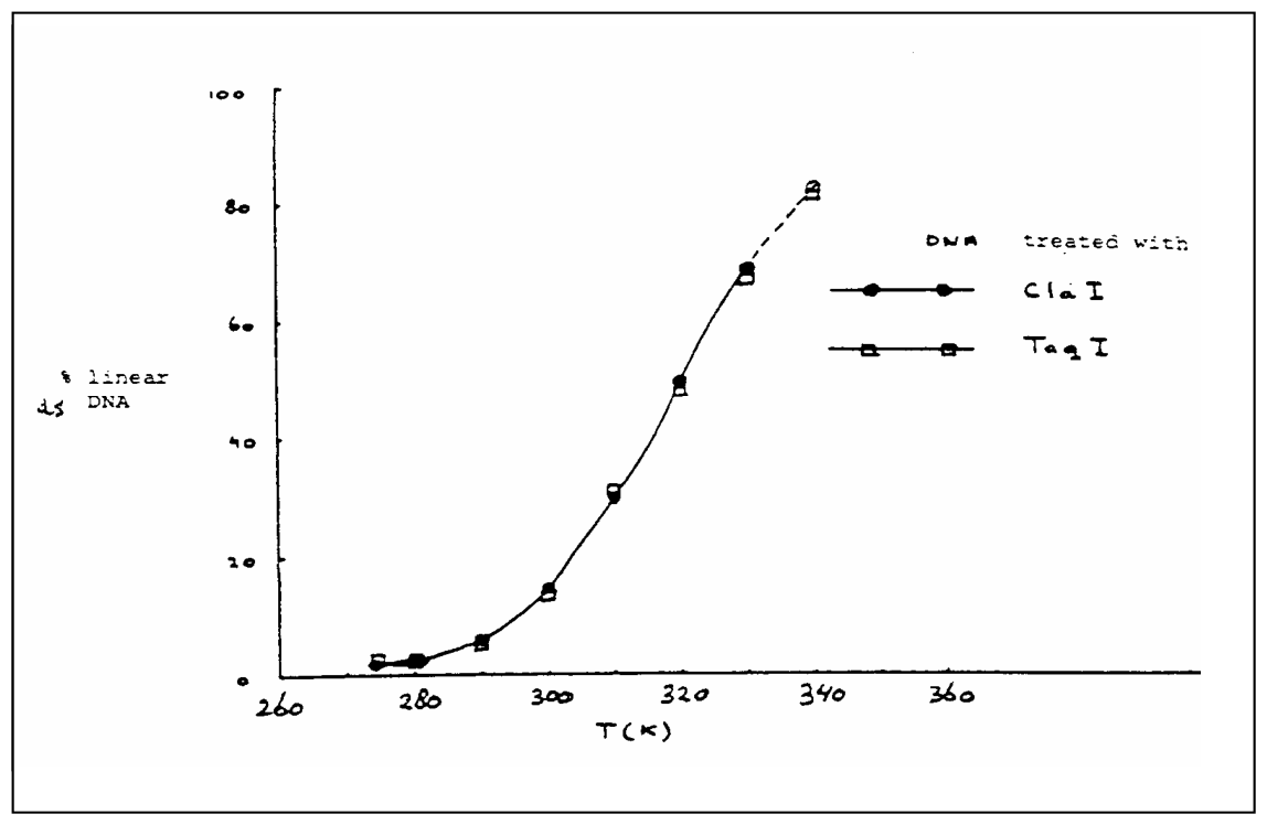
In Fig. 1 the percentage of linear DNA is given as a function of temperature, measured in a solution of 0.15 M \(\ce{NaCl}\) buffered with citrate at pH = 6.5. With Taq I as cleavage enzyme, the same curve is obtained.
6.6 Is the reaction as written endothermic or exothermic? Explain your answer.
6.7 Show, considering the information given, which of the two base sequences of the answer to 6.4 is the correct one.
6.8 What would look the curve for Taq I like if the recognition pattern would have been the other possibility of d)?
A large DNA molecule is cut into fragments with the aid of Cla I. One fragment is isolated, purified and mixed in the ratio of 1:1 with phage DNA which was also cleaved with Cla I. Thereby recombinant molecules can be formed through the reaction:

6.9 Would the enthalpy of this reaction be positive, negative or about zero? Explain your answer.
6.10 Which combination of temperature, DNA concentration and ionic strength (high or low in each case) will give the maximum percentage of recombinant molecules?
Official English text of the problem.
Answers
- 6.1: The complementary strand, written \(5'\rightarrow3'\), is also \(5'\text{-AT}\downarrow\text{CGAT-}3'\). Cla I produces two-nucleotide 5'-CG cohesive ends.
- 6.2: Approximately 24 occurrences in one strand: \(10^5/4^6\approx24.4\).
- 6.3: Four nucleotides per strand, corresponding to a four-base-pair recognition site.
- 6.4: \(5'\text{-T}\downarrow\text{CGA-}3'\) and \(5'\text{-GAT}\downarrow\text{C-}3'\).
- 6.5: Cutting the single site opens the circular duplex into one linear molecule with a 5'-CG overhang at each end, terminal 5'-phosphorylated C residues, and terminal 3'-hydroxyl T residues.
- 6.6: Circular \(\rightarrow\) linear is endothermic, \(\Delta H>0\).
- 6.7: Taq I recognizes \(5'\text{-TCGA-}3'\) and cleaves \(T\downarrow C\).
- 6.8: The alternative would give a similar rising curve shifted toward lower temperatures.
- 6.9: Negative enthalpy, \(\Delta H<0\), for association of the open, cleaved molecules through their cohesive ends.
- 6.10: Low temperature, low DNA concentration, and high ionic strength. Dilution suppresses formation of chains containing several DNA molecules; excessive dilution would also suppress the initial phage–fragment association.
Solution
6.1
The base symbols denote residues of adenine, A (Nc1ncnc2[nH]cnc12); thymine, T (Cc1c[nH]c(=O)[nH]c1=O); cytosine, C (Nc1cc[nH]c(=O)n1); and guanine, G (Nc1nc2[nH]cnc2c(=O)[nH]1). The SMILES in this sentence describe the corresponding free nucleobases.
A pairs with T, and C pairs with G. The strands run antiparallel, so the aligned recognition site and cleavage positions are
\[ \begin{array}{rccccccccl} 5'&A&T&\downarrow&C&G&&A&T&3'\\ 3'&T&A&&G&C&\uparrow&T&A&5' \end{array} \]
Reading the lower strand from its own 5' end gives
\[ \boxed{5'\text{-AT}\downarrow\text{CGAT-}3'}. \]
Thus ATCGAT is a reverse-complement palindrome. The staggered cuts leave two-nucleotide 5'-CG overhangs.
Each cleavage hydrolyses a phosphodiester linkage, producing a 3'-hydroxyl end and a 5'-phosphate end. A balanced representation, with \(R\) and \(R'\) standing for the sugar-containing DNA portions, is
\[ \ce{R-O-P(=O)(O^-)-O-R' + H2O -> R-OH + HO-P(=O)(O^-)-O-R'}. \]
This equation uses one possible protonation state of the terminal phosphate; the buffer controls subsequent proton exchange. Each double-strand cut consumes two water molecules. Water has SMILES O; the generic phosphodiester and phosphate monoester groups have SMILES *OP(=O)([O-])O* and *OP(=O)([O-])O, respectively, where * denotes an unspecified DNA portion.
For structural identification, the recognition sequences can be represented as isolated 5'-phosphorylated, 3'-hydroxy oligodeoxynucleotides. Their SMILES below encode the natural sugar stereochemistry and use neutral phosphate acid forms for notation. They do not encode duplex pairing or helicity. Complete SMILES for the phage and recombinant DNA cannot be specified because their remaining sequences are not given.
5'-Phosphorylated DNA hexamer d(ATCGAT):
O=P(O)(O)OC[C@H]1O[C@@H](n2cnc3c(N)ncnc32)C[C@@H]1OP(=O)(O)OC[C@H]1O[C@@H](n2cc(C)c(=O)[nH]c2=O)C[C@@H]1OP(=O)(O)OC[C@H]1O[C@@H](n2ccc(N)nc2=O)C[C@@H]1OP(=O)(O)OC[C@H]1O[C@@H](n2cnc3c(=O)[nH]c(N)nc32)C[C@@H]1OP(=O)(O)OC[C@H]1O[C@@H](n2cnc3c(N)ncnc32)C[C@@H]1OP(=O)(O)OC[C@H]1O[C@@H](n2cc(C)c(=O)[nH]c2=O)C[C@@H]1O
6.2
At a specified starting position, the probability of the particular six-base sequence is
\[ p=\left(\frac14\right)^6=\frac1{4096}. \]
For a linear strand containing \(N=10^5\) nucleotides, there are \(N-6+1\) possible starting positions. Therefore,
\[ \mathbb E[n] =(10^5-5)\frac1{4096} =24.4128\ldots \approx\boxed{24\text{ occurrences}}. \]
Neglecting the five terminal positions gives \(24.4141\), essentially the same result. The mean spacing is approximately \(4096\) base pairs.
There is no factor of two: the question asks about one strand, and the complementary occurrence identifies the same duplex site.
6.3
For a particular recognition sequence of length \(m\), the occurrence probability is \(4^{-m}\). In a long DNA molecule, the mean fragment length is consequently approximately \(4^m\) base pairs.
Thus,
\[ 4^m=256=4^4, \qquad \boxed{m=4}. \]
6.4
A terminal 3'-T and terminal 5'-C mean that cleavage occurs at
\[ 5'\text{-}\cdots T\downarrow C\cdots\text{-}3'. \]
A palindromic four-base recognition site has its first and fourth bases complementary, and its second and third bases complementary.
There are two possible placements of \(T\downarrow C\):
| Position of \(T\downarrow C\) | Recognition sequence | Aligned complementary strand |
|---|---|---|
| First–second bases | \(5'\text{-T}\downarrow\text{CGA-}3'\) | \(3'\text{-AGC}\uparrow\text{T-}5'\) |
| Third–fourth bases | \(5'\text{-GAT}\downarrow\text{C-}3'\) | \(3'\text{-C}\uparrow\text{TAG-}5'\) |
It cannot occupy the middle two positions because T and C are not complementary.
The two candidates therefore produce different cohesive ends:
- TCGA: two-nucleotide 5'-CG overhangs.
- GATC: two-nucleotide 3'-AT overhangs.
5'-Phosphorylated DNA tetramer d(TCGA):
O=P(O)(O)OC[C@H]1O[C@@H](n2cc(C)c(=O)[nH]c2=O)C[C@@H]1OP(=O)(O)OC[C@H]1O[C@@H](n2ccc(N)nc2=O)C[C@@H]1OP(=O)(O)OC[C@H]1O[C@@H](n2cnc3c(=O)[nH]c(N)nc32)C[C@@H]1OP(=O)(O)OC[C@H]1O[C@@H](n2cnc3c(N)ncnc32)C[C@@H]1O
5'-Phosphorylated DNA tetramer d(GATC):
O=P(O)(O)OC[C@H]1O[C@@H](n2cnc3c(=O)[nH]c(N)nc32)C[C@@H]1OP(=O)(O)OC[C@H]1O[C@@H](n2cnc3c(N)ncnc32)C[C@@H]1OP(=O)(O)OC[C@H]1O[C@@H](n2cc(C)c(=O)[nH]c2=O)C[C@@H]1OP(=O)(O)OC[C@H]1O[C@@H](n2ccc(N)nc2=O)C[C@@H]1O
6.5
The requested drawing should show the following.
For the original circular molecule, draw two continuous antiparallel circular strands. At the unique recognition site, place the paired bases and staggered cleavage positions exactly as in 6.1. The intact circle has no free 3' or 5' termini; these labels indicate strand direction.
For the linear molecule, open both backbones at those positions and separate the cohesive ends. Reading the resulting duplex from left to right:
- The upper strand starts \(5'\text{-pCGAT}\cdots\) and ends \(\cdots\text{AT-}3'\).
- The lower strand starts \(3'\text{-TA}\cdots\) and ends \(\cdots\text{TAGC-p-}5'\).
At the left end, the upper strand’s initial CG projects beyond the lower strand. At the right end, the lower strand’s terminal GC, read left to right, projects beyond the upper strand. Read in its own \(5'\rightarrow3'\) direction, that overhang is also CG.
Thus every 5' terminus is a phosphorylated C, and every 3' terminus is a T bearing a free hydroxyl group.
After cleavage, these complementary overhangs can pair again to produce a circular, annealed molecule with two backbone nicks. This is the circular species participating in the temperature-dependent equilibrium:
\[ \ce{DNA_{circular,annealed} <=> DNA_{linear}}. \]
Annealing alone does not restore the hydrolysed covalent bonds; covalent sealing would require an additional ligation step.
6.6
The fraction of linear DNA increases as the temperature increases. Heating therefore favours the forward direction,
\[ \ce{DNA_{circular,annealed} -> DNA_{linear}}, \]
so this direction is endothermic:
\[ \boxed{\Delta H>0}. \]
More formally, if \(f\) is the fraction of linear molecules,
\[ K=\frac{[\text{linear DNA}]}{[\text{circular DNA}]} =\frac{f}{1-f}. \]
The graph shows \(K\) increasing with temperature. The van ’t Hoff relation,
\[ \frac{d\ln K}{dT}=\frac{\Delta H^\circ}{RT^2}, \]
then gives \(\Delta H^\circ>0\).
Physically, opening the annealed circle disrupts the base pairing and stacking at its cohesive junction. This conclusion concerns end dissociation, not the initial phosphodiester hydrolysis.
6.7
Cla I produces 5'-CG overhangs. Closing their junction forms two G–C base pairs.
Of the two Taq I candidates:
- \(T\downarrow CGA\) produces the same 5'-CG overhangs.
- \(GAT\downarrow C\) produces 3'-AT overhangs, which would form two A–T pairs.
The identical temperature curves therefore select, within the model of the problem,
\[ \boxed{5'\text{-T}\downarrow\text{CGA-}3'}. \]
Indeed, this recognition sequence lies within the Cla I site:
\[ 5'\text{-A}\underbrace{\text{T}\downarrow\text{CGA}}_{\text{Taq I site}}\text{T-}3'. \]
Both enzymes consequently cut the same bonds at this site.
6.8
The alternative \(GAT\downarrow C\) site would produce AT cohesive ends. Their annealing would form two A–T pairs instead of two G–C pairs.
A–T pairs have two hydrogen bonds each, whereas G–C pairs have three. In the intended comparison, the AT junction is less stable and dissociates at a lower temperature.
The requested curve should therefore be described as a similar increasing, approximately sigmoidal curve shifted to the left:
- At a given temperature in the transition region, the percentage of linear DNA would be higher.
- The temperature at which 50% is linear would be lower.
The supplied information does not determine the numerical shift.
6.9
Let \(P\) denote the open phage DNA, \(F\) the open fragment, and \(R\) the intended circular recombinant containing one of each:
\[ \ce{P_{linear} + F_{linear} <=> R_{circular,annealed}}. \]
Each molecule supplies two compatible cohesive ends. Formation of the recombinant circle produces two cohesive junctions, each containing two G–C pairs: four newly formed G–C pairs in total.
Base pairing and stacking release heat, so
\[ \boxed{\Delta H<0}. \]
There is no release of water in this annealing reaction. The backbone nicks remain unless a subsequent covalent ligation step is performed.
6.10
The intended qualitative combination is
\[ \boxed{\text{low temperature; low DNA concentration; high ionic strength}.} \]
- Low temperature: Cohesive-end association is exothermic, so cooling favours the associated recombinant.
- Low DNA concentration: After one phage end joins one fragment end, the remaining two ends are tethered within the same combined molecule and can close it into a circle. At high concentration, they can instead encounter additional DNA molecules and form longer chains. Dilution suppresses these additional intermolecular encounters relative to intramolecular closure.
- High ionic strength: Salt, such as sodium chloride (
[Na+].[Cl-]), screens electrostatic repulsion between the negatively charged phosphate backbones and stabilizes the paired cohesive ends.
The concentration argument must therefore include both ends of each molecule. Applying mass action only to the first bimolecular association would overlook chain formation.
“Low concentration” does not mean unlimited dilution: excessive dilution suppresses the initial phage–fragment encounter and favours separate self-circularization. The useful condition is sufficiently dilute to suppress multimer formation while still permitting the initial association.
The solution was checked against the official solution.
IChO 1986 · Theorieaufgabe 6
Restriktionsendonukleasen Cla I und Taq I und rekombinante DNA
In der rekombinanten DNA-Technologie können spezifische Endonukleasen die Phosphorsäureesterbindung in jedem der beiden Stränge erkennen und hydrolysieren. Cla I hydrolysiert beispielsweise die Bindung zwischen zwei Nukleotiden in der Sequenz:
6.1 Geben Sie die Basensequenz des komplementären Strangs in 5' - 3'-Richtung an und kennzeichnen Sie mit Pfeilen die Stellen, an denen die Hydrolyse durch Cla I stattfinden würde.
6.2 Wie oft kommt diese Sequenz durchschnittlich in einem Strang eines DNA-Moleküls mit \(10^5\) Basenpaaren vor? Sie können annehmen, dass die vier Basen gleich häufig vorkommen und in den beiden Ketten zufällig verteilt sind.
Taq I hydrolysiert ein langes doppelsträngiges DNA-Molekül zu Fragmenten, die im Durchschnitt 256 Basenpaare lang sind. Das 3'-Ende dieser durch Spaltung erhaltenen Fragmente erweist sich als Thymin(T)-Ende und das 5'-Ende als Cytosin(C)-Ende.
6.3 Wie lang ist die von Taq I erkannte Sequenz?
6.4 Geben Sie die beiden möglichen Basensequenzen (in 5' - 3'-Richtung) an, die das Erkennungsmuster für Taq I bilden (dieses muss offensichtlich eine gewisse Symmetrie aufweisen).
Die DNA eines Phagen, die als geschlossener Ring vorliegt, enthält in jedem der beiden Stränge nur eine 5'-pApTpCpGpApT-3'-Sequenz. Nach der Behandlung mit ClaI stellt sich ein Gleichgewicht ein:
6.5 Zeichnen Sie schematisch die ringförmigen und linearen Moleküle. Geben Sie die an die Spaltstelle angrenzenden Basen in beiden Strängen an. Kennzeichnen Sie auch die 3'- und 5'-Enden.
In Abb. 1 ist der prozentuale Anteil linearer DNA in Abhängigkeit von der Temperatur dargestellt, gemessen in einer mit Citrat auf pH = 6.5 gepufferten Lösung von 0.15 M \(\ce{NaCl}\). Mit Taq I als Spaltungsenzym erhält man dieselbe Kurve.
6.6 Ist die Reaktion in der angegebenen Richtung endotherm oder exotherm? Begründen Sie Ihre Antwort.
6.7 Zeigen Sie unter Berücksichtigung der gegebenen Informationen, welche der beiden Basensequenzen aus der Antwort auf 6.4 die richtige ist.
6.8 Wie würde die Kurve für Taq I aussehen, wenn das Erkennungsmuster der anderen Möglichkeit aus d) entspräche?
Ein großes DNA-Molekül wird mithilfe von Cla I in Fragmente geschnitten. Ein Fragment wird isoliert, gereinigt und im Verhältnis 1:1 mit Phagen-DNA gemischt, die ebenfalls mit Cla I gespalten wurde. Dabei können durch folgende Reaktion rekombinante Moleküle gebildet werden:
6.9 Wäre die Enthalpie dieser Reaktion positiv, negativ oder ungefähr null? Begründen Sie Ihre Antwort.
6.10 Welche Kombination aus Temperatur, DNA-Konzentration und Ionenstärke (jeweils hoch oder niedrig) ergibt den maximalen prozentualen Anteil rekombinanter Moleküle?
Aufgabentext aus dem offiziellen englischen Text übersetzt.
Antworten
- 6.1: Der komplementäre Strang lautet, in \(5'\rightarrow3'\)-Richtung geschrieben, ebenfalls \(5'\text{-AT}\downarrow\text{CGAT-}3'\). Cla I erzeugt kohäsive 5'-CG-Enden aus zwei Nukleotiden.
- 6.2: Ungefähr 24 Vorkommen in einem Strang: \(10^5/4^6\approx24.4\).
- 6.3: Vier Nukleotide pro Strang, entsprechend einer Erkennungsstelle aus vier Basenpaaren.
- 6.4: \(5'\text{-T}\downarrow\text{CGA-}3'\) und \(5'\text{-GAT}\downarrow\text{C-}3'\).
- 6.5: Die Spaltung an der einzigen Schnittstelle öffnet den ringförmigen Doppelstrang zu einem linearen Molekül mit einem 5'-CG-Überhang an jedem Ende, terminalen 5'-phosphorylierten C-Resten und terminalen T-Resten mit 3'-Hydroxylgruppe.
- 6.6: Ringförmig \(\rightarrow\) linear ist endotherm, \(\Delta H>0\).
- 6.7: Taq I erkennt \(5'\text{-TCGA-}3'\) und spaltet bei \(T\downarrow C\).
- 6.8: Die Alternative würde eine ähnliche ansteigende Kurve ergeben, die zu niedrigeren Temperaturen verschoben ist.
- 6.9: Negative Enthalpie, \(\Delta H<0\), für die Assoziation der offenen, gespaltenen Moleküle über ihre kohäsiven Enden.
- 6.10: Niedrige Temperatur, niedrige DNA-Konzentration und hohe Ionenstärke. Verdünnung unterdrückt die Bildung von Ketten aus mehreren DNA-Molekülen; eine zu starke Verdünnung würde auch die anfängliche Assoziation zwischen Phagen-DNA und Fragment unterdrücken.
Lösung
6.1
Die Basensymbole bezeichnen Reste von Adenin, A (Nc1ncnc2[nH]cnc12); Thymin, T (Cc1c[nH]c(=O)[nH]c1=O); Cytosin, C (Nc1cc[nH]c(=O)n1); und Guanin, G (Nc1nc2[nH]cnc2c(=O)[nH]1). Die SMILES in diesem Satz beschreiben die entsprechenden freien Nukleobasen.
A paart mit T und C paart mit G. Die Stränge verlaufen antiparallel, sodass die einander gegenübergestellten Stränge an der Erkennungsstelle und die Spaltpositionen wie folgt aussehen:
\[ \begin{array}{rccccccccl} 5'&A&T&\downarrow&C&G&&A&T&3'\\ 3'&T&A&&G&C&\uparrow&T&A&5' \end{array} \]
Liest man den unteren Strang von seinem eigenen 5'-Ende aus, erhält man
\[ \boxed{5'\text{-AT}\downarrow\text{CGAT-}3'}. \]
Somit ist ATCGAT ein Palindrom bezüglich der umgekehrten Komplementärsequenz. Die versetzten Schnitte hinterlassen 5'-CG-Überhänge aus zwei Nukleotiden.
Bei jeder Spaltung wird eine Phosphodiesterbindung hydrolysiert, wobei ein 3'-Hydroxylende und ein 5'-Phosphatende entstehen. Eine ausgeglichene Darstellung, in der \(R\) und \(R'\) für die zuckerhaltigen DNA-Abschnitte stehen, lautet
\[ \ce{R-O-P(=O)(O^-)-O-R' + H2O -> R-OH + HO-P(=O)(O^-)-O-R'}. \]
Diese Gleichung verwendet einen möglichen Protonierungszustand des terminalen Phosphats; der Puffer reguliert den anschließenden Protonenaustausch. Jeder Doppelstrangschnitt verbraucht zwei Wassermoleküle. Wasser hat die SMILES O; die allgemeinen Phosphodiester- und Phosphatmonoestergruppen haben die SMILES *OP(=O)([O-])O* beziehungsweise *OP(=O)([O-])O, wobei * einen nicht näher bestimmten DNA-Abschnitt bezeichnet.
Zur strukturellen Identifizierung können die Erkennungssequenzen als isolierte 5'-phosphorylierte Oligodesoxynukleotide mit 3'-Hydroxylgruppe dargestellt werden. Ihre unten angegebenen SMILES kodieren die natürliche Stereochemie der Zucker und verwenden zur Darstellung neutrale Säureformen der Phosphatgruppen. Sie kodieren weder die Paarung zum Doppelstrang noch die Helizität. Vollständige SMILES für die Phagen-DNA und die rekombinante DNA können nicht angegeben werden, da ihre übrigen Sequenzen nicht bekannt sind.
5'-Phosphoryliertes DNA-Hexamer d(ATCGAT):
O=P(O)(O)OC[C@H]1O[C@@H](n2cnc3c(N)ncnc32)C[C@@H]1OP(=O)(O)OC[C@H]1O[C@@H](n2cc(C)c(=O)[nH]c2=O)C[C@@H]1OP(=O)(O)OC[C@H]1O[C@@H](n2ccc(N)nc2=O)C[C@@H]1OP(=O)(O)OC[C@H]1O[C@@H](n2cnc3c(=O)[nH]c(N)nc32)C[C@@H]1OP(=O)(O)OC[C@H]1O[C@@H](n2cnc3c(N)ncnc32)C[C@@H]1OP(=O)(O)OC[C@H]1O[C@@H](n2cc(C)c(=O)[nH]c2=O)C[C@@H]1O
6.2
An einer bestimmten Startposition beträgt die Wahrscheinlichkeit für die betreffende Sequenz aus sechs Basen
\[ p=\left(\frac14\right)^6=\frac1{4096}. \]
Für einen linearen Strang mit \(N=10^5\) Nukleotiden gibt es \(N-6+1\) mögliche Startpositionen. Daher gilt
\[ \mathbb E[n] =(10^5-5)\frac1{4096} =24.4128\ldots \approx\boxed{24\text{ Vorkommen}}. \]
Vernachlässigt man die fünf endständigen Positionen, erhält man \(24.4141\), also praktisch dasselbe Ergebnis. Der mittlere Abstand beträgt ungefähr \(4096\) Basenpaare.
Ein Faktor zwei tritt nicht auf: Die Frage bezieht sich auf einen Strang, und das komplementäre Vorkommen kennzeichnet dieselbe Stelle im Doppelstrang.
6.3
Für eine bestimmte Erkennungssequenz der Länge \(m\) beträgt die Wahrscheinlichkeit ihres Auftretens \(4^{-m}\). In einem langen DNA-Molekül beträgt die mittlere Fragmentlänge folglich ungefähr \(4^m\) Basenpaare.
Somit gilt
\[ 4^m=256=4^4, \qquad \boxed{m=4}. \]
6.4
Ein terminales 3'-T und ein terminales 5'-C bedeuten, dass die Spaltung an folgender Stelle erfolgt:
\[ 5'\text{-}\cdots T\downarrow C\cdots\text{-}3'. \]
Bei einer palindromischen Erkennungsstelle aus vier Basen sind die erste und vierte Base sowie die zweite und dritte Base jeweils komplementär.
Für \(T\downarrow C\) gibt es zwei mögliche Positionen:
| Position von \(T\downarrow C\) | Erkennungssequenz | Gegenübergestellter komplementärer Strang |
|---|---|---|
| Erste–zweite Base | \(5'\text{-T}\downarrow\text{CGA-}3'\) | \(3'\text{-AGC}\uparrow\text{T-}5'\) |
| Dritte–vierte Base | \(5'\text{-GAT}\downarrow\text{C-}3'\) | \(3'\text{-C}\uparrow\text{TAG-}5'\) |
Die beiden mittleren Positionen kommen nicht infrage, da T und C nicht komplementär sind.
Die beiden Kandidaten erzeugen daher unterschiedliche kohäsive Enden:
- TCGA: 5'-CG-Überhänge aus zwei Nukleotiden.
- GATC: 3'-AT-Überhänge aus zwei Nukleotiden.
5'-Phosphoryliertes DNA-Tetramer d(TCGA):
O=P(O)(O)OC[C@H]1O[C@@H](n2cc(C)c(=O)[nH]c2=O)C[C@@H]1OP(=O)(O)OC[C@H]1O[C@@H](n2ccc(N)nc2=O)C[C@@H]1OP(=O)(O)OC[C@H]1O[C@@H](n2cnc3c(=O)[nH]c(N)nc32)C[C@@H]1OP(=O)(O)OC[C@H]1O[C@@H](n2cnc3c(N)ncnc32)C[C@@H]1O
5'-Phosphoryliertes DNA-Tetramer d(GATC):
O=P(O)(O)OC[C@H]1O[C@@H](n2cnc3c(=O)[nH]c(N)nc32)C[C@@H]1OP(=O)(O)OC[C@H]1O[C@@H](n2cnc3c(N)ncnc32)C[C@@H]1OP(=O)(O)OC[C@H]1O[C@@H](n2cc(C)c(=O)[nH]c2=O)C[C@@H]1OP(=O)(O)OC[C@H]1O[C@@H](n2ccc(N)nc2=O)C[C@@H]1O
6.5
Die geforderte Zeichnung sollte Folgendes zeigen.
Zeichnen Sie für das ursprüngliche ringförmige Molekül zwei durchgehende antiparallele ringförmige Stränge. Tragen Sie an der einzigen Erkennungsstelle die gepaarten Basen und die versetzten Spaltpositionen genau wie in 6.1 ein. Der intakte Ring besitzt keine freien 3'- oder 5'-Enden; diese Beschriftungen geben die Strangrichtung an.
Öffnen Sie für das lineare Molekül beide Rückgrate an diesen Positionen und trennen Sie die kohäsiven Enden. Liest man den entstehenden Doppelstrang von links nach rechts, gilt:
- Der obere Strang beginnt mit \(5'\text{-pCGAT}\cdots\) und endet mit \(\cdots\text{AT-}3'\).
- Der untere Strang beginnt mit \(3'\text{-TA}\cdots\) und endet mit \(\cdots\text{TAGC-p-}5'\).
Am linken Ende ragt das anfängliche CG des oberen Strangs über den unteren Strang hinaus. Am rechten Ende ragt das terminale GC des unteren Strangs, von links nach rechts gelesen, über den oberen Strang hinaus. In seiner eigenen \(5'\rightarrow3'\)-Richtung gelesen lautet auch dieser Überhang CG.
Somit ist jedes 5'-Ende ein phosphoryliertes C und jedes 3'-Ende ein T mit einer freien Hydroxylgruppe.
Nach der Spaltung können sich diese komplementären Überhänge erneut paaren und ein ringförmiges, hybridisiertes Molekül mit zwei Einzelstrangunterbrechungen im Rückgrat bilden. Dies ist die ringförmige Spezies, die am temperaturabhängigen Gleichgewicht teilnimmt:
\[ \ce{DNA_{circular,annealed} <=> DNA_{linear}}. \]
Die Hybridisierung allein stellt die hydrolysierten kovalenten Bindungen nicht wieder her; ein kovalenter Verschluss würde einen zusätzlichen Ligationsschritt erfordern.
6.6
Der Anteil linearer DNA nimmt mit steigender Temperatur zu. Erwärmen begünstigt daher die Hinreaktion,
\[ \ce{DNA_{circular,annealed} -> DNA_{linear}}, \]
somit ist diese Richtung endotherm:
\[ \boxed{\Delta H>0}. \]
Formal gilt, wenn \(f\) der Anteil linearer Moleküle ist,
\[ K=\frac{[\text{lineare DNA}]}{[\text{ringförmige DNA}]} =\frac{f}{1-f}. \]
Die Kurve zeigt, dass \(K\) mit der Temperatur zunimmt. Die van-’t-Hoff-Beziehung,
\[ \frac{d\ln K}{dT}=\frac{\Delta H^\circ}{RT^2}, \]
ergibt dann \(\Delta H^\circ>0\).
Physikalisch betrachtet werden beim Öffnen des hybridisierten Rings die Basenpaarung und die Basenstapelung an der Verbindungsstelle seiner kohäsiven Enden aufgebrochen. Diese Schlussfolgerung betrifft die Dissoziation der Enden, nicht die anfängliche Phosphodiesterhydrolyse.
6.7
Cla I erzeugt 5'-CG-Überhänge. Beim Schließen ihrer Verbindungsstelle entstehen zwei G–C-Basenpaare.
Für die beiden Taq-I-Kandidaten gilt:
- \(T\downarrow CGA\) erzeugt dieselben 5'-CG-Überhänge.
- \(GAT\downarrow C\) erzeugt 3'-AT-Überhänge, die zwei A–T-Paare bilden würden.
Die identischen Temperaturkurven ergeben daher im Rahmen des Modells der Aufgabe die Auswahl
\[ \boxed{5'\text{-T}\downarrow\text{CGA-}3'}. \]
Tatsächlich liegt diese Erkennungssequenz innerhalb der Cla-I-Stelle:
\[ 5'\text{-A}\underbrace{\text{T}\downarrow\text{CGA}}_{\text{Taq-I-Stelle}}\text{T-}3'. \]
Beide Enzyme spalten folglich an dieser Stelle dieselben Bindungen.
6.8
Die alternative \(GAT\downarrow C\)-Stelle würde kohäsive AT-Enden erzeugen. Bei ihrer Hybridisierung würden zwei A–T-Paare anstelle von zwei G–C-Paaren entstehen.
A–T-Paare besitzen jeweils zwei Wasserstoffbrückenbindungen, G–C-Paare hingegen drei. Im beabsichtigten Vergleich ist die AT-Verbindungsstelle weniger stabil und dissoziiert bei einer niedrigeren Temperatur.
Die geforderte Kurve sollte daher als ähnliche ansteigende, annähernd sigmoidale, nach links verschobene Kurve beschrieben werden:
- Bei einer gegebenen Temperatur im Übergangsbereich wäre der prozentuale Anteil linearer DNA höher.
- Die Temperatur, bei der 50% linear vorliegen, wäre niedriger.
Aus den gegebenen Informationen lässt sich die Verschiebung nicht quantitativ bestimmen.
6.9
Es bezeichne \(P\) die offene Phagen-DNA, \(F\) das offene Fragment und \(R\) die angestrebte ringförmige rekombinante DNA, die jeweils eines von beiden enthält:
\[ \ce{P_{linear} + F_{linear} <=> R_{circular,annealed}}. \]
Jedes Molekül stellt zwei kompatible kohäsive Enden bereit. Bei der Bildung des rekombinanten Rings entstehen zwei Verbindungsstellen kohäsiver Enden, die jeweils zwei G–C-Paare enthalten: insgesamt vier neu gebildete G–C-Paare.
Basenpaarung und Basenstapelung setzen Wärme frei, daher gilt
\[ \boxed{\Delta H<0}. \]
Bei dieser Hybridisierungsreaktion wird kein Wasser freigesetzt. Die Einzelstrangunterbrechungen im Rückgrat bleiben bestehen, sofern nicht anschließend ein kovalenter Ligationsschritt durchgeführt wird.
6.10
Die gesuchte qualitative Kombination lautet
\[ \boxed{\text{niedrige Temperatur; niedrige DNA-Konzentration; hohe Ionenstärke}.} \]
- Niedrige Temperatur: Die Assoziation kohäsiver Enden ist exotherm, daher begünstigt Abkühlen die assoziierte rekombinante DNA.
- Niedrige DNA-Konzentration: Nachdem sich ein Ende der Phagen-DNA mit einem Ende des Fragments verbunden hat, sind die beiden verbleibenden Enden innerhalb desselben vereinigten Moleküls miteinander verbunden und können dieses zu einem Ring schließen. Bei hoher Konzentration können sie stattdessen auf weitere DNA-Moleküle treffen und längere Ketten bilden. Verdünnung unterdrückt diese zusätzlichen intermolekularen Begegnungen gegenüber dem intramolekularen Ringschluss.
- Hohe Ionenstärke: Salz, beispielsweise Natriumchlorid (
[Na+].[Cl-]), schirmt die elektrostatische Abstoßung zwischen den negativ geladenen Phosphatrückgraten ab und stabilisiert die gepaarten kohäsiven Enden.
Die Argumentation zur Konzentration muss daher beide Enden jedes Moleküls berücksichtigen. Würde man das Massenwirkungsgesetz nur auf die erste bimolekulare Assoziation anwenden, bliebe die Kettenbildung unberücksichtigt.
„Niedrige Konzentration“ bedeutet nicht unbegrenzte Verdünnung: Eine zu starke Verdünnung unterdrückt die anfängliche Begegnung zwischen Phagen-DNA und Fragment und begünstigt den getrennten Ringschluss der einzelnen Moleküle. Zweckmäßig ist eine Verdünnung, die ausreicht, um die Multimerbildung zu unterdrücken, zugleich aber noch die anfängliche Assoziation ermöglicht.
Die Lösung wurde mit der offiziellen Lösung abgeglichen.
IChO 1986 · Teori sorusu 6
Restriksiyon endonükleazları Cla I ve Taq I ve rekombinant DNA
Rekombinant DNA teknolojisinde özgül endonükleazlar, her iki zincirdeki fosforik asit ester bağını tanıyabilir ve hidroliz edebilir. Örneğin Cla I, aşağıdaki dizide iki nükleotit arasındaki bağı hidroliz eder:
6.1 Tamamlayıcı zincirin baz dizisini 5' - 3' yönünde yazınız ve Cla I ile hidrolizin gerçekleşeceği yeri oklarla gösteriniz.
6.2 Bu dizi, \(10^5\) baz çiftinden oluşan bir DNA molekülünün bir zincirinde ortalama kaç kez bulunur? Dört bazın eşit sıklıkta bulunduğunu ve iki zincire rastgele dağıldığını varsayabilirsiniz.
Taq I, uzun bir çift zincirli DNA molekülünü, ortalama uzunluğu 256 baz çifti olan parçalara hidroliz eder. Kesim sonucunda oluşan bu parçaların 3' ucunun bir timin(T) ucu, 5' ucunun ise bir sitozin(C) ucu olduğu belirlenmiştir.
6.3 Taq I tarafından tanınan dizinin uzunluğu nedir?
6.4 Taq I için tanıma örüntüsünü oluşturan iki olası baz dizisini (5' - 3' yönünde) yazınız (bunların belirli bir simetriye sahip olması gerektiği açıktır).
Kapalı bir halka biçiminde bulunan bir fajın DNA'sı, iki zincirinin her birinde yalnızca bir 5'-pApTpCpGpApT-3' dizisi içerir. ClaI ile işlemden sonra şu denge kurulur:
6.5 Halkasal ve doğrusal moleküllerin şematik bir çizimini yapınız. Her iki zincirde kesim yerine komşu bazları gösteriniz. Ayrıca 3' ve 5' uçlarını da belirtiniz.
Şekil 1'de, pH = 6.5'te sitrat ile tamponlanmış 0.15 M \(\ce{NaCl}\) çözeltisinde ölçülen doğrusal DNA yüzdesi, sıcaklığın bir fonksiyonu olarak verilmiştir. Kesim enzimi olarak Taq I kullanıldığında da aynı eğri elde edilir.
6.6 Tepkime, yazıldığı yönde endotermik midir, ekzotermik midir? Yanıtınızı açıklayınız.
6.7 Verilen bilgileri dikkate alarak 6.4 sorusunun yanıtındaki iki baz dizisinden hangisinin doğru olduğunu gösteriniz.
6.8 Tanıma örüntüsü d)'deki diğer olasılık olsaydı, Taq I için eğri nasıl görünürdü?
Büyük bir DNA molekülü, Cla I yardımıyla parçalara kesilir. Parçalardan biri ayrılır, saflaştırılır ve yine Cla I ile kesilmiş faj DNA'sı ile 1:1 oranında karıştırılır. Böylece aşağıdaki tepkime yoluyla rekombinant moleküller oluşabilir:
6.9 Bu tepkimenin entalpisi pozitif mi, negatif mi, yoksa yaklaşık sıfır mı olur? Yanıtınızı açıklayınız.
6.10 Sıcaklık, DNA derişimi ve iyonik şiddetin hangi birleşimi (her biri için yüksek veya düşük), en yüksek rekombinant molekül yüzdesini verir?
Soru metni resmi İngilizce metinden çevrilmiştir.
Cevaplar
- 6.1: \(5'\rightarrow3'\) yönünde yazılan tamamlayıcı zincir de \(5'\text{-AT}\downarrow\text{CGAT-}3'\) biçimindedir. Cla I, iki nükleotit uzunluğunda 5'-CG yapışkan uçları oluşturur.
- 6.2: Bir zincirde yaklaşık 24 kez bulunur: \(10^5/4^6\approx24.4\).
- 6.3: Zincir başına dört nükleotit; bu, dört baz çiftlik bir tanıma bölgesine karşılık gelir.
- 6.4: \(5'\text{-T}\downarrow\text{CGA-}3'\) ve \(5'\text{-GAT}\downarrow\text{C-}3'\).
- 6.5: Tek bölgedeki kesim, halkasal çift zincirli yapıyı her iki ucunda birer 5'-CG çıkıntısı bulunan tek bir doğrusal moleküle açar; uçlardaki C kalıntıları 5' konumunda fosforillenmiştir ve uçlardaki T kalıntıları 3'-hidroksil grupları taşır.
- 6.6: Halkasal \(\rightarrow\) doğrusal dönüşümü endotermiktir, \(\Delta H>0\).
- 6.7: Taq I, \(5'\text{-TCGA-}3'\) dizisini tanır ve \(T\downarrow C\) noktasında keser.
- 6.8: Diğer olasılık, daha düşük sıcaklıklara kaymış, benzer bir yükselen eğri verir.
- 6.9: Açık, kesilmiş moleküllerin yapışkan uçları aracılığıyla birleşmesi için entalpi negatiftir, \(\Delta H<0\).
- 6.10: Düşük sıcaklık, düşük DNA derişimi ve yüksek iyonik şiddet. Seyreltme, birkaç DNA molekülü içeren zincirlerin oluşumunu baskılar; aşırı seyreltme ise başlangıçtaki faj–parça birleşmesini de baskılar.
Çözüm
6.1
Baz simgeleri adenin, A (Nc1ncnc2[nH]cnc12); timin, T (Cc1c[nH]c(=O)[nH]c1=O); sitozin, C (Nc1cc[nH]c(=O)n1); ve guanin, G (Nc1nc2[nH]cnc2c(=O)[nH]1) kalıntılarını gösterir. Bu cümledeki SMILES gösterimleri, karşılık gelen serbest nükleobazları tanımlar.
A, T ile; C ise G ile eşleşir. Zincirler antiparalel uzandığından, hizalanmış tanıma bölgesi ve kesim konumları şöyledir:
\[ \begin{array}{rccccccccl} 5'&A&T&\downarrow&C&G&&A&T&3'\\ 3'&T&A&&G&C&\uparrow&T&A&5' \end{array} \]
Alt zincir kendi 5' ucundan okunursa şu dizi elde edilir:
\[ \boxed{5'\text{-AT}\downarrow\text{CGAT-}3'}. \]
Dolayısıyla ATCGAT, ters tamamlayıcı bir palindromdur. Birbirine göre kaymış konumlardaki kesimler, iki nükleotit uzunluğunda 5'-CG çıkıntıları bırakır.
Her kesim bir fosfodiester bağını hidroliz ederek bir 3'-hidroksil ucu ve bir 5'-fosfat ucu oluşturur. \(R\) ve \(R'\), DNA'nın şeker içeren kısımlarını göstermek üzere, denkleştirilmiş bir gösterim şöyledir:
\[ \ce{R-O-P(=O)(O^-)-O-R' + H2O -> R-OH + HO-P(=O)(O^-)-O-R'}. \]
Bu denklemde uç fosfat grubunun olası protonlanma durumlarından biri kullanılmıştır; daha sonraki proton alışverişini tampon kontrol eder. Her çift zincir kesimi iki su molekülü tüketir. Suyun SMILES gösterimi O'dur; genel fosfodiester ve fosfat monoester gruplarının SMILES gösterimleri sırasıyla *OP(=O)([O-])O* ve *OP(=O)([O-])O şeklindedir; burada *, DNA'nın belirtilmemiş bir kısmını gösterir.
Yapısal tanımlama amacıyla tanıma dizileri, izole edilmiş 5'-fosforillenmiş, 3'-hidroksi oligodeoksinükleotitler olarak gösterilebilir. Aşağıdaki SMILES gösterimleri, doğal şeker stereokimyasını kodlar ve gösterim için fosfat gruplarının nötr asit biçimlerini kullanır. Bunlar çift zincir eşleşmesini veya sarmallığı kodlamaz. Faj DNA'sının ve rekombinant DNA'nın geri kalan dizileri verilmediğinden, bunların tam SMILES gösterimleri belirtilemez.
5'-Fosforillenmiş DNA heksameri d(ATCGAT):
O=P(O)(O)OC[C@H]1O[C@@H](n2cnc3c(N)ncnc32)C[C@@H]1OP(=O)(O)OC[C@H]1O[C@@H](n2cc(C)c(=O)[nH]c2=O)C[C@@H]1OP(=O)(O)OC[C@H]1O[C@@H](n2ccc(N)nc2=O)C[C@@H]1OP(=O)(O)OC[C@H]1O[C@@H](n2cnc3c(=O)[nH]c(N)nc32)C[C@@H]1OP(=O)(O)OC[C@H]1O[C@@H](n2cnc3c(N)ncnc32)C[C@@H]1OP(=O)(O)OC[C@H]1O[C@@H](n2cc(C)c(=O)[nH]c2=O)C[C@@H]1O
6.2
Belirli bir başlangıç konumunda, söz konusu altı bazlık dizinin bulunma olasılığı şöyledir:
\[ p=\left(\frac14\right)^6=\frac1{4096}. \]
\(N=10^5\) nükleotit içeren doğrusal bir zincirde \(N-6+1\) olası başlangıç konumu vardır. Dolayısıyla,
\[ \mathbb E[n] =(10^5-5)\frac1{4096} =24.4128\ldots \approx\boxed{24\text{ kez}}. \]
Uçtaki beş konumun ihmal edilmesi \(24.4141\) sonucunu verir; bu da esasen aynı sonuçtur. Ortalama aralık yaklaşık \(4096\) baz çiftidir.
İki çarpanı yoktur: soru bir zincirle ilgilidir ve tamamlayıcı zincirdeki karşılık gelen dizi, aynı çift zincirli bölgeyi tanımlar.
6.3
Uzunluğu \(m\) olan belirli bir tanıma dizisinin bulunma olasılığı \(4^{-m}\)'dir. Bu nedenle, uzun bir DNA molekülünde ortalama parça uzunluğu yaklaşık \(4^m\) baz çiftidir.
Böylece,
\[ 4^m=256=4^4, \qquad \boxed{m=4}. \]
6.4
Uçlarda 3'-T ve 5'-C bulunması, kesimin şu noktada gerçekleştiği anlamına gelir:
\[ 5'\text{-}\cdots T\downarrow C\cdots\text{-}3'. \]
Palindromik, dört bazlık bir tanıma bölgesinde birinci ve dördüncü bazlar birbirini tamamlar; ikinci ve üçüncü bazlar da birbirini tamamlar.
\(T\downarrow C\) için iki olası yerleşim vardır:
| \(T\downarrow C\)'nin konumu | Tanıma dizisi | Hizalanmış tamamlayıcı zincir |
|---|---|---|
| Birinci–ikinci bazlar | \(5'\text{-T}\downarrow\text{CGA-}3'\) | \(3'\text{-AGC}\uparrow\text{T-}5'\) |
| Üçüncü–dördüncü bazlar | \(5'\text{-GAT}\downarrow\text{C-}3'\) | \(3'\text{-C}\uparrow\text{TAG-}5'\) |
T ve C birbirini tamamlamadığından ortadaki iki konumda bulunamaz.
Dolayısıyla iki aday farklı yapışkan uçlar oluşturur:
- TCGA: iki nükleotit uzunluğunda 5'-CG çıkıntıları.
- GATC: iki nükleotit uzunluğunda 3'-AT çıkıntıları.
5'-Fosforillenmiş DNA tetrameri d(TCGA):
O=P(O)(O)OC[C@H]1O[C@@H](n2cc(C)c(=O)[nH]c2=O)C[C@@H]1OP(=O)(O)OC[C@H]1O[C@@H](n2ccc(N)nc2=O)C[C@@H]1OP(=O)(O)OC[C@H]1O[C@@H](n2cnc3c(=O)[nH]c(N)nc32)C[C@@H]1OP(=O)(O)OC[C@H]1O[C@@H](n2cnc3c(N)ncnc32)C[C@@H]1O
5'-Fosforillenmiş DNA tetrameri d(GATC):
O=P(O)(O)OC[C@H]1O[C@@H](n2cnc3c(=O)[nH]c(N)nc32)C[C@@H]1OP(=O)(O)OC[C@H]1O[C@@H](n2cnc3c(N)ncnc32)C[C@@H]1OP(=O)(O)OC[C@H]1O[C@@H](n2cc(C)c(=O)[nH]c2=O)C[C@@H]1OP(=O)(O)OC[C@H]1O[C@@H](n2ccc(N)nc2=O)C[C@@H]1O
6.5
İstenen çizim aşağıdakileri göstermelidir.
Başlangıçtaki halkasal molekül için iki kesintisiz, antiparalel halkasal zincir çiziniz. Tek tanıma bölgesinde, eşleşmiş bazları ve birbirine göre kaymış kesim konumlarını tam olarak 6.1'deki gibi yerleştiriniz. Bütünlüğü bozulmamış halkada serbest 3' veya 5' uçları yoktur; bu etiketler zincir yönünü gösterir.
Doğrusal molekül için her iki omurgayı bu konumlardan açınız ve yapışkan uçları birbirinden ayırınız. Oluşan çift zincirli yapı soldan sağa okunduğunda:
- Üst zincir \(5'\text{-pCGAT}\cdots\) ile başlar ve \(\cdots\text{AT-}3'\) ile biter.
- Alt zincir \(3'\text{-TA}\cdots\) ile başlar ve \(\cdots\text{TAGC-p-}5'\) ile biter.
Sol uçta, üst zincirin başlangıcındaki CG, alt zincirin ötesine uzanır. Sağ uçta, alt zincirin soldan sağa okunan uç GC dizisi, üst zincirin ötesine uzanır. Kendi \(5'\rightarrow3'\) yönünde okunduğunda bu çıkıntı da CG'dir.
Dolayısıyla her 5' ucu fosforillenmiş bir C, her 3' ucu ise serbest bir hidroksil grubu taşıyan bir T'dir.
Kesimden sonra bu tamamlayıcı çıkıntılar yeniden eşleşerek omurgalarında iki tek zincir kesiği bulunan, uçları eşleşmiş halkasal bir molekül oluşturabilir. Sıcaklığa bağlı dengeye katılan halkasal tür budur:
\[ \ce{DNA_{circular,annealed} <=> DNA_{linear}}. \]
Uçların eşleşmesi tek başına hidroliz edilmiş kovalent bağları yeniden oluşturmaz; bu kesiklerin kovalent olarak kapatılması için ek bir ligasyon basamağı gerekir.
6.6
Sıcaklık arttıkça doğrusal DNA'nın oranı artar. Dolayısıyla ısıtma, ileri yönü destekler:
\[ \ce{DNA_{circular,annealed} -> DNA_{linear}}, \]
bu nedenle bu yön endotermiktir:
\[ \boxed{\Delta H>0}. \]
Daha biçimsel olarak, \(f\) doğrusal moleküllerin oranı ise,
\[ K=\frac{[\text{doğrusal DNA}]}{[\text{halkasal DNA}]} =\frac{f}{1-f}. \]
Grafik, \(K\)'nın sıcaklıkla arttığını gösterir. Bu durumda van ’t Hoff bağıntısı,
\[ \frac{d\ln K}{dT}=\frac{\Delta H^\circ}{RT^2}, \]
\(\Delta H^\circ>0\) sonucunu verir.
Fiziksel açıdan, uçları eşleşmiş halkanın açılması, yapışkan uçların birleşme bölgesindeki baz eşleşmesini ve istiflenmesini bozar. Bu sonuç, başlangıçtaki fosfodiester hidroliziyle değil, uçların ayrışmasıyla ilgilidir.
6.7
Cla I, 5'-CG çıkıntıları oluşturur. Bu uçların birleşme bölgesinin kapanmasıyla iki G–C baz çifti oluşur.
İki Taq I adayından:
- \(T\downarrow CGA\), aynı 5'-CG çıkıntılarını oluşturur.
- \(GAT\downarrow C\), iki A–T çifti oluşturacak 3'-AT çıkıntılarını üretir.
Dolayısıyla özdeş sıcaklık eğrileri, sorunun modeli çerçevesinde şu diziyi seçtirir:
\[ \boxed{5'\text{-T}\downarrow\text{CGA-}3'}. \]
Nitekim bu tanıma dizisi, Cla I bölgesinin içinde yer alır:
\[ 5'\text{-A}\underbrace{\text{T}\downarrow\text{CGA}}_{\text{Taq I bölgesi}}\text{T-}3'. \]
Sonuç olarak her iki enzim de bu bölgede aynı bağları keser.
6.8
Diğer olasılık olan \(GAT\downarrow C\) bölgesi, AT yapışkan uçları oluştururdu. Bunların eşleşmesiyle iki G–C çifti yerine iki A–T çifti oluşurdu.
Her A–T çiftinde iki hidrojen bağı bulunurken, her G–C çiftinde üç hidrojen bağı bulunur. Amaçlanan karşılaştırmada AT birleşme bölgesi daha az kararlıdır ve daha düşük sıcaklıkta ayrışır.
Bu nedenle istenen eğri, sola kaymış, benzer biçimde artan, yaklaşık sigmoid bir eğri olarak tanımlanmalıdır:
- Geçiş bölgesindeki belirli bir sıcaklıkta, doğrusal DNA yüzdesi daha yüksek olurdu.
- %50'sinin doğrusal olduğu sıcaklık daha düşük olurdu.
Verilen bilgiler, kaymanın sayısal değerini belirlemeye yeterli değildir.
6.9
\(P\) açık faj DNA'sını, \(F\) açık parçayı ve \(R\) her birinden bir tane içeren, amaçlanan halkasal rekombinantı göstersin:
\[ \ce{P_{linear} + F_{linear} <=> R_{circular,annealed}}. \]
Her molekül iki uyumlu yapışkan uç sağlar. Rekombinant halkanın oluşumu, her biri iki G–C çifti içeren iki yapışkan uç birleşme bölgesi meydana getirir: toplamda dört yeni G–C çifti oluşur.
Baz eşleşmesi ve istiflenmesi ısı açığa çıkarır; dolayısıyla
\[ \boxed{\Delta H<0}. \]
Bu eşleşme tepkimesinde su açığa çıkmaz. Sonrasında bir kovalent ligasyon basamağı gerçekleştirilmedikçe omurgadaki tek zincir kesikleri kalır.
6.10
Amaçlanan nitel birleşim şöyledir:
\[ \boxed{\text{düşük sıcaklık; düşük DNA derişimi; yüksek iyonik şiddet}.} \]
- Düşük sıcaklık: Yapışkan uçların birleşmesi ekzotermik olduğundan soğutma, birleşmiş rekombinantın oluşumunu destekler.
- Düşük DNA derişimi: Fajın bir ucu parçanın bir ucuyla birleştikten sonra, kalan iki uç aynı birleşik molekül içinde birbirine bağlıdır ve molekülü bir halka oluşturacak biçimde kapatabilir. Yüksek derişimde ise bu uçlar başka DNA molekülleriyle karşılaşarak daha uzun zincirler oluşturabilir. Seyreltme, molekül içi kapanmaya kıyasla bu ek moleküller arası karşılaşmaları baskılar.
- Yüksek iyonik şiddet: Sodyum klorür (
[Na+].[Cl-]) gibi tuzlar, negatif yüklü fosfat omurgaları arasındaki elektrostatik itmeyi perdeler ve eşleşmiş yapışkan uçları kararlı hâle getirir.
Bu nedenle derişime ilişkin gerekçelendirmede her molekülün her iki ucu da dikkate alınmalıdır. Kütle etkisi yasasını yalnızca ilk iki moleküllü birleşmeye uygulamak, zincir oluşumunu gözden kaçırır.
“Düşük derişim”, sınırsız seyreltme anlamına gelmez: aşırı seyreltme, başlangıçtaki faj–parça karşılaşmasını baskılar ve ayrı ayrı kendi üzerine halkalaşmayı destekler. Uygun koşul, ilk birleşmeye hâlâ izin verirken multimer oluşumunu baskılayacak kadar seyreltik olmaktır.
Çözüm, resmi çözümle karşılaştırılarak kontrol edildi.
IChO 1986 · Problema teórico 6
Endonucleasas de restricción Cla I y Taq I y ADN recombinante
En la tecnología del ADN recombinante, endonucleasas específicas pueden reconocer e hidrolizar el enlace éster fosfórico en cada una de las dos hebras. Cla I, por ejemplo, hidroliza el enlace entre dos nucleótidos de la secuencia:
6.1 Indique la secuencia de bases de la hebra complementaria en la dirección 5' - 3' y señale con flechas el lugar donde se produciría la hidrólisis por Cla I.
6.2 ¿Cuántas veces, en promedio, aparecerá esta secuencia en una hebra de una molécula de ADN de \(10^5\) pares de bases? Puede suponer que las cuatro bases aparecen con la misma frecuencia y que se distribuyen aleatoriamente en las dos cadenas.
Taq I hidroliza una molécula larga de ADN de doble hebra y genera fragmentos cuya longitud media es de 256 pares de bases. El extremo 3' de estos fragmentos obtenidos por escisión resulta ser un extremo de timina (T), y el extremo 5', un extremo de citosina (C).
6.3 ¿Qué longitud tiene la secuencia reconocida por Taq I?
6.4 Indique las dos posibles secuencias de bases (en la dirección 5' - 3') que constituyen el patrón de reconocimiento de Taq I (evidentemente, deben presentar cierta simetría).
El ADN de un fago, que se encuentra en forma de círculo cerrado, contiene una única secuencia 5'-pApTpCpGpApT-3' en cada una de las dos hebras. Tras el tratamiento con ClaI se establece un equilibrio:
6.5 Dibuje un esquema de las moléculas circular y lineal. Indique las bases adyacentes al sitio de escisión en ambas hebras. Indique también los extremos 3' y 5'.
En la Fig. 1 se muestra el porcentaje de ADN lineal en función de la temperatura, medido en una disolución de \(\ce{NaCl}\) 0.15 M tamponada con citrato a pH = 6.5. Con Taq I como enzima de escisión se obtiene la misma curva.
6.6 ¿Es endotérmica o exotérmica la reacción tal como está escrita? Explique su respuesta.
6.7 Determine, considerando la información proporcionada, cuál de las dos secuencias de bases de la respuesta a 6.4 es la correcta.
6.8 ¿Qué aspecto tendría la curva correspondiente a Taq I si el patrón de reconocimiento hubiera sido la otra posibilidad de d)?
Una molécula grande de ADN se corta en fragmentos con ayuda de Cla I. Uno de los fragmentos se aísla, se purifica y se mezcla en una proporción de 1:1 con ADN de fago que también se ha escindido con Cla I. De este modo pueden formarse moléculas recombinantes mediante la reacción:
6.9 ¿Sería la entalpía de esta reacción positiva, negativa o aproximadamente cero? Explique su respuesta.
6.10 ¿Qué combinación de temperatura, concentración de ADN y fuerza iónica (alta o baja en cada caso) proporcionará el máximo porcentaje de moléculas recombinantes?
Enunciado traducido del texto oficial en inglés.
Respuestas
- 6.1: La hebra complementaria, escrita en el sentido \(5'\rightarrow3'\), es también \(5'\text{-AT}\downarrow\text{CGAT-}3'\). Cla I produce extremos cohesivos 5'-CG de dos nucleótidos.
- 6.2: Aproximadamente 24 apariciones en una hebra: \(10^5/4^6\approx24.4\).
- 6.3: Cuatro nucleótidos por hebra, correspondientes a un sitio de reconocimiento de cuatro pares de bases.
- 6.4: \(5'\text{-T}\downarrow\text{CGA-}3'\) y \(5'\text{-GAT}\downarrow\text{C-}3'\).
- 6.5: El corte en el único sitio abre el dúplex circular y lo convierte en una molécula lineal con un saliente 5'-CG en cada extremo, residuos terminales de C fosforilados en 5' y residuos terminales de T con un grupo hidroxilo en 3'.
- 6.6: La transformación circular \(\rightarrow\) lineal es endotérmica, \(\Delta H>0\).
- 6.7: Taq I reconoce \(5'\text{-TCGA-}3'\) y escinde \(T\downarrow C\).
- 6.8: La alternativa daría una curva ascendente similar desplazada hacia temperaturas más bajas.
- 6.9: Entalpía negativa, \(\Delta H<0\), para la asociación de las moléculas abiertas y escindidas a través de sus extremos cohesivos.
- 6.10: Temperatura baja, concentración baja de ADN y fuerza iónica alta. La dilución suprime la formación de cadenas que contienen varias moléculas de ADN; una dilución excesiva también suprimiría la asociación inicial entre el fago y el fragmento.
Solución
6.1
Los símbolos de las bases representan residuos de adenina, A (Nc1ncnc2[nH]cnc12); timina, T (Cc1c[nH]c(=O)[nH]c1=O); citosina, C (Nc1cc[nH]c(=O)n1); y guanina, G (Nc1nc2[nH]cnc2c(=O)[nH]1). Los SMILES de esta oración describen las correspondientes bases nitrogenadas libres.
A se aparea con T, y C se aparea con G. Las hebras son antiparalelas, de modo que el sitio de reconocimiento alineado y las posiciones de escisión son
\[ \begin{array}{rccccccccl} 5'&A&T&\downarrow&C&G&&A&T&3'\\ 3'&T&A&&G&C&\uparrow&T&A&5' \end{array} \]
Al leer la hebra inferior desde su propio extremo 5' se obtiene
\[ \boxed{5'\text{-AT}\downarrow\text{CGAT-}3'}. \]
Por tanto, ATCGAT es un palíndromo de complemento inverso. Los cortes escalonados dejan salientes 5'-CG de dos nucleótidos.
Cada escisión hidroliza un enlace fosfodiéster y produce un extremo 3'-hidroxilo y un extremo 5'-fosfato. Una representación ajustada, en la que \(R\) y \(R'\) representan las porciones de ADN que contienen el azúcar, es
\[ \ce{R-O-P(=O)(O^-)-O-R' + H2O -> R-OH + HO-P(=O)(O^-)-O-R'}. \]
Esta ecuación utiliza uno de los posibles estados de protonación del fosfato terminal; el tampón controla el intercambio posterior de protones. Cada corte de doble hebra consume dos moléculas de agua. El SMILES del agua es O; los grupos genéricos fosfodiéster y monoéster de fosfato tienen los SMILES *OP(=O)([O-])O* y *OP(=O)([O-])O, respectivamente, donde * representa una porción de ADN no especificada.
Para su identificación estructural, las secuencias de reconocimiento pueden representarse como oligodesoxinucleótidos aislados fosforilados en 5' y con un grupo hidroxilo en 3'. Sus SMILES, que se muestran a continuación, codifican la estereoquímica natural del azúcar y utilizan las formas ácidas neutras de los grupos fosfato como notación. No codifican el apareamiento del dúplex ni su helicidad. No pueden especificarse los SMILES completos del ADN del fago y del ADN recombinante porque no se proporcionan sus secuencias restantes.
Hexámero de ADN d(ATCGAT) fosforilado en 5':
O=P(O)(O)OC[C@H]1O[C@@H](n2cnc3c(N)ncnc32)C[C@@H]1OP(=O)(O)OC[C@H]1O[C@@H](n2cc(C)c(=O)[nH]c2=O)C[C@@H]1OP(=O)(O)OC[C@H]1O[C@@H](n2ccc(N)nc2=O)C[C@@H]1OP(=O)(O)OC[C@H]1O[C@@H](n2cnc3c(=O)[nH]c(N)nc32)C[C@@H]1OP(=O)(O)OC[C@H]1O[C@@H](n2cnc3c(N)ncnc32)C[C@@H]1OP(=O)(O)OC[C@H]1O[C@@H](n2cc(C)c(=O)[nH]c2=O)C[C@@H]1O
6.2
En una posición inicial determinada, la probabilidad de que aparezca esa secuencia concreta de seis bases es
\[ p=\left(\frac14\right)^6=\frac1{4096}. \]
Para una hebra lineal que contiene \(N=10^5\) nucleótidos, hay \(N-6+1\) posiciones iniciales posibles. Por tanto,
\[ \mathbb E[n] =(10^5-5)\frac1{4096} =24.4128\ldots \approx\boxed{24\text{ apariciones}}. \]
Si se desprecian las cinco posiciones terminales, se obtiene \(24.4141\), esencialmente el mismo resultado. La separación media es de aproximadamente \(4096\) pares de bases.
No hay un factor de dos: la pregunta se refiere a una hebra, y la aparición complementaria identifica el mismo sitio del dúplex.
6.3
Para una secuencia de reconocimiento concreta de longitud \(m\), la probabilidad de aparición es \(4^{-m}\). En una molécula larga de ADN, la longitud media de los fragmentos es, por consiguiente, de aproximadamente \(4^m\) pares de bases.
Así,
\[ 4^m=256=4^4, \qquad \boxed{m=4}. \]
6.4
Una T terminal en 3' y una C terminal en 5' significan que la escisión se produce en
\[ 5'\text{-}\cdots T\downarrow C\cdots\text{-}3'. \]
En un sitio de reconocimiento palindrómico de cuatro bases, la primera y la cuarta bases son complementarias, al igual que la segunda y la tercera.
Hay dos ubicaciones posibles para \(T\downarrow C\):
| Posición de \(T\downarrow C\) | Secuencia de reconocimiento | Hebra complementaria alineada |
|---|---|---|
| Primera–segunda bases | \(5'\text{-T}\downarrow\text{CGA-}3'\) | \(3'\text{-AGC}\uparrow\text{T-}5'\) |
| Tercera–cuarta bases | \(5'\text{-GAT}\downarrow\text{C-}3'\) | \(3'\text{-C}\uparrow\text{TAG-}5'\) |
No puede ocupar las dos posiciones centrales porque T y C no son complementarias.
Por tanto, las dos secuencias candidatas producen extremos cohesivos diferentes:
- TCGA: salientes 5'-CG de dos nucleótidos.
- GATC: salientes 3'-AT de dos nucleótidos.
Tetrámero de ADN d(TCGA) fosforilado en 5':
O=P(O)(O)OC[C@H]1O[C@@H](n2cc(C)c(=O)[nH]c2=O)C[C@@H]1OP(=O)(O)OC[C@H]1O[C@@H](n2ccc(N)nc2=O)C[C@@H]1OP(=O)(O)OC[C@H]1O[C@@H](n2cnc3c(=O)[nH]c(N)nc32)C[C@@H]1OP(=O)(O)OC[C@H]1O[C@@H](n2cnc3c(N)ncnc32)C[C@@H]1O
Tetrámero de ADN d(GATC) fosforilado en 5':
O=P(O)(O)OC[C@H]1O[C@@H](n2cnc3c(=O)[nH]c(N)nc32)C[C@@H]1OP(=O)(O)OC[C@H]1O[C@@H](n2cnc3c(N)ncnc32)C[C@@H]1OP(=O)(O)OC[C@H]1O[C@@H](n2cc(C)c(=O)[nH]c2=O)C[C@@H]1OP(=O)(O)OC[C@H]1O[C@@H](n2ccc(N)nc2=O)C[C@@H]1O
6.5
El dibujo solicitado debe mostrar lo siguiente.
Para la molécula circular original, dibuje dos hebras circulares continuas y antiparalelas. En el único sitio de reconocimiento, coloque las bases apareadas y las posiciones de escisión escalonadas exactamente como en 6.1. El círculo intacto no tiene extremos 3' ni 5' libres; estas etiquetas indican la dirección de las hebras.
Para la molécula lineal, abra ambos esqueletos en esas posiciones y separe los extremos cohesivos. Al leer el dúplex resultante de izquierda a derecha:
- La hebra superior comienza con \(5'\text{-pCGAT}\cdots\) y termina con \(\cdots\text{AT-}3'\).
- La hebra inferior comienza con \(3'\text{-TA}\cdots\) y termina con \(\cdots\text{TAGC-p-}5'\).
En el extremo izquierdo, el CG inicial de la hebra superior sobresale más allá de la hebra inferior. En el extremo derecho, el GC terminal de la hebra inferior, leído de izquierda a derecha, sobresale más allá de la hebra superior. Leído en su propio sentido \(5'\rightarrow3'\), ese saliente también es CG.
Así, cada extremo 5' es una C fosforilada, y cada extremo 3' es una T que porta un grupo hidroxilo libre.
Tras la escisión, estos salientes complementarios pueden aparearse de nuevo para producir una molécula circular hibridada con dos interrupciones en el esqueleto. Esta es la especie circular que participa en el equilibrio dependiente de la temperatura:
\[ \ce{DNA_{circular,annealed} <=> DNA_{linear}}. \]
La hibridación por sí sola no restablece los enlaces covalentes hidrolizados; el sellado covalente requeriría una etapa adicional de ligación.
6.6
La fracción de ADN lineal aumenta al aumentar la temperatura. Por tanto, el calentamiento favorece el sentido directo,
\[ \ce{DNA_{circular,annealed} -> DNA_{linear}}, \]
de modo que este sentido es endotérmico:
\[ \boxed{\Delta H>0}. \]
Más formalmente, si \(f\) es la fracción de moléculas lineales,
\[ K=\frac{[\text{ADN lineal}]}{[\text{ADN circular}]} =\frac{f}{1-f}. \]
La gráfica muestra que \(K\) aumenta con la temperatura. La relación de van ’t Hoff,
\[ \frac{d\ln K}{dT}=\frac{\Delta H^\circ}{RT^2}, \]
da entonces \(\Delta H^\circ>0\).
Desde el punto de vista físico, la apertura del círculo hibridado rompe el apareamiento y el apilamiento de las bases en su unión cohesiva. Esta conclusión se refiere a la disociación de los extremos, no a la hidrólisis inicial de los enlaces fosfodiéster.
6.7
Cla I produce salientes 5'-CG. El cierre de su unión forma dos pares de bases G–C.
De las dos secuencias candidatas para Taq I:
- \(T\downarrow CGA\) produce los mismos salientes 5'-CG.
- \(GAT\downarrow C\) produce salientes 3'-AT, que formarían dos pares A–T.
Por tanto, las curvas de temperatura idénticas permiten seleccionar, dentro del modelo del problema,
\[ \boxed{5'\text{-T}\downarrow\text{CGA-}3'}. \]
En efecto, esta secuencia de reconocimiento se encuentra dentro del sitio de Cla I:
\[ 5'\text{-A}\underbrace{\text{T}\downarrow\text{CGA}}_{\text{sitio de Taq I}}\text{T-}3'. \]
Por consiguiente, ambas enzimas cortan los mismos enlaces en este sitio.
6.8
El sitio alternativo \(GAT\downarrow C\) produciría extremos cohesivos AT. Su hibridación formaría dos pares A–T en lugar de dos pares G–C.
Los pares A–T tienen dos enlaces de hidrógeno cada uno, mientras que los pares G–C tienen tres. En la comparación que plantea el problema, la unión AT es menos estable y se disocia a una temperatura más baja.
Por tanto, la curva solicitada debe describirse como una curva creciente similar, aproximadamente sigmoidal, desplazada hacia la izquierda:
- A una temperatura dada en la región de transición, el porcentaje de ADN lineal sería mayor.
- La temperatura a la que el 50% es lineal sería menor.
La información proporcionada no permite determinar el desplazamiento numérico.
6.9
Sea \(P\) el ADN abierto del fago, \(F\) el fragmento abierto y \(R\) el recombinante circular deseado que contiene uno de cada:
\[ \ce{P_{linear} + F_{linear} <=> R_{circular,annealed}}. \]
Cada molécula aporta dos extremos cohesivos compatibles. La formación del círculo recombinante produce dos uniones cohesivas, cada una de las cuales contiene dos pares G–C: en total, cuatro pares G–C recién formados.
El apareamiento y el apilamiento de las bases liberan calor, por lo que
\[ \boxed{\Delta H<0}. \]
No se libera agua en esta reacción de hibridación. Las interrupciones del esqueleto permanecen a menos que se lleve a cabo una etapa posterior de ligación covalente.
6.10
La combinación cualitativa buscada es
\[ \boxed{\text{temperatura baja; concentración baja de ADN; fuerza iónica alta}.} \]
- Temperatura baja: La asociación de los extremos cohesivos es exotérmica, por lo que el enfriamiento favorece el recombinante asociado.
- Concentración baja de ADN: Después de que un extremo del fago se una a un extremo del fragmento, los dos extremos restantes quedan conectados dentro de la misma molécula combinada y pueden cerrarla formando un círculo. A concentraciones altas, pueden encontrarse, en cambio, con otras moléculas de ADN y formar cadenas más largas. La dilución suprime estos encuentros intermoleculares adicionales en relación con el cierre intramolecular.
- Fuerza iónica alta: La sal, como el cloruro de sodio (
[Na+].[Cl-]), apantalla la repulsión electrostática entre los esqueletos de fosfato con carga negativa y estabiliza los extremos cohesivos apareados.
Por tanto, el argumento sobre la concentración debe incluir ambos extremos de cada molécula. Aplicar la ley de acción de masas únicamente a la primera asociación bimolecular pasaría por alto la formación de cadenas.
«Concentración baja» no significa dilución ilimitada: una dilución excesiva suprime el encuentro inicial entre el fago y el fragmento y favorece la autocircularización por separado. La condición útil es una dilución suficiente para suprimir la formación de multímeros, pero que siga permitiendo la asociación inicial.
La solución se comprobó con la solución oficial.
IChO 1986 · 理论题 6
限制性内切核酸酶 Cla I 和 Taq I 与重组 DNA
在重组 DNA 技术中,特异性内切核酸酶能够识别并水解两条链中各自的磷酸酯键。例如,Cla I 水解下列序列中两个核苷酸之间的键:
6.1 给出互补链沿 5' - 3' 方向的碱基序列,并用箭头标出 Cla I 发生水解的位置。
6.2 在一个含有 \(10^5\) 个碱基对的 DNA 分子的一条链中,该序列平均会出现多少次?可假设四种碱基出现的频率相同,并且在两条链中随机分布。
Taq I 将一个长的双链 DNA 分子水解为平均长度为 256 个碱基对的片段。经切割处理后,这些片段的 3' 端为胸腺嘧啶(T)端,5' 端为胞嘧啶(C)端。
6.3 Taq I 识别的序列有多长?
6.4 给出构成 Taq I 识别模式的两种可能的碱基序列(沿 5' - 3' 方向)(显然必须具有某种对称性)。
某噬菌体的 DNA 呈闭合环状,其两条链中各仅含有一个 5'-pApTpCpGpApT-3' 序列。经 ClaI 处理后,建立如下平衡:
6.5 画出环状分子和线性分子的示意图。标出两条链中切割位点相邻的碱基,同时标出 3' 端和 5' 端。
图 1 给出了线性 DNA 百分比随温度变化的关系,测定所用溶液为含 0.15 M \(\ce{NaCl}\)、以柠檬酸盐缓冲至 pH = 6.5 的溶液。以 Taq I 作为切割酶时,得到相同的曲线。
6.6 按所写方向进行的反应是吸热反应还是放热反应?解释你的答案。
6.7 根据所给信息,说明 6.4 的答案中哪一种碱基序列是正确的。
6.8 如果识别模式是 d) 中的另一种可能序列,Taq I 的曲线会是什么样的?
利用 Cla I 将一个大的 DNA 分子切割成片段。分离并纯化其中一个片段,再将其与同样经 Cla I 切割的噬菌体 DNA 按 1:1 的比例混合。由此可通过下列反应形成重组分子:
6.9 该反应的焓变为正、为负还是约为零?解释你的答案。
6.10 温度、DNA 浓度和离子强度采用怎样的组合(每项均选择高或低),可使重组分子的百分比最大?
题目译自官方英文原文。
答案
- 6.1: 按 \(5'\rightarrow3'\) 方向书写的互补链同样为 \(5'\text{-AT}\downarrow\text{CGAT-}3'\)。Cla I 产生含两个核苷酸的 5'-CG 黏性末端。
- 6.2: 在一条链中约出现 24 次:\(10^5/4^6\approx24.4\)。
- 6.3: 每条链上四个核苷酸,对应一个由四个碱基对组成的识别位点。
- 6.4: \(5'\text{-T}\downarrow\text{CGA-}3'\) 和 \(5'\text{-GAT}\downarrow\text{C-}3'\)。
- 6.5: 切割唯一的位点,使环状双链打开成为一个线性分子,其两端各有一个 5'-CG 突出端,5' 末端为磷酸化的 C 残基,3' 末端为带羟基的 T 残基。
- 6.6: 环状 \(\rightarrow\) 线性是吸热过程,\(\Delta H>0\)。
- 6.7: Taq I 识别 \(5'\text{-TCGA-}3'\),并在 \(T\downarrow C\) 处切割。
- 6.8: 另一种序列会给出一条相似的上升曲线,但向低温方向移动。
- 6.9: 被切割而打开的分子通过黏性末端缔合时,焓变为负,\(\Delta H<0\)。
- 6.10: 低温、低 DNA 浓度和高离子强度。 稀释会抑制由多个 DNA 分子组成的链的形成;过度稀释也会抑制最初的噬菌体 DNA 与片段的缔合。
解答
6.1
碱基符号分别表示腺嘌呤残基 A(Nc1ncnc2[nH]cnc12)、胸腺嘧啶残基 T(Cc1c[nH]c(=O)[nH]c1=O)、胞嘧啶残基 C(Nc1cc[nH]c(=O)n1)和鸟嘌呤残基 G(Nc1nc2[nH]cnc2c(=O)[nH]1)。本句中的 SMILES 描述的是相应的游离核碱基。
A 与 T 配对,C 与 G 配对。两条链反向平行,因此对齐后的识别位点和切割位置为
\[ \begin{array}{rccccccccl} 5'&A&T&\downarrow&C&G&&A&T&3'\\ 3'&T&A&&G&C&\uparrow&T&A&5' \end{array} \]
从下方链自身的 5' 端开始读取,得到
\[ \boxed{5'\text{-AT}\downarrow\text{CGAT-}3'}. \]
因此,ATCGAT 是一个反向互补回文序列。交错切割留下含两个核苷酸的 5'-CG 突出端。
每次切割均水解一个磷酸二酯键,产生一个 3'-羟基末端和一个 5'-磷酸末端。以 \(R\) 和 \(R'\) 表示 DNA 中含糖的部分,可用下列配平的反应式表示:
\[ \ce{R-O-P(=O)(O^-)-O-R' + H2O -> R-OH + HO-P(=O)(O^-)-O-R'}. \]
该反应式采用末端磷酸基团的一种可能的质子化状态;缓冲液控制随后的质子交换。每次双链切割消耗两个水分子。水的 SMILES 为 O;通用的磷酸二酯基团和磷酸单酯基团的 SMILES 分别为 *OP(=O)([O-])O* 和 *OP(=O)([O-])O,其中 * 表示未具体指定的 DNA 部分。
为明确结构,可将识别序列表示为独立的 5'-磷酸化、3'-羟基寡聚脱氧核苷酸。下列 SMILES 编码了天然糖的立体化学,并采用电中性的磷酸酸式形式进行表示。它们不编码双链配对或螺旋结构。由于未给出噬菌体 DNA 和重组 DNA 的其余序列,因此无法指定它们的完整 SMILES。
5'-磷酸化 DNA 六聚体 d(ATCGAT):
O=P(O)(O)OC[C@H]1O[C@@H](n2cnc3c(N)ncnc32)C[C@@H]1OP(=O)(O)OC[C@H]1O[C@@H](n2cc(C)c(=O)[nH]c2=O)C[C@@H]1OP(=O)(O)OC[C@H]1O[C@@H](n2ccc(N)nc2=O)C[C@@H]1OP(=O)(O)OC[C@H]1O[C@@H](n2cnc3c(=O)[nH]c(N)nc32)C[C@@H]1OP(=O)(O)OC[C@H]1O[C@@H](n2cnc3c(N)ncnc32)C[C@@H]1OP(=O)(O)OC[C@H]1O[C@@H](n2cc(C)c(=O)[nH]c2=O)C[C@@H]1O
6.2
在一个指定的起始位置,出现该特定六碱基序列的概率为
\[ p=\left(\frac14\right)^6=\frac1{4096}. \]
对于含有 \(N=10^5\) 个核苷酸的线性链,可能的起始位置共有 \(N-6+1\) 个。因此,
\[ \mathbb E[n] =(10^5-5)\frac1{4096} =24.4128\ldots \approx\boxed{24\text{ 次}}. \]
忽略末端的五个位置,得到 \(24.4141\),结果基本相同。平均间距约为 \(4096\) 个碱基对。
不需要乘以二:题目问的是一条链中的出现次数,而互补链上相应的序列对应的是同一个双链位点。
6.3
对于长度为 \(m\) 的某一特定识别序列,其出现概率为 \(4^{-m}\)。因此,在一个长 DNA 分子中,平均片段长度约为 \(4^m\) 个碱基对。
于是,
\[ 4^m=256=4^4, \qquad \boxed{m=4}. \]
6.4
3' 末端为 T、5' 末端为 C,意味着切割发生在
\[ 5'\text{-}\cdots T\downarrow C\cdots\text{-}3'. \]
一个回文式四碱基识别位点的第一个和第四个碱基互补,第二个和第三个碱基互补。
\(T\downarrow C\) 有两种可能的位置:
| \(T\downarrow C\) 的位置 | 识别序列 | 对齐的互补链 |
|---|---|---|
| 第一至第二个碱基 | \(5'\text{-T}\downarrow\text{CGA-}3'\) | \(3'\text{-AGC}\uparrow\text{T-}5'\) |
| 第三至第四个碱基 | \(5'\text{-GAT}\downarrow\text{C-}3'\) | \(3'\text{-C}\uparrow\text{TAG-}5'\) |
它不能占据中间的两个位置,因为 T 和 C 不互补。
因此,这两个候选序列产生不同的黏性末端:
- TCGA: 含两个核苷酸的 5'-CG 突出端。
- GATC: 含两个核苷酸的 3'-AT 突出端。
5'-磷酸化 DNA 四聚体 d(TCGA):
O=P(O)(O)OC[C@H]1O[C@@H](n2cc(C)c(=O)[nH]c2=O)C[C@@H]1OP(=O)(O)OC[C@H]1O[C@@H](n2ccc(N)nc2=O)C[C@@H]1OP(=O)(O)OC[C@H]1O[C@@H](n2cnc3c(=O)[nH]c(N)nc32)C[C@@H]1OP(=O)(O)OC[C@H]1O[C@@H](n2cnc3c(N)ncnc32)C[C@@H]1O
5'-磷酸化 DNA 四聚体 d(GATC):
O=P(O)(O)OC[C@H]1O[C@@H](n2cnc3c(=O)[nH]c(N)nc32)C[C@@H]1OP(=O)(O)OC[C@H]1O[C@@H](n2cnc3c(N)ncnc32)C[C@@H]1OP(=O)(O)OC[C@H]1O[C@@H](n2cc(C)c(=O)[nH]c2=O)C[C@@H]1OP(=O)(O)OC[C@H]1O[C@@H](n2ccc(N)nc2=O)C[C@@H]1O
6.5
所要求的示意图应展示以下内容。
对于原始环状分子,画出两条连续且反向平行的环状链。在唯一的识别位点,严格按照 6.1 中的方式标出配对碱基和交错切割位置。完整的环没有游离的 3' 或 5' 末端;这些标记表示链的方向。
对于线性分子,在这些位置将两条骨架打开,并将黏性末端分开。从左向右读取所得双链:
- 上方链以 \(5'\text{-pCGAT}\cdots\) 开始,以 \(\cdots\text{AT-}3'\) 结束。
- 下方链以 \(3'\text{-TA}\cdots\) 开始,以 \(\cdots\text{TAGC-p-}5'\) 结束。
在左端,上方链起始的 CG 超出下方链。在右端,下方链末端从左向右读为 GC 的部分超出上方链。沿其自身的 \(5'\rightarrow3'\) 方向读取时,该突出端同样为 CG。
因此,每个 5' 末端都是磷酸化的 C,每个 3' 末端都是带有游离羟基的 T。
切割后,这些互补的突出端可以重新配对,形成一个经退火形成、骨架上有两个切口的环状分子。参与温度依赖性平衡的环状物种就是这种分子:
\[ \ce{DNA_{circular,annealed} <=> DNA_{linear}}. \]
仅靠退火不会恢复已被水解的共价键;共价封口还需要额外的连接步骤。
6.6
线性 DNA 的比例随温度升高而增加。因此,升温有利于正向过程,
\[ \ce{DNA_{circular,annealed} -> DNA_{linear}}, \]
所以这一方向是吸热的:
\[ \boxed{\Delta H>0}. \]
更严格地说,若 \(f\) 为线性分子的比例,则
\[ K=\frac{[\text{线性 DNA}]}{[\text{环状 DNA}]} =\frac{f}{1-f}. \]
图像表明 \(K\) 随温度升高而增大。根据范特霍夫关系式,
\[ \frac{d\ln K}{dT}=\frac{\Delta H^\circ}{RT^2}, \]
可得 \(\Delta H^\circ>0\)。
从物理上看,打开退火形成的环会破坏其黏性连接处的碱基配对和堆积作用。这一结论涉及的是末端解离,而非最初的磷酸二酯键水解。
6.7
Cla I 产生 5'-CG 突出端。使它们的连接处闭合会形成两个 G–C 碱基对。
在 Taq I 的两个候选序列中:
- \(T\downarrow CGA\) 产生相同的 5'-CG 突出端。
- \(GAT\downarrow C\) 产生 3'-AT 突出端,它们会形成两个 A–T 碱基对。
因此,在本题的模型范围内,相同的温度曲线确定了下列序列:
\[ \boxed{5'\text{-T}\downarrow\text{CGA-}3'}. \]
事实上,这个识别序列位于 Cla I 位点内部:
\[ 5'\text{-A}\underbrace{\text{T}\downarrow\text{CGA}}_{\text{Taq I 位点}}\text{T-}3'. \]
因此,两种酶在此位点切割的是相同的键。
6.8
另一种可能的 \(GAT\downarrow C\) 位点会产生 AT 黏性末端。它们退火时会形成两个 A–T 碱基对,而不是两个 G–C 碱基对。
每个 A–T 碱基对含有两个氢键,而每个 G–C 碱基对含有三个氢键。在题目所设定的比较中,AT 连接处的稳定性较低,会在较低温度下解离。
因此,所要求的曲线应描述为一条相似的、向左移动的近似 S 形上升曲线:
- 在过渡区内的某一给定温度下,线性 DNA 的百分比会更高。
- 线性 DNA 占 50% 时的温度会更低。
所给信息无法确定移动幅度的数值。
6.9
令 \(P\) 表示打开的噬菌体 DNA,\(F\) 表示打开的片段,\(R\) 表示各含一个上述分子的目标环状重组体:
\[ \ce{P_{linear} + F_{linear} <=> R_{circular,annealed}}. \]
每个分子提供两个相容的黏性末端。重组环的形成产生两个黏性连接处,每处含有两个 G–C 碱基对:总共新形成四个 G–C 碱基对。
碱基配对和堆积作用释放热量,因此
\[ \boxed{\Delta H<0}. \]
这一退火反应不释放水。除非随后进行共价连接步骤,否则骨架上的切口仍然存在。
6.10
题目所要求的定性组合为
\[ \boxed{\text{低温;低 DNA 浓度;高离子强度}.} \]
- 低温: 黏性末端的缔合是放热过程,因此降温有利于形成缔合的重组体。
- 低 DNA 浓度: 当噬菌体 DNA 的一个末端与片段的一个末端结合后,剩余的两个末端被限制在同一个组合分子内,可以使其闭合成环。在高浓度下,它们则可能遇到其他 DNA 分子并形成更长的链。相对于分子内闭环,稀释会抑制这些额外的分子间相遇。
- 高离子强度: 盐,例如氯化钠(
[Na+].[Cl-]),可屏蔽带负电的磷酸骨架之间的静电排斥,并稳定已配对的黏性末端。
因此,关于浓度的论证必须考虑每个分子的两个末端。若只对最初的双分子缔合应用质量作用定律,就会忽略链的形成。
“低浓度”并不意味着无限稀释:过度稀释会抑制最初的噬菌体 DNA 与片段的相遇,并有利于两者各自环化。有效条件是充分稀释以抑制多聚体形成,同时仍允许最初的缔合发生。
解答已与官方解答核对。
IChO 1986 · Теоретическая задача 6
Эндонуклеазы рестрикции Cla I и Taq I и рекомбинантная ДНК
В технологии рекомбинантной ДНК специфические эндонуклеазы способны распознавать и гидролизовать фосфоэфирную связь в каждой из двух цепей. Например, Cla I гидролизует связь между двумя нуклеотидами в последовательности:
6.1 Приведите последовательность оснований комплементарной цепи в направлении 5' - 3' и укажите стрелками места гидролиза под действием Cla I.
6.2 Сколько раз в среднем эта последовательность будет встречаться в одной цепи молекулы ДНК, содержащей \(10^5\) пар оснований? Можно считать, что все четыре основания встречаются одинаково часто и случайным образом распределены в обеих цепях.
Taq I гидролизует длинную двухцепочечную молекулу ДНК до фрагментов, длина которых в среднем составляет 256 пар оснований. Оказывается, что образующиеся при расщеплении фрагменты имеют тиминовые (T) 3'-концы и цитозиновые (C) 5'-концы.
6.3 Какова длина последовательности, распознаваемой Taq I?
6.4 Приведите две возможные последовательности оснований (в направлении 5' - 3'), образующие сайт узнавания Taq I (он, очевидно, должен обладать некоторой симметрией).
ДНК фага, существующая в форме замкнутого кольца, содержит лишь одну последовательность 5'-pApTpCpGpApT-3' в каждой из двух цепей. После обработки ClaI устанавливается равновесие:
6.5 Схематически изобразите кольцевую и линейную молекулы. Укажите основания, прилегающие к месту расщепления в обеих цепях. Укажите также 3'- и 5'-концы.
На рис. 1 представлена зависимость процентного содержания линейной ДНК от температуры, измеренная в растворе 0.15 M \(\ce{NaCl}\) с цитратным буфером при pH = 6.5. При использовании Taq I в качестве расщепляющего фермента получается такая же кривая.
6.6 Является ли реакция в записанном направлении эндотермической или экзотермической? Объясните свой ответ.
6.7 Используя приведённую информацию, покажите, какая из двух последовательностей оснований в ответе на 6.4 является правильной.
6.8 Как выглядела бы кривая для Taq I, если бы сайт узнавания соответствовал другому варианту из d)?
Большую молекулу ДНК разрезают на фрагменты с помощью Cla I. Один фрагмент выделяют, очищают и смешивают в соотношении 1:1 с ДНК фага, также расщеплённой Cla I. При этом в результате реакции могут образовываться рекомбинантные молекулы:
6.9 Будет ли энтальпия этой реакции положительной, отрицательной или приблизительно равной нулю? Объясните свой ответ.
6.10 Какое сочетание температуры, концентрации ДНК и ионной силы (высоких или низких в каждом случае) обеспечит максимальное процентное содержание рекомбинантных молекул?
Условие переведено с официального английского текста.
Ответы
- 6.1: Комплементарная цепь, записанная в направлении \(5'\rightarrow3'\), также имеет вид \(5'\text{-AT}\downarrow\text{CGAT-}3'\). Cla I образует двухнуклеотидные липкие 5'-CG-концы.
- 6.2: Приблизительно 24 вхождения в одной цепи: \(10^5/4^6\approx24.4\).
- 6.3: Четыре нуклеотида в каждой цепи, что соответствует сайту узнавания длиной четыре пары оснований.
- 6.4: \(5'\text{-T}\downarrow\text{CGA-}3'\) и \(5'\text{-GAT}\downarrow\text{C-}3'\).
- 6.5: Разрезание в единственном сайте превращает кольцевую двухцепочечную ДНК в одну линейную молекулу с выступающим участком 5'-CG на каждом конце, концевыми 5'-фосфорилированными остатками C и концевыми остатками T с 3'-гидроксильными группами.
- 6.6: Переход кольцевая \(\rightarrow\) линейная является эндотермическим, \(\Delta H>0\).
- 6.7: Taq I распознаёт \(5'\text{-TCGA-}3'\) и расщепляет связь \(T\downarrow C\).
- 6.8: Альтернативный вариант дал бы аналогичную возрастающую кривую, сдвинутую в сторону более низких температур.
- 6.9: Отрицательная энтальпия, \(\Delta H<0\), для ассоциации разомкнутых, расщеплённых молекул посредством их липких концов.
- 6.10: Низкая температура, низкая концентрация ДНК и высокая ионная сила. Разбавление подавляет образование цепочек, содержащих несколько молекул ДНК; чрезмерное разбавление подавляло бы также первоначальную ассоциацию ДНК фага с фрагментом.
Решение
6.1
Символы оснований обозначают остатки аденина, A (Nc1ncnc2[nH]cnc12); тимина, T (Cc1c[nH]c(=O)[nH]c1=O); цитозина, C (Nc1cc[nH]c(=O)n1); и гуанина, G (Nc1nc2[nH]cnc2c(=O)[nH]1). Записи SMILES в этом предложении описывают соответствующие свободные азотистые основания.
A образует пару с T, а C — с G. Цепи антипараллельны, поэтому сайт узнавания и места расщепления при совмещении цепей имеют вид
\[ \begin{array}{rccccccccl} 5'&A&T&\downarrow&C&G&&A&T&3'\\ 3'&T&A&&G&C&\uparrow&T&A&5' \end{array} \]
Чтение нижней цепи от её собственного 5'-конца даёт
\[ \boxed{5'\text{-AT}\downarrow\text{CGAT-}3'}. \]
Таким образом, ATCGAT — обратно-комплементарный палиндром. Смещённые относительно друг друга разрезы оставляют двухнуклеотидные выступающие участки 5'-CG.
При каждом расщеплении гидролизуется фосфодиэфирная связь с образованием 3'-гидроксильного и 5'-фосфатного концов. Сбалансированное уравнение, в котором \(R\) и \(R'\) обозначают содержащие сахар участки ДНК, имеет вид
\[ \ce{R-O-P(=O)(O^-)-O-R' + H2O -> R-OH + HO-P(=O)(O^-)-O-R'}. \]
В этом уравнении использовано одно из возможных состояний протонирования концевой фосфатной группы; последующий обмен протонами регулируется буфером. На каждый двухцепочечный разрез расходуются две молекулы воды. SMILES воды — O; обобщённые фосфодиэфирная и фосфомоноэфирная группы имеют SMILES *OP(=O)([O-])O* и *OP(=O)([O-])O соответственно, где * обозначает неуказанный участок ДНК.
Для структурной идентификации последовательности узнавания можно представить как отдельные 5'-фосфорилированные олигодезоксинуклеотиды с 3'-гидроксильной группой. Приведённые ниже записи SMILES кодируют природную стереохимию сахара и используют для записи нейтральные кислотные формы фосфатных групп. Они не кодируют спаривание цепей или спиральную структуру. Полные SMILES ДНК фага и рекомбинантной ДНК указать невозможно, поскольку остальные их последовательности не приведены.
5'-Фосфорилированный гексамер ДНК d(ATCGAT):
O=P(O)(O)OC[C@H]1O[C@@H](n2cnc3c(N)ncnc32)C[C@@H]1OP(=O)(O)OC[C@H]1O[C@@H](n2cc(C)c(=O)[nH]c2=O)C[C@@H]1OP(=O)(O)OC[C@H]1O[C@@H](n2ccc(N)nc2=O)C[C@@H]1OP(=O)(O)OC[C@H]1O[C@@H](n2cnc3c(=O)[nH]c(N)nc32)C[C@@H]1OP(=O)(O)OC[C@H]1O[C@@H](n2cnc3c(N)ncnc32)C[C@@H]1OP(=O)(O)OC[C@H]1O[C@@H](n2cc(C)c(=O)[nH]c2=O)C[C@@H]1O
6.2
Для заданной начальной позиции вероятность конкретной последовательности из шести оснований равна
\[ p=\left(\frac14\right)^6=\frac1{4096}. \]
В линейной цепи, содержащей \(N=10^5\) нуклеотидов, имеется \(N-6+1\) возможных начальных позиций. Следовательно,
\[ \mathbb E[n] =(10^5-5)\frac1{4096} =24.4128\ldots \approx\boxed{24\text{ вхождения}}. \]
Если пренебречь пятью концевыми позициями, получается \(24.4141\), то есть практически тот же результат. Среднее расстояние между вхождениями составляет приблизительно \(4096\) пар оснований.
Множитель два отсутствует: в вопросе речь идёт об одной цепи, а комплементарное вхождение соответствует тому же сайту двухцепочечной ДНК.
6.3
Для конкретной последовательности узнавания длиной \(m\) вероятность вхождения равна \(4^{-m}\). Следовательно, в длинной молекуле ДНК средняя длина фрагмента составляет приблизительно \(4^m\) пар оснований.
Таким образом,
\[ 4^m=256=4^4, \qquad \boxed{m=4}. \]
6.4
Концевой 3'-T и концевой 5'-C означают, что расщепление происходит в положении
\[ 5'\text{-}\cdots T\downarrow C\cdots\text{-}3'. \]
В палиндромном сайте узнавания из четырёх оснований первое и четвёртое основания комплементарны, как и второе и третье.
Возможны два расположения \(T\downarrow C\):
| Положение \(T\downarrow C\) | Последовательность узнавания | Совмещённая комплементарная цепь |
|---|---|---|
| Первое–второе основания | \(5'\text{-T}\downarrow\text{CGA-}3'\) | \(3'\text{-AGC}\uparrow\text{T-}5'\) |
| Третье–четвёртое основания | \(5'\text{-GAT}\downarrow\text{C-}3'\) | \(3'\text{-C}\uparrow\text{TAG-}5'\) |
Эта пара не может занимать две средние позиции, поскольку T и C некомплементарны.
Следовательно, два возможных варианта образуют разные липкие концы:
- TCGA: двухнуклеотидные выступающие участки 5'-CG.
- GATC: двухнуклеотидные выступающие участки 3'-AT.
5'-Фосфорилированный тетрамер ДНК d(TCGA):
O=P(O)(O)OC[C@H]1O[C@@H](n2cc(C)c(=O)[nH]c2=O)C[C@@H]1OP(=O)(O)OC[C@H]1O[C@@H](n2ccc(N)nc2=O)C[C@@H]1OP(=O)(O)OC[C@H]1O[C@@H](n2cnc3c(=O)[nH]c(N)nc32)C[C@@H]1OP(=O)(O)OC[C@H]1O[C@@H](n2cnc3c(N)ncnc32)C[C@@H]1O
5'-Фосфорилированный тетрамер ДНК d(GATC):
O=P(O)(O)OC[C@H]1O[C@@H](n2cnc3c(=O)[nH]c(N)nc32)C[C@@H]1OP(=O)(O)OC[C@H]1O[C@@H](n2cnc3c(N)ncnc32)C[C@@H]1OP(=O)(O)OC[C@H]1O[C@@H](n2cc(C)c(=O)[nH]c2=O)C[C@@H]1OP(=O)(O)OC[C@H]1O[C@@H](n2ccc(N)nc2=O)C[C@@H]1O
6.5
На требуемой схеме следует показать следующее.
Для исходной кольцевой молекулы изобразите две непрерывные антипараллельные кольцевые цепи. В единственном сайте узнавания расположите спаренные основания и смещённые места расщепления в точности как в 6.1. У неповреждённого кольца нет свободных 3'- или 5'-концов; эти обозначения указывают направление цепей.
Для линейной молекулы разомкните оба сахарофосфатных остова в этих положениях и разъедините липкие концы. При чтении полученной двухцепочечной ДНК слева направо:
- Верхняя цепь начинается с \(5'\text{-pCGAT}\cdots\) и заканчивается на \(\cdots\text{AT-}3'\).
- Нижняя цепь начинается с \(3'\text{-TA}\cdots\) и заканчивается на \(\cdots\text{TAGC-p-}5'\).
На левом конце начальный участок CG верхней цепи выступает за пределы нижней цепи. На правом конце концевой участок GC нижней цепи, читаемый слева направо, выступает за пределы верхней цепи. При чтении в собственном направлении \(5'\rightarrow3'\) этот выступающий участок также имеет последовательность CG.
Таким образом, каждый 5'-конец представляет собой фосфорилированный C, а каждый 3'-конец — T со свободной гидроксильной группой.
После расщепления эти комплементарные выступающие участки могут снова спариваться с образованием кольцевой молекулы со спаренными концами и двумя одноцепочечными разрывами остова. Именно эта кольцевая форма участвует в зависящем от температуры равновесии:
\[ \ce{DNA_{circular,annealed} <=> DNA_{linear}}. \]
Сам по себе отжиг не восстанавливает гидролизованные ковалентные связи; для ковалентного замыкания потребовалась бы дополнительная стадия лигирования.
6.6
Доля линейной ДНК возрастает с повышением температуры. Следовательно, нагревание благоприятствует прямому направлению,
\[ \ce{DNA_{circular,annealed} -> DNA_{linear}}, \]
поэтому это направление является эндотермическим:
\[ \boxed{\Delta H>0}. \]
Более формально, если \(f\) — доля линейных молекул, то
\[ K=\frac{[\text{линейная ДНК}]}{[\text{кольцевая ДНК}]} =\frac{f}{1-f}. \]
График показывает, что \(K\) возрастает с температурой. Тогда из соотношения Вант-Гоффа,
\[ \frac{d\ln K}{dT}=\frac{\Delta H^\circ}{RT^2}, \]
следует \(\Delta H^\circ>0\).
С физической точки зрения раскрытие кольца со спаренными концами нарушает спаривание оснований и стэкинг-взаимодействия в месте соединения липких концов. Этот вывод относится к диссоциации концов, а не к первоначальному гидролизу фосфодиэфирных связей.
6.7
Cla I образует выступающие участки 5'-CG. При их соединении образуются две пары оснований G–C.
Из двух возможных вариантов для Taq I:
- \(T\downarrow CGA\) образует такие же выступающие участки 5'-CG.
- \(GAT\downarrow C\) образует выступающие участки 3'-AT, которые образовали бы две пары A–T.
Следовательно, в рамках модели задачи одинаковые температурные кривые позволяют выбрать
\[ \boxed{5'\text{-T}\downarrow\text{CGA-}3'}. \]
Действительно, эта последовательность узнавания находится внутри сайта Cla I:
\[ 5'\text{-A}\underbrace{\text{T}\downarrow\text{CGA}}_{\text{сайт Taq I}}\text{T-}3'. \]
Следовательно, оба фермента расщепляют одни и те же связи в этом сайте.
6.8
Альтернативный сайт \(GAT\downarrow C\) давал бы липкие концы AT. При их отжиге образовывались бы две пары A–T вместо двух пар G–C.
Каждая пара A–T содержит две водородные связи, тогда как каждая пара G–C — три. В предполагаемом сравнении соединение AT менее устойчиво и диссоциирует при более низкой температуре.
Поэтому искомую кривую следует описать как аналогичную возрастающую, приблизительно сигмоидальную кривую, сдвинутую влево:
- При заданной температуре в области перехода процентное содержание линейной ДНК было бы выше.
- Температура, при которой 50% ДНК находится в линейной форме, была бы ниже.
Приведённая информация не позволяет определить численную величину сдвига.
6.9
Пусть \(P\) обозначает разомкнутую ДНК фага, \(F\) — разомкнутый фрагмент, а \(R\) — целевую кольцевую рекомбинантную молекулу, содержащую по одной молекуле каждого из них:
\[ \ce{P_{linear} + F_{linear} <=> R_{circular,annealed}}. \]
Каждая молекула предоставляет два совместимых липких конца. При образовании рекомбинантного кольца возникают два соединения липких концов, каждое из которых содержит две пары G–C: всего образуются четыре новые пары G–C.
Спаривание оснований и стэкинг сопровождаются выделением теплоты, поэтому
\[ \boxed{\Delta H<0}. \]
В этой реакции отжига вода не выделяется. Одноцепочечные разрывы остова сохраняются, если затем не проводится стадия ковалентного лигирования.
6.10
Предполагаемое качественное сочетание условий:
\[ \boxed{\text{низкая температура; низкая концентрация ДНК; высокая ионная сила}.} \]
- Низкая температура: Ассоциация липких концов экзотермична, поэтому охлаждение способствует образованию ассоциированной рекомбинантной молекулы.
- Низкая концентрация ДНК: После соединения одного конца ДНК фага с одним концом фрагмента два оставшихся конца оказываются связанными в пределах одной объединённой молекулы и могут замкнуть её в кольцо. При высокой концентрации они могут вместо этого встретить дополнительные молекулы ДНК и образовать более длинные цепочки. Разбавление подавляет эти дополнительные межмолекулярные встречи по сравнению с внутримолекулярным замыканием.
- Высокая ионная сила: Соль, например хлорид натрия (
[Na+].[Cl-]), экранирует электростатическое отталкивание между отрицательно заряженными фосфатными группами остовов и стабилизирует спаренные липкие концы.
Следовательно, рассуждение о концентрации должно учитывать оба конца каждой молекулы. Применение закона действующих масс только к первой бимолекулярной ассоциации оставило бы без внимания образование цепочек.
«Низкая концентрация» не означает неограниченного разбавления: чрезмерное разбавление подавляет первоначальную встречу ДНК фага с фрагментом и способствует их раздельной самоциклизации. Подходящие условия — достаточно сильное разбавление, чтобы подавить образование мультимеров, но при этом сохранить возможность первоначальной ассоциации.
Решение проверено по официальному решению.
IChO 1986 · مسألة نظرية 6
نوكليازات التقييد الداخلية Cla I وTaq I والدنا المؤتلف
في تقنية الدنا المؤتلف، تستطيع نوكليازات داخلية نوعية التعرّف على الرابطة الإسترية الفوسفاتية في كلٍّ من السلسلتين وحلمأتها. فعلى سبيل المثال، يحلمئ Cla I الرابطة بين نوكليوتيدين في التتابع:
6.1 أعطِ تتابع القواعد في السلسلة المتممة في الاتجاه 5' - 3'، وبيّن بالأسهم موضع حدوث الحلمأة بواسطة Cla I.
6.2 كم مرة، في المتوسط، سيظهر هذا التتابع في إحدى سلسلتي جزيء دنا يتألف من \(10^5\) زوج من القواعد؟ يمكنك افتراض أن القواعد الأربع تتكرر بالتواتر نفسه وأنها تتوزع عشوائيًا في السلسلتين.
يحلمئ Taq I جزيئًا طويلًا من الدنا مزدوج السلسلة إلى قطع يبلغ متوسط طولها 256 زوجًا من القواعد. ويتبيّن أن النهاية 3' لهذه القطع الناتجة عن القطع هي نهاية ثايمين(T)، وأن النهاية 5' هي نهاية سيتوزين(C).
6.3 ما طول التتابع الذي يتعرّف عليه Taq I؟
6.4 أعطِ تتابعي القواعد الممكنين (في الاتجاه 5' - 3') اللذين يشكّلان نمط التعرّف الخاص بـ Taq I (ومن الواضح أنه يجب أن يتّصف بقدر من التناظر).
يحتوي دنا عاثية، يوجد على هيئة حلقة مغلقة، على تتابع واحد فقط من النوع 5'-pApTpCpGpApT-3' في كلٍّ من السلسلتين. وبعد المعالجة بـ ClaI، يستقر الاتزان:
6.5 ارسم رسمًا تخطيطيًا للجزيئات الحلقية والخطية. بيّن القواعد المجاورة لموضع القطع في كلتا السلسلتين. وبيّن أيضًا النهايتين 3' و5'.
في الشكل 1، تُعطى النسبة المئوية للدنا الخطي بدلالة درجة الحرارة، وقد قِيست في محلول من \(\ce{NaCl}\) تركيزه 0.15 M، منظم بالسترات عند pH = 6.5. ويُحصل على المنحنى نفسه عند استخدام Taq I إنزيمًا للقطع.
6.6 هل التفاعل كما هو مكتوب ماصّ للحرارة أم طارد لها؟ فسّر إجابتك.
6.7 بيّن، بالاستناد إلى المعلومات المعطاة، أيّ تتابعي القواعد الواردين في إجابة 6.4 هو الصحيح.
6.8 كيف سيكون شكل المنحنى الخاص بـ Taq I لو كان نمط التعرّف هو الاحتمال الآخر في d)؟
يُقطع جزيء دنا كبير إلى قطع بمساعدة Cla I. وتُعزل إحدى القطع وتُنقّى وتُمزج بنسبة 1:1 مع دنا عاثية قُطع أيضًا بواسطة Cla I. وبذلك يمكن أن تتكوّن جزيئات مؤتلفة من خلال التفاعل:
6.9 هل سيكون تغيّر الإنثالبي لهذا التفاعل موجبًا أم سالبًا أم قريبًا من الصفر؟ فسّر إجابتك.
6.10 أيّ توليفة من درجة الحرارة وتركيز الدنا والقوة الأيونية (مرتفعة أو منخفضة في كل حالة) ستعطي أعلى نسبة مئوية من الجزيئات المؤتلفة؟
نص المسألة مترجم عن النص الإنجليزي الرسمي.
الإجابات
- 6.1: السلسلة المتممة، عند كتابتها في الاتجاه \(5'\rightarrow3'\)، هي أيضًا \(5'\text{-AT}\downarrow\text{CGAT-}3'\). يُنتج Cla I نهايات متماسكة 5'-CG تتألف من نوكليوتيدين.
- 6.2: نحو 24 ظهورًا في سلسلة واحدة: \(10^5/4^6\approx24.4\).
- 6.3: أربعة نوكليوتيدات في كل سلسلة، تقابل موقع تعرّف يتألف من أربعة أزواج من القواعد.
- 6.4: \(5'\text{-T}\downarrow\text{CGA-}3'\) و\(5'\text{-GAT}\downarrow\text{C-}3'\).
- 6.5: يؤدي قطع الموقع الوحيد إلى فتح الجزيء الحلقي مزدوج السلسلة وتحويله إلى جزيء خطي واحد ذي بروز 5'-CG عند كل نهاية، وبقايا C طرفية مفسفرة عند 5'، وبقايا T طرفية تحمل مجموعة هيدروكسيل عند 3'.
- 6.6: التحوّل من الحلقي \(\rightarrow\) الخطي ماصّ للحرارة، \(\Delta H>0\).
- 6.7: يتعرّف Taq I على \(5'\text{-TCGA-}3'\) ويقطع عند \(T\downarrow C\).
- 6.8: سيعطي الاحتمال البديل منحنى صاعدًا مشابهًا منزاحًا نحو درجات حرارة أقل.
- 6.9: تغيّر الإنثالبي سالب، \(\Delta H<0\)، لاقتران الجزيئات المفتوحة المقطوعة عبر نهاياتها المتماسكة.
- 6.10: درجة حرارة منخفضة، وتركيز دنا منخفض، وقوة أيونية مرتفعة. يثبّط التخفيف تكوّن سلاسل تحتوي على عدة جزيئات دنا؛ كما أن التخفيف المفرط يثبّط الاقتران الأولي بين دنا العاثية والقطعة.
الحل
6.1
تدل رموز القواعد على بقايا الأدينين، A (Nc1ncnc2[nH]cnc12)؛ والثايمين، T (Cc1c[nH]c(=O)[nH]c1=O)؛ والسيتوزين، C (Nc1cc[nH]c(=O)n1)؛ والغوانين، G (Nc1nc2[nH]cnc2c(=O)[nH]1). وتصف صيغ SMILES في هذه الجملة القواعد النووية الحرة المقابلة.
تتزاوج A مع T، وتتزاوج C مع G. وتسير السلسلتان في اتجاهين متعاكسين، لذا يكون موقع التعرّف ومواضع القطع عند محاذاتهما كما يأتي:
\[ \begin{array}{rccccccccl} 5'&A&T&\downarrow&C&G&&A&T&3'\\ 3'&T&A&&G&C&\uparrow&T&A&5' \end{array} \]
تعطي قراءة السلسلة السفلى بدءًا من نهايتها 5':
\[ \boxed{5'\text{-AT}\downarrow\text{CGAT-}3'}. \]
إذن ATCGAT هو تتابع متناظر يطابق متمّمه المعكوس. وتترك القطوع المتزاحة بروزات 5'-CG تتألف من نوكليوتيدين.
يحلمئ كل قطع رابطة فوسفوديستر، منتجًا نهاية هيدروكسيل عند 3' ونهاية فوسفات عند 5'. ويمكن تمثيل ذلك بمعادلة موزونة، حيث يرمز \(R\) و\(R'\) إلى جزأي الدنا المحتويين على السكر، كما يأتي:
\[ \ce{R-O-P(=O)(O^-)-O-R' + H2O -> R-OH + HO-P(=O)(O^-)-O-R'}. \]
تستخدم هذه المعادلة إحدى حالات البروتنة الممكنة للفوسفات الطرفي؛ ويتحكم المحلول المنظم في التبادل اللاحق للبروتونات. ويستهلك كل قطع في السلسلتين جزيئَي ماء. صيغة SMILES للماء هي O؛ وصيغتا SMILES العامتان لمجموعتي الفوسفوديستر وأحادي إستر الفوسفات هما *OP(=O)([O-])O* و*OP(=O)([O-])O، على الترتيب، حيث يدل * على جزء غير محدد من الدنا.
لتحديد البنية، يمكن تمثيل تتابعات التعرّف على هيئة قليلات ديوكسي النوكليوتيد المعزولة، المفسفرة عند 5' والحاملة لمجموعة هيدروكسيل عند 3'. وترمّز صيغ SMILES الواردة أدناه الكيمياء الفراغية الطبيعية للسكر، وتستخدم الصيغ الحمضية المتعادلة للفوسفات في التمثيل. وهي لا ترمّز تزاوج السلسلتين أو البنية الحلزونية. ولا يمكن تحديد صيغ SMILES كاملة لدنا العاثية والدنا المؤتلف لأن بقية تتابعاتهما غير معطاة.
سداسي وحدات الدنا d(ATCGAT) المفسفر عند 5':
O=P(O)(O)OC[C@H]1O[C@@H](n2cnc3c(N)ncnc32)C[C@@H]1OP(=O)(O)OC[C@H]1O[C@@H](n2cc(C)c(=O)[nH]c2=O)C[C@@H]1OP(=O)(O)OC[C@H]1O[C@@H](n2ccc(N)nc2=O)C[C@@H]1OP(=O)(O)OC[C@H]1O[C@@H](n2cnc3c(=O)[nH]c(N)nc32)C[C@@H]1OP(=O)(O)OC[C@H]1O[C@@H](n2cnc3c(N)ncnc32)C[C@@H]1OP(=O)(O)OC[C@H]1O[C@@H](n2cc(C)c(=O)[nH]c2=O)C[C@@H]1O
6.2
عند موضع بداية محدد، يكون احتمال ظهور التتابع المحدد المؤلف من ست قواعد:
\[ p=\left(\frac14\right)^6=\frac1{4096}. \]
في سلسلة خطية تحتوي على \(N=10^5\) نوكليوتيد، يوجد \(N-6+1\) موضع بداية ممكنًا. ومن ثم،
\[ \mathbb E[n] =(10^5-5)\frac1{4096} =24.4128\ldots \approx\boxed{24\text{ ظهورًا}}. \]
يؤدي إهمال المواضع الطرفية الخمسة إلى \(24.4141\)، وهي النتيجة نفسها عمليًا. ويبلغ متوسط التباعد نحو \(4096\) زوجًا من القواعد.
لا يوجد عامل ضرب يساوي اثنين: فالسؤال يتعلق بسلسلة واحدة، وظهور التتابع المتمم يحدد الموقع نفسه في الجزيء مزدوج السلسلة.
6.3
لتتابع تعرّف محدد طوله \(m\)، يكون احتمال الظهور \(4^{-m}\). وفي جزيء دنا طويل، يكون متوسط طول القطع تبعًا لذلك نحو \(4^m\) زوجًا من القواعد.
إذن،
\[ 4^m=256=4^4, \qquad \boxed{m=4}. \]
6.4
وجود T طرفية عند 3' وC طرفية عند 5' يعني أن القطع يحدث عند
\[ 5'\text{-}\cdots T\downarrow C\cdots\text{-}3'. \]
في موقع تعرّف متناظر مؤلف من أربع قواعد، تكون القاعدتان الأولى والرابعة متتمتين، وتكون القاعدتان الثانية والثالثة متتمتين.
هناك موضعان ممكنان لـ \(T\downarrow C\):
| موضع \(T\downarrow C\) | تتابع التعرّف | السلسلة المتممة المحاذية |
|---|---|---|
| القاعدتان الأولى والثانية | \(5'\text{-T}\downarrow\text{CGA-}3'\) | \(3'\text{-AGC}\uparrow\text{T-}5'\) |
| القاعدتان الثالثة والرابعة | \(5'\text{-GAT}\downarrow\text{C-}3'\) | \(3'\text{-C}\uparrow\text{TAG-}5'\) |
ولا يمكن أن يشغل الموضعين الأوسطين لأن T وC ليستا متتمتين.
لذلك يُنتج التتابعان المرشحان نهايات متماسكة مختلفة:
- TCGA: بروزات 5'-CG تتألف من نوكليوتيدين.
- GATC: بروزات 3'-AT تتألف من نوكليوتيدين.
رباعي وحدات الدنا d(TCGA) المفسفر عند 5':
O=P(O)(O)OC[C@H]1O[C@@H](n2cc(C)c(=O)[nH]c2=O)C[C@@H]1OP(=O)(O)OC[C@H]1O[C@@H](n2ccc(N)nc2=O)C[C@@H]1OP(=O)(O)OC[C@H]1O[C@@H](n2cnc3c(=O)[nH]c(N)nc32)C[C@@H]1OP(=O)(O)OC[C@H]1O[C@@H](n2cnc3c(N)ncnc32)C[C@@H]1O
رباعي وحدات الدنا d(GATC) المفسفر عند 5':
O=P(O)(O)OC[C@H]1O[C@@H](n2cnc3c(=O)[nH]c(N)nc32)C[C@@H]1OP(=O)(O)OC[C@H]1O[C@@H](n2cnc3c(N)ncnc32)C[C@@H]1OP(=O)(O)OC[C@H]1O[C@@H](n2cc(C)c(=O)[nH]c2=O)C[C@@H]1OP(=O)(O)OC[C@H]1O[C@@H](n2ccc(N)nc2=O)C[C@@H]1O
6.5
ينبغي أن يبيّن الرسم المطلوب ما يأتي.
بالنسبة إلى الجزيء الحلقي الأصلي، ارسم سلسلتين حلقيتين متصلتين ومتعاكستي الاتجاه. وعند موقع التعرّف الوحيد، ضع القواعد المتزاوجة ومواضع القطع المتزاحة تمامًا كما في 6.1. لا توجد في الحلقة السليمة نهايات حرة عند 3' أو 5'؛ فهاتان العلامتان تدلان على اتجاه السلسلة.
بالنسبة إلى الجزيء الخطي، افتح كلا العمودين الفقريين عند تلك المواضع وافصل النهايات المتماسكة. وعند قراءة الجزيء الناتج مزدوج السلسلة من اليسار إلى اليمين:
- تبدأ السلسلة العليا بـ \(5'\text{-pCGAT}\cdots\) وتنتهي بـ \(\cdots\text{AT-}3'\).
- تبدأ السلسلة السفلى بـ \(3'\text{-TA}\cdots\) وتنتهي بـ \(\cdots\text{TAGC-p-}5'\).
عند النهاية اليسرى، يبرز CG في بداية السلسلة العليا متجاوزًا السلسلة السفلى. وعند النهاية اليمنى، يبرز GC الطرفي في السلسلة السفلى، عند قراءته من اليسار إلى اليمين، متجاوزًا السلسلة العليا. وعند قراءة ذلك البروز في اتجاهه الخاص \(5'\rightarrow3'\)، يكون هو أيضًا CG.
إذن كل نهاية 5' هي C مفسفرة، وكل نهاية 3' هي T تحمل مجموعة هيدروكسيل حرة.
بعد القطع، تستطيع هذه البروزات المتتمة أن تتزاوج مجددًا لتُنتج جزيئًا حلقيًا ملدنًا ذا شقّين في العمود الفقري. وهذا هو النوع الحلقي المشارك في الاتزان المعتمد على درجة الحرارة:
\[ \ce{DNA_{circular,annealed} <=> DNA_{linear}}. \]
لا يعيد التلدين وحده تكوين الروابط التساهمية التي خضعت للحلمأة؛ إذ يتطلب إغلاق الشقوق تساهميًا خطوة ربط إضافية.
6.6
يزداد كسر الدنا الخطي مع ارتفاع درجة الحرارة. ولذلك يرجّح التسخين الاتجاه الأمامي،
\[ \ce{DNA_{circular,annealed} -> DNA_{linear}}, \]
ومن ثم يكون هذا الاتجاه ماصًّا للحرارة:
\[ \boxed{\Delta H>0}. \]
وبصياغة أدق، إذا كان \(f\) هو كسر الجزيئات الخطية، فإن
\[ K=\frac{[\text{الدنا الخطي}]}{[\text{الدنا الحلقي}]} =\frac{f}{1-f}. \]
يُظهر الرسم البياني أن \(K\) يزداد مع درجة الحرارة. وعندئذ تعطي علاقة فان ’ت هوف،
\[ \frac{d\ln K}{dT}=\frac{\Delta H^\circ}{RT^2}, \]
النتيجة \(\Delta H^\circ>0\).
من الناحية الفيزيائية، يخلّ فتح الحلقة الملدنة بتزاوج القواعد وتراصّها عند وصلة نهاياتها المتماسكة. ويتعلق هذا الاستنتاج بـ تفكك النهايات، لا بالحلمأة الأولية للفوسفوديستر.
6.7
يُنتج Cla I بروزات 5'-CG. ويؤدي إغلاق وصلتها إلى تكوين زوجين من القواعد G–C.
ومن بين التتابعين المرشحين لـ Taq I:
- يُنتج \(T\downarrow CGA\) بروزات 5'-CG نفسها.
- يُنتج \(GAT\downarrow C\) بروزات 3'-AT، التي ستكوّن زوجين من A–T.
لذلك فإن تطابق منحنيَي درجة الحرارة يحدد، في إطار نموذج المسألة، التتابع
\[ \boxed{5'\text{-T}\downarrow\text{CGA-}3'}. \]
وبالفعل، يقع تتابع التعرّف هذا داخل موقع Cla I:
\[ 5'\text{-A}\underbrace{\text{T}\downarrow\text{CGA}}_{\text{موقع Taq I}}\text{T-}3'. \]
وبالتالي يقطع الإنزيمان الروابط نفسها في هذا الموقع.
6.8
سيُنتج الموقع البديل \(GAT\downarrow C\) نهايات متماسكة AT. وسيؤدي تلدينها إلى تكوين زوجين من A–T بدلًا من زوجين من G–C.
يحتوي كل زوج A–T على رابطتين هيدروجينيتين، بينما يحتوي كل زوج G–C على ثلاث روابط. وفي المقارنة المقصودة، تكون وصلة AT أقل استقرارًا وتتفكك عند درجة حرارة أقل.
لذلك ينبغي وصف المنحنى المطلوب بأنه منحنى متزايد مشابه، ذو شكل سيني تقريبًا، ومنزاح إلى اليسار:
- عند درجة حرارة معينة في منطقة الانتقال، ستكون النسبة المئوية للدنا الخطي أعلى.
- ستكون درجة الحرارة التي تكون عندها نسبة 50% خطية أقل.
لا تحدد المعلومات المعطاة مقدار الانزياح عدديًا.
6.9
ليرمز \(P\) إلى دنا العاثية المفتوح، و\(F\) إلى القطعة المفتوحة، و\(R\) إلى الجزيء المؤتلف الحلقي المقصود الذي يحتوي على واحد من كل منهما:
\[ \ce{P_{linear} + F_{linear} <=> R_{circular,annealed}}. \]
يوفر كل جزيء نهايتين متماسكتين متوافقتين. ويُنتج تكوّن الحلقة المؤتلفة وصلتين من النهايات المتماسكة، تحتوي كل منهما على زوجين من G–C: أي أربعة أزواج G–C متكوّنة حديثًا في المجموع.
يحرر تزاوج القواعد وتراصّها حرارة، لذا
\[ \boxed{\Delta H<0}. \]
لا يتحرر ماء في تفاعل التلدين هذا. وتبقى الشقوق في العمود الفقري ما لم تُجرَ خطوة ربط تساهمي لاحقة.
6.10
التوليفة النوعية المقصودة هي
\[ \boxed{\text{درجة حرارة منخفضة؛ تركيز دنا منخفض؛ قوة أيونية مرتفعة}.} \]
- درجة حرارة منخفضة: اقتران النهايات المتماسكة طارد للحرارة، ولذلك يرجّح التبريد الجزيء المؤتلف المقترن.
- تركيز دنا منخفض: بعد أن تنضم إحدى نهايتي دنا العاثية إلى إحدى نهايتي القطعة، تصبح النهايتان المتبقيتان مربوطتين ضمن الجزيء الموحّد نفسه، ويمكنهما إغلاقه ليشكّل حلقة. وعند التركيز المرتفع، يمكنهما بدلًا من ذلك مصادفة جزيئات دنا إضافية وتكوين سلاسل أطول. ويثبّط التخفيف هذه اللقاءات الإضافية بين الجزيئات مقارنة بالإغلاق داخل الجزيء.
- قوة أيونية مرتفعة: يحجب الملح، مثل كلوريد الصوديوم (
[Na+].[Cl-])، التنافر الكهروستاتيكي بين العمودين الفقريين الفوسفاتيين سالبَي الشحنة، ويثبّت النهايات المتماسكة المتزاوجة.
لذلك يجب أن تشمل الحجة المتعلقة بالتركيز نهايتَي كل جزيء. فتطبيق قانون فعل الكتلة على الاقتران الأول ثنائي الجزيء فقط سيغفل تكوّن السلاسل.
«التركيز المنخفض» لا يعني التخفيف غير المحدود: فالتخفيف المفرط يثبّط اللقاء الأولي بين دنا العاثية والقطعة، ويرجّح التحلّق الذاتي المنفصل. والشرط المفيد هو أن يكون المحلول مخففًا بما يكفي لتثبيط تكوّن متعددات الوحدات، مع السماح بالاقتران الأولي.
تمت مراجعة الحل بمقارنته بالحل الرسمي.