IChO 2022 · Theory problem 1
Rapid and Visual Nucleic Acid Testing for COVID-19
Fast and simple methods for early detection of COVID-19 are urgently needed. Gold nanoparticles-aided detection is one of the promising methods. Gold nanoparticles are widely used as a visible readout for test strips due to their high molar extinction coefficients (molar absorptivity). The color appearance of gold nanoparticles is closely associated with their sizes and dispersity. Generally, the larger the gold nanoparticles, the more reddish the color is. Once the nanoparticles are aggregated, the color shifts from red to blue.
When the surface of gold nanoparticles is modified with two kinds of single-stranded nucleic acid fragments \(a\) and \(b\), the gold nanoparticles would aggregate in the presence of the target nucleic acid (\(a’b’\)), causing the color change of the solution from red to blue in several minutes (as indicated below). Based on this principle, the target nucleic acids collected from the coronavirus in a sample can be detected.
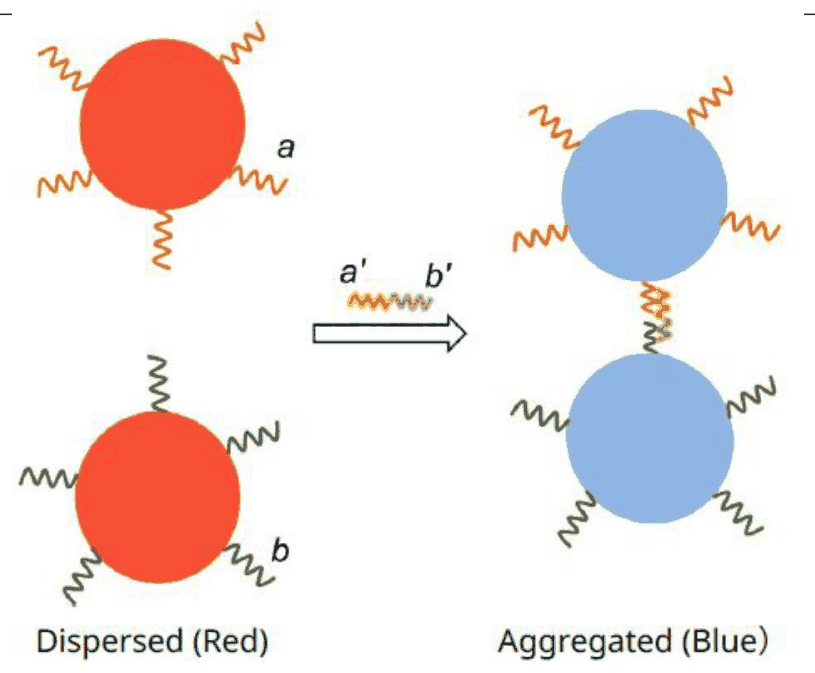
1.1 Indicate the absorption band region of the dispersed gold nanoparticles with an appropriate letter (a-f in the color wheel of Figure 0.1 in General Instructions).
The Color Wheel
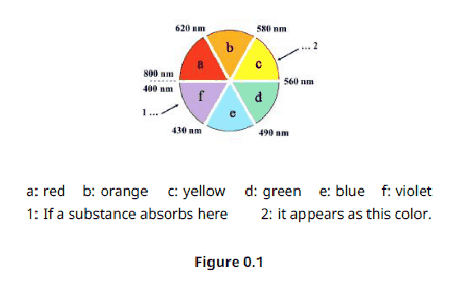
Figure 0.1
1.2 Indicate the change in the absorption spectrum of the gold nanoparticles once they are aggregated. Compared to the absorption wavelength of the dispersed gold nanoparticles, the wavelength of the aggregated nanoparticles
(a) becomes longer
(b) becomes shorter
(c) does not change
Gold nanoparticles are composed of gold atoms that are closely packed as solid gold (density \(\rho = 19.3\ \mathrm{g\,cm^{-3}}\)).
1.3 Calculate how many gold atoms (\(N\)) are there in a spherical gold nanoparticle with a diameter of 30.0 nm.
The synthesis of gold nanoparticles depends on the redox reaction between hydrogen tetrachloroaurate(III) (\(\ce{HAuCl4 * 3H2O}\), \(MW = 394\)) and reducing substances (such as sodium citrate). 5.2 mg of \(\ce{HAuCl4 * 3H2O}\) was completely converted into uniform spherical gold nanoparticles with a diameter of 30.0 nm in a 100.0 mL reaction solution. The absorbance of the resulting red solution was 0.800 measured at 530 nm by a UV-Vis spectrometer.
1.4 Calculate the molar extinction coefficient (molar absorptivity) of the resulting gold nanoparticles solution at 530 nm per mol of gold nanoparticles. The optical path of the cuvette used is 1 cm. If you failed to get the number of gold atoms (\(N\)) in 1.3 , use \(N = 1.00 \times 10^5\).
The standard addition method was used for colorimetric detection of the target nucleic acid. The original throat swab sample was collected and divided equally into two portions. After addition of the probe solutions and water (as indicated below), the absorbance of the two resulting solutions was measured at 600 nm separately.
| No. | Volume of throat swab sample (mL) | Volume of solution of gold nanoparticles modified with nucleic acid fragments (mL) | Volume of standard solution containing \(2.0\ \mu\mathrm{g\,mL^{-1}}\) target nucleic acid (mL) | Volume of water (mL) | Absorbance |
|---|---|---|---|---|---|
| 1 | 0.10 | 0.80 | 0.00 | 0.10 | 0.400 |
| 2 | 0.10 | 0.80 | 0.10 | 0.00 | 0.900 |
1.5 Calculate the concentration of the viral nucleic acids in the original throat swab sample.
Official English text of the problem.
Answers
- 1.1: d, the green region, approximately \(490\text{–}560\ \mathrm{nm}\).
- 1.2: (a) becomes longer: the absorption band undergoes a red shift.
- 1.3: \(\boxed{N = 8.34\times10^5\ \text{gold atoms per nanoparticle}}\).
- 1.4: \(\boxed{\varepsilon_{530}=5.1\times10^9\ \mathrm{L\,mol^{-1}\,cm^{-1}}}\), per mole of nanoparticles.
- 1.5: \(\boxed{C_{\text{original}}=1.6\ \mu\mathrm{g\,mL^{-1}}}\).
Solution
1.1
The observed color is complementary to the absorbed color. On the supplied color wheel, red lies opposite green. Therefore, the dispersed nanoparticles, which appear red, absorb in the green region, d, approximately \(490\text{–}560\ \mathrm{nm}\). The stated measurement wavelength of \(530\ \mathrm{nm}\) lies in this region.
1.2
The aggregated nanoparticles appear blue. On the supplied wheel, blue is complementary to orange, corresponding to approximately \(580\text{–}620\ \mathrm{nm}\).
Thus, aggregation shifts the absorption from the green region toward longer wavelengths:
\[ \lambda_{\mathrm{aggregated}}>\lambda_{\mathrm{dispersed}}. \]
The answer is (a) becomes longer.
1.3
Use the standard constants
\[ M(\ce{Au})=197.0\ \mathrm{g\,mol^{-1}}, \qquad N_{\mathrm A}=6.022\times10^{23}\ \mathrm{mol^{-1}}. \]
The nanoparticle radius is
\[ r=\frac{30.0}{2}\ \mathrm{nm} =15.0\ \mathrm{nm} =1.50\times10^{-6}\ \mathrm{cm}. \]
Its volume and mass are
\[ V_{\mathrm p} =\frac43\pi r^3 =\frac43\pi(1.50\times10^{-6}\ \mathrm{cm})^3 =1.4137\times10^{-17}\ \mathrm{cm^3}, \]
\[ m_{\mathrm p} =\rho V_{\mathrm p} =(19.3\ \mathrm{g\,cm^{-3}}) (1.4137\times10^{-17}\ \mathrm{cm^3}) =2.7285\times10^{-16}\ \mathrm g. \]
Therefore, the number of gold atoms in one nanoparticle is
\[ N=\frac{m_{\mathrm p}}{M(\ce{Au})}N_{\mathrm A} =\frac{2.7285\times10^{-16}\ \mathrm g} {197.0\ \mathrm{g\,mol^{-1}}} (6.022\times10^{23}\ \mathrm{mol^{-1}}). \]
\[ \boxed{N=8.34\times10^5\ \text{atoms per nanoparticle}}. \]
1.4
Each formula unit of the precursor contains one gold atom. Complete conversion therefore gives
\[ n_{\ce{Au}} =\frac{5.2\times10^{-3}\ \mathrm g} {394\ \mathrm{g\,mol^{-1}}} =1.3198\times10^{-5}\ \mathrm{mol}. \]
Because each nanoparticle contains \(N\) gold atoms, the amount of nanoparticles is
\[ n_{\mathrm{NP}}=\frac{n_{\ce{Au}}}{N} =\frac{1.3198\times10^{-5}}{8.34\times10^5} \approx1.582\times10^{-11}\ \mathrm{mol}. \]
Their molar concentration in \(100.0\ \mathrm{mL}\) is consequently
\[ c_{\mathrm{NP}} =\frac{1.582\times10^{-11}\ \mathrm{mol}} {0.1000\ \mathrm L} =1.582\times10^{-10}\ \mathrm{mol\,L^{-1}}. \]
Using the Beer–Lambert law,
\[ A=\varepsilon c_{\mathrm{NP}}l, \]
\[ \varepsilon_{530} =\frac{0.800} {(1.582\times10^{-10}\ \mathrm{mol\,L^{-1}}) (1\ \mathrm{cm})} \approx5.06\times10^9\ \mathrm{L\,mol^{-1}\,cm^{-1}}. \]
Rounding to two significant figures, consistent with the supplied precursor mass,
\[ \boxed{\varepsilon_{530} =5.1\times10^9\ \mathrm{L\,mol^{-1}\,cm^{-1}}}. \]
This coefficient is expressed per mole of nanoparticles, so the concentration used must count particles.
1.5
Both measured solutions have the same final volume:
\[ V_f=1.00\ \mathrm{mL}. \]
Let \(C_x\) be the concentration in the original sample. Each solution contains \(V_x=0.10\ \mathrm{mL}\) of that sample. The second solution additionally contains \(V_s=0.10\ \mathrm{mL}\) of standard with concentration \(C_s=2.0\ \mu\mathrm{g\,mL^{-1}}\).
Using the linear absorbance response assumed in this standard-addition determination, with a common proportionality constant \(k\),
\[ A_1=k\frac{C_xV_x}{V_f}, \qquad A_2=k\frac{C_xV_x+C_sV_s}{V_f}. \]
Subtracting and taking a ratio eliminates both \(k\) and \(V_f\):
\[ \frac{A_1}{A_2-A_1} =\frac{C_xV_x}{C_sV_s}. \]
Hence,
\[ C_x =\frac{A_1}{A_2-A_1}\frac{C_sV_s}{V_x} =\frac{0.400}{0.900-0.400} \frac{(2.0\ \mu\mathrm{g\,mL^{-1}})(0.10\ \mathrm{mL})} {0.10\ \mathrm{mL}}. \]
\[ \boxed{C_x=1.6\ \mu\mathrm{g\,mL^{-1}}}. \]
The solution was checked against the official solution.
IChO 2022 · Theorieaufgabe 1
Schneller und visueller Nukleinsäurenachweis für COVID-19
Schnelle und einfache Methoden zur Früherkennung von COVID-19 werden dringend benötigt. Der Nachweis mithilfe von Goldnanopartikeln gehört zu den vielversprechenden Methoden. Goldnanopartikel werden aufgrund ihrer hohen molaren Extinktionskoeffizienten (molaren Absorptionskoeffizienten) häufig als sichtbare Anzeige auf Teststreifen eingesetzt. Die Farbe von Goldnanopartikeln hängt eng mit ihrer Größe und ihrem Dispersitätsgrad zusammen. Im Allgemeinen gilt: Je größer die Goldnanopartikel sind, desto rötlicher ist ihre Farbe. Sobald die Nanopartikel aggregieren, schlägt die Farbe von Rot nach Blau um.
Wenn die Oberfläche von Goldnanopartikeln mit zwei Arten einzelsträngiger Nukleinsäurefragmente \(a\) und \(b\) modifiziert wird, aggregieren die Goldnanopartikel in Gegenwart der Zielnukleinsäure (\(a’b’\)), wodurch die Farbe der Lösung innerhalb weniger Minuten von Rot nach Blau umschlägt (wie unten dargestellt). Auf der Grundlage dieses Prinzips lassen sich die aus dem Coronavirus in einer Probe gewonnenen Zielnukleinsäuren nachweisen.
1.1 Geben Sie den Bereich der Absorptionsbande der dispergierten Goldnanopartikel mit dem entsprechenden Buchstaben an (a-f im Farbkreis in Abbildung 0.1 der Allgemeinen Hinweise).
Der Farbkreis
Abbildung 0.1
1.2 Geben Sie an, wie sich das Absorptionsspektrum der Goldnanopartikel verändert, sobald sie aggregieren. Im Vergleich zur Absorptionswellenlänge der dispergierten Goldnanopartikel ist die Wellenlänge der aggregierten Nanopartikel
(a) länger
(b) kürzer
(c) unverändert
Goldnanopartikel bestehen aus Goldatomen, die wie in festem Gold dicht gepackt sind (Dichte \(\rho = 19.3\ \mathrm{g\,cm^{-3}}\)).
1.3 Berechnen Sie, wie viele Goldatome (\(N\)) sich in einem kugelförmigen Goldnanopartikel mit einem Durchmesser von 30.0 nm befinden.
Die Synthese von Goldnanopartikeln beruht auf der Redoxreaktion zwischen Tetrachlorogoldsäure(III) (\(\ce{HAuCl4 * 3H2O}\), \(MW = 394\)) und reduzierenden Substanzen (wie Natriumcitrat). 5.2 mg \(\ce{HAuCl4 * 3H2O}\) wurden in einer Reaktionslösung mit einem Volumen von 100.0 mL vollständig in einheitliche kugelförmige Goldnanopartikel mit einem Durchmesser von 30.0 nm umgewandelt. Die mit einem UV-Vis-Spektrometer bei 530 nm gemessene Extinktion der entstandenen roten Lösung betrug 0.800.
1.4 Berechnen Sie den molaren Extinktionskoeffizienten (molaren Absorptionskoeffizienten) der entstandenen Goldnanopartikellösung bei 530 nm, bezogen auf ein Mol Goldnanopartikel. Die optische Weglänge der verwendeten Küvette beträgt 1 cm. Falls Sie die Anzahl der Goldatome (\(N\)) in 1.3 nicht bestimmen konnten, verwenden Sie \(N = 1.00 \times 10^5\).
Für den kolorimetrischen Nachweis der Zielnukleinsäure wurde die Standardadditionsmethode verwendet. Die ursprüngliche Rachenabstrichprobe wurde entnommen und in zwei gleiche Portionen aufgeteilt. Nach Zugabe der Sondenlösungen und von Wasser (wie unten angegeben) wurde die Extinktion der beiden entstandenen Lösungen jeweils bei 600 nm gemessen.
| Nr. | Volumen der Rachenabstrichprobe (mL) | Volumen der Lösung von mit Nukleinsäurefragmenten modifizierten Goldnanopartikeln (mL) | Volumen der Standardlösung mit \(2.0\ \mu\mathrm{g\,mL^{-1}}\) Zielnukleinsäure (mL) | Volumen des Wassers (mL) | Extinktion |
|---|---|---|---|---|---|
| 1 | 0.10 | 0.80 | 0.00 | 0.10 | 0.400 |
| 2 | 0.10 | 0.80 | 0.10 | 0.00 | 0.900 |
1.5 Berechnen Sie die Konzentration der viralen Nukleinsäuren in der ursprünglichen Rachenabstrichprobe.
Aufgabentext aus dem offiziellen englischen Text übersetzt.
Antworten
- 1.1: d, der grüne Bereich, ungefähr \(490\text{–}560\ \mathrm{nm}\).
- 1.2: (a) länger: Die Absorptionsbande erfährt eine Rotverschiebung.
- 1.3: \(\boxed{N = 8.34\times10^5\ \text{Goldatome pro Nanopartikel}}\).
- 1.4: \(\boxed{\varepsilon_{530}=5.1\times10^9\ \mathrm{L\,mol^{-1}\,cm^{-1}}}\), bezogen auf ein Mol Nanopartikel.
- 1.5: \(\boxed{C_{\text{ursprünglich}}=1.6\ \mu\mathrm{g\,mL^{-1}}}\).
Lösung
1.1
Die beobachtete Farbe ist die Komplementärfarbe der absorbierten Farbe. Im vorgegebenen Farbkreis liegt Rot gegenüber von Grün. Daher absorbieren die dispergierten Nanopartikel, die rot erscheinen, im grünen Bereich, d, bei ungefähr \(490\text{–}560\ \mathrm{nm}\). Die angegebene Messwellenlänge von \(530\ \mathrm{nm}\) liegt in diesem Bereich.
1.2
Die aggregierten Nanopartikel erscheinen blau. Im vorgegebenen Farbkreis ist Orange die Komplementärfarbe von Blau; Orange entspricht ungefähr \(580\text{–}620\ \mathrm{nm}\).
Die Aggregation verschiebt die Absorption somit vom grünen Bereich zu längeren Wellenlängen:
\[ \lambda_{\mathrm{aggregated}}>\lambda_{\mathrm{dispersed}}. \]
Die Antwort lautet (a) länger.
1.3
Wir verwenden die üblichen Konstanten
\[ M(\ce{Au})=197.0\ \mathrm{g\,mol^{-1}}, \qquad N_{\mathrm A}=6.022\times10^{23}\ \mathrm{mol^{-1}}. \]
Der Radius des Nanopartikels beträgt
\[ r=\frac{30.0}{2}\ \mathrm{nm} =15.0\ \mathrm{nm} =1.50\times10^{-6}\ \mathrm{cm}. \]
Sein Volumen und seine Masse betragen
\[ V_{\mathrm p} =\frac43\pi r^3 =\frac43\pi(1.50\times10^{-6}\ \mathrm{cm})^3 =1.4137\times10^{-17}\ \mathrm{cm^3}, \]
\[ m_{\mathrm p} =\rho V_{\mathrm p} =(19.3\ \mathrm{g\,cm^{-3}}) (1.4137\times10^{-17}\ \mathrm{cm^3}) =2.7285\times10^{-16}\ \mathrm g. \]
Die Anzahl der Goldatome in einem Nanopartikel ist daher
\[ N=\frac{m_{\mathrm p}}{M(\ce{Au})}N_{\mathrm A} =\frac{2.7285\times10^{-16}\ \mathrm g} {197.0\ \mathrm{g\,mol^{-1}}} (6.022\times10^{23}\ \mathrm{mol^{-1}}). \]
\[ \boxed{N=8.34\times10^5\ \text{Atome pro Nanopartikel}}. \]
1.4
Jede Formeleinheit des Ausgangsstoffs enthält ein Goldatom. Bei vollständiger Umsetzung ergibt sich daher
\[ n_{\ce{Au}} =\frac{5.2\times10^{-3}\ \mathrm g} {394\ \mathrm{g\,mol^{-1}}} =1.3198\times10^{-5}\ \mathrm{mol}. \]
Da jeder Nanopartikel \(N\) Goldatome enthält, beträgt die Stoffmenge der Nanopartikel
\[ n_{\mathrm{NP}}=\frac{n_{\ce{Au}}}{N} =\frac{1.3198\times10^{-5}}{8.34\times10^5} \approx1.582\times10^{-11}\ \mathrm{mol}. \]
Ihre Stoffmengenkonzentration in \(100.0\ \mathrm{mL}\) beträgt folglich
\[ c_{\mathrm{NP}} =\frac{1.582\times10^{-11}\ \mathrm{mol}} {0.1000\ \mathrm L} =1.582\times10^{-10}\ \mathrm{mol\,L^{-1}}. \]
Mit dem Lambert-Beer-Gesetz gilt
\[ A=\varepsilon c_{\mathrm{NP}}l, \]
\[ \varepsilon_{530} =\frac{0.800} {(1.582\times10^{-10}\ \mathrm{mol\,L^{-1}}) (1\ \mathrm{cm})} \approx5.06\times10^9\ \mathrm{L\,mol^{-1}\,cm^{-1}}. \]
Auf zwei signifikante Stellen gerundet, entsprechend der angegebenen Masse des Ausgangsstoffs, ergibt sich
\[ \boxed{\varepsilon_{530} =5.1\times10^9\ \mathrm{L\,mol^{-1}\,cm^{-1}}}. \]
Dieser Koeffizient ist auf ein Mol Nanopartikel bezogen; die verwendete Konzentration muss sich daher auf die Anzahl der Partikel beziehen.
1.5
Beide gemessenen Lösungen haben dasselbe Endvolumen:
\[ V_f=1.00\ \mathrm{mL}. \]
Sei \(C_x\) die Konzentration in der ursprünglichen Probe. Jede Lösung enthält \(V_x=0.10\ \mathrm{mL}\) dieser Probe. Die zweite Lösung enthält zusätzlich \(V_s=0.10\ \mathrm{mL}\) der Standardlösung mit der Konzentration \(C_s=2.0\ \mu\mathrm{g\,mL^{-1}}\).
Mit der bei dieser Standardadditionsbestimmung angenommenen linearen Abhängigkeit der Extinktion von der Konzentration und einer gemeinsamen Proportionalitätskonstanten \(k\) gilt
\[ A_1=k\frac{C_xV_x}{V_f}, \qquad A_2=k\frac{C_xV_x+C_sV_s}{V_f}. \]
Durch Subtraktion und anschließende Bildung eines Verhältnisses werden sowohl \(k\) als auch \(V_f\) eliminiert:
\[ \frac{A_1}{A_2-A_1} =\frac{C_xV_x}{C_sV_s}. \]
Daraus folgt
\[ C_x =\frac{A_1}{A_2-A_1}\frac{C_sV_s}{V_x} =\frac{0.400}{0.900-0.400} \frac{(2.0\ \mu\mathrm{g\,mL^{-1}})(0.10\ \mathrm{mL})} {0.10\ \mathrm{mL}}. \]
\[ \boxed{C_x=1.6\ \mu\mathrm{g\,mL^{-1}}}. \]
Die Lösung wurde mit der offiziellen Lösung abgeglichen.
IChO 2022 · Teori sorusu 1
COVID-19 için Hızlı ve Görsel Nükleik Asit Testi
COVID-19'un erken tespiti için hızlı ve basit yöntemlere acilen ihtiyaç duyulmaktadır. Altın nanoparçacıkların yardımıyla tespit, umut vadeden yöntemlerden biridir. Altın nanoparçacıklar, yüksek molar sönüm katsayıları (molar absorptivite) nedeniyle test şeritlerinde görsel bir gösterge olarak yaygın biçimde kullanılır. Altın nanoparçacıkların görünen rengi, boyutları ve dağılma durumlarıyla yakından ilişkilidir. Genel olarak, altın nanoparçacıklar ne kadar büyükse renk o kadar kırmızımsıdır. Nanoparçacıklar agregat oluşturduğunda renk kırmızıdan maviye döner.
Altın nanoparçacıkların yüzeyi, iki tür tek zincirli nükleik asit parçası olan \(a\) ve \(b\) ile modifiye edildiğinde, altın nanoparçacıklar hedef nükleik asidin (\(a’b’\)) varlığında agregat oluşturur ve çözeltinin rengi birkaç dakika içinde kırmızıdan maviye döner (aşağıda gösterildiği gibi). Bu ilkeye dayanarak, bir örnekteki koronavirüsten elde edilen hedef nükleik asitler tespit edilebilir.
1.1 Dağılmış altın nanoparçacıkların soğurma bandının bulunduğu bölgeyi uygun bir harfle (Genel Talimatlar bölümündeki Şekil 0.1'in renk çemberinde a-f) belirtiniz.
Renk Çemberi
Şekil 0.1
1.2 Altın nanoparçacıklar agregat oluşturduğunda soğurma spektrumunda meydana gelen değişimi belirtiniz. Dağılmış altın nanoparçacıkların soğurma dalga boyuna kıyasla, agregat oluşturmuş nanoparçacıkların soğurma dalga boyu
(a) uzar
(b) kısalır
(c) değişmez
Altın nanoparçacıklar, katı altındaki gibi sıkı paketlenmiş altın atomlarından oluşur (yoğunluk \(\rho = 19.3\ \mathrm{g\,cm^{-3}}\)).
1.3 Çapı 30.0 nm olan küresel bir altın nanoparçacıkta kaç altın atomu (\(N\)) bulunduğunu hesaplayınız.
Altın nanoparçacıkların sentezi, hidrojen tetrakloroaurat(III) (\(\ce{HAuCl4 * 3H2O}\), \(MW = 394\)) ile indirgen maddeler (sodyum sitrat gibi) arasındaki redoks tepkimesine dayanır. 5.2 mg \(\ce{HAuCl4 * 3H2O}\), 100.0 mL tepkime çözeltisinde tamamen, çapı 30.0 nm olan eş boyutlu küresel altın nanoparçacıklara dönüştürülmüştür. Elde edilen kırmızı çözeltinin absorbansı, bir UV-Vis spektrometresiyle 530 nm'de 0.800 olarak ölçülmüştür.
1.4 Elde edilen altın nanoparçacık çözeltisinin 530 nm'deki molar sönüm katsayısını (molar absorptivitesini), altın nanoparçacıkların molü başına hesaplayınız. Kullanılan küvetin optik yol uzunluğu 1 cm'dir. 1.3'te altın atomlarının sayısını (\(N\)) bulamadıysanız, \(N = 1.00 \times 10^5\) kullanınız.
Hedef nükleik asidin kolorimetrik tespiti için standart ekleme yöntemi kullanılmıştır. Başlangıçtaki boğaz sürüntüsü örneği alınmış ve iki eşit kısma bölünmüştür. Prob çözeltileri ve su eklendikten sonra (aşağıda gösterildiği gibi), elde edilen iki çözeltinin absorbansı 600 nm'de ayrı ayrı ölçülmüştür.
| No. | Boğaz sürüntüsü örneğinin hacmi (mL) | Nükleik asit parçalarıyla modifiye edilmiş altın nanoparçacık çözeltisinin hacmi (mL) | \(2.0\ \mu\mathrm{g\,mL^{-1}}\) hedef nükleik asit içeren standart çözeltinin hacmi (mL) | Suyun hacmi (mL) | Absorbans |
|---|---|---|---|---|---|
| 1 | 0.10 | 0.80 | 0.00 | 0.10 | 0.400 |
| 2 | 0.10 | 0.80 | 0.10 | 0.00 | 0.900 |
1.5 Başlangıçtaki boğaz sürüntüsü örneğinde bulunan viral nükleik asitlerin derişimini hesaplayınız.
Soru metni resmi İngilizce metinden çevrilmiştir.
Cevaplar
- 1.1: d, yeşil bölge, yaklaşık \(490\text{–}560\ \mathrm{nm}\).
- 1.2: (a) uzar: soğurma bandı kırmızıya kayar.
- 1.3: \(\boxed{N = 8.34\times10^5\ \text{nanoparçacık başına altın atomu}}\).
- 1.4: \(\boxed{\varepsilon_{530}=5.1\times10^9\ \mathrm{L\,mol^{-1}\,cm^{-1}}}\), nanoparçacıkların molü başına.
- 1.5: \(\boxed{C_{\text{başlangıç}}=1.6\ \mu\mathrm{g\,mL^{-1}}}\).
Çözüm
1.1
Gözlenen renk, soğurulan rengin tamamlayıcısıdır. Verilen renk çemberinde kırmızı, yeşilin karşısında yer alır. Dolayısıyla, kırmızı görünen dağılmış nanoparçacıklar, yaklaşık \(490\text{–}560\ \mathrm{nm}\) aralığındaki yeşil bölgede, d, soğurma yapar. Belirtilen \(530\ \mathrm{nm}\) ölçüm dalga boyu bu bölgede yer alır.
1.2
Agregat oluşturmuş nanoparçacıklar mavi görünür. Verilen çemberde mavi, yaklaşık \(580\text{–}620\ \mathrm{nm}\) aralığına karşılık gelen turuncunun tamamlayıcısıdır.
Bu nedenle agregat oluşumu, soğurmayı yeşil bölgeden daha uzun dalga boylarına kaydırır:
\[ \lambda_{\mathrm{aggregated}}>\lambda_{\mathrm{dispersed}}. \]
Cevap (a) uzar seçeneğidir.
1.3
Standart sabitleri kullanalım:
\[ M(\ce{Au})=197.0\ \mathrm{g\,mol^{-1}}, \qquad N_{\mathrm A}=6.022\times10^{23}\ \mathrm{mol^{-1}}. \]
Nanoparçacığın yarıçapı şöyledir:
\[ r=\frac{30.0}{2}\ \mathrm{nm} =15.0\ \mathrm{nm} =1.50\times10^{-6}\ \mathrm{cm}. \]
Hacmi ve kütlesi şöyledir:
\[ V_{\mathrm p} =\frac43\pi r^3 =\frac43\pi(1.50\times10^{-6}\ \mathrm{cm})^3 =1.4137\times10^{-17}\ \mathrm{cm^3}, \]
\[ m_{\mathrm p} =\rho V_{\mathrm p} =(19.3\ \mathrm{g\,cm^{-3}}) (1.4137\times10^{-17}\ \mathrm{cm^3}) =2.7285\times10^{-16}\ \mathrm g. \]
Dolayısıyla, bir nanoparçacıktaki altın atomlarının sayısı şöyledir:
\[ N=\frac{m_{\mathrm p}}{M(\ce{Au})}N_{\mathrm A} =\frac{2.7285\times10^{-16}\ \mathrm g} {197.0\ \mathrm{g\,mol^{-1}}} (6.022\times10^{23}\ \mathrm{mol^{-1}}). \]
\[ \boxed{N=8.34\times10^5\ \text{nanoparçacık başına atom}}. \]
1.4
Başlangıç maddesinin her formül birimi bir altın atomu içerir. Dolayısıyla tam dönüşüm sonucunda şu elde edilir:
\[ n_{\ce{Au}} =\frac{5.2\times10^{-3}\ \mathrm g} {394\ \mathrm{g\,mol^{-1}}} =1.3198\times10^{-5}\ \mathrm{mol}. \]
Her nanoparçacık \(N\) altın atomu içerdiğinden, nanoparçacıkların madde miktarı şöyledir:
\[ n_{\mathrm{NP}}=\frac{n_{\ce{Au}}}{N} =\frac{1.3198\times10^{-5}}{8.34\times10^5} \approx1.582\times10^{-11}\ \mathrm{mol}. \]
Buna göre, \(100.0\ \mathrm{mL}\) içindeki molar derişimleri şöyledir:
\[ c_{\mathrm{NP}} =\frac{1.582\times10^{-11}\ \mathrm{mol}} {0.1000\ \mathrm L} =1.582\times10^{-10}\ \mathrm{mol\,L^{-1}}. \]
Beer–Lambert yasasını kullanarak,
\[ A=\varepsilon c_{\mathrm{NP}}l, \]
\[ \varepsilon_{530} =\frac{0.800} {(1.582\times10^{-10}\ \mathrm{mol\,L^{-1}}) (1\ \mathrm{cm})} \approx5.06\times10^9\ \mathrm{L\,mol^{-1}\,cm^{-1}}. \]
Verilen başlangıç maddesi kütlesiyle tutarlı olacak şekilde iki anlamlı basamağa yuvarlayarak,
\[ \boxed{\varepsilon_{530} =5.1\times10^9\ \mathrm{L\,mol^{-1}\,cm^{-1}}}. \]
Bu katsayı nanoparçacıkların molü başına ifade edildiğinden, kullanılan derişim parçacık sayısına dayanmalıdır.
1.5
Ölçüm yapılan her iki çözeltinin son hacmi aynıdır:
\[ V_f=1.00\ \mathrm{mL}. \]
Başlangıçtaki örneğin derişimi \(C_x\) olsun. Her çözelti, bu örnekten \(V_x=0.10\ \mathrm{mL}\) içerir. İkinci çözelti ayrıca \(V_s=0.10\ \mathrm{mL}\) hacminde, derişimi \(C_s=2.0\ \mu\mathrm{g\,mL^{-1}}\) olan standart çözelti içerir.
Bu standart ekleme tayininde varsayılan doğrusal absorbans yanıtını, ortak bir \(k\) orantı sabitiyle kullanarak,
\[ A_1=k\frac{C_xV_x}{V_f}, \qquad A_2=k\frac{C_xV_x+C_sV_s}{V_f}. \]
Çıkarma işlemi yapıp oran alarak hem \(k\) hem de \(V_f\) elenir:
\[ \frac{A_1}{A_2-A_1} =\frac{C_xV_x}{C_sV_s}. \]
Böylece,
\[ C_x =\frac{A_1}{A_2-A_1}\frac{C_sV_s}{V_x} =\frac{0.400}{0.900-0.400} \frac{(2.0\ \mu\mathrm{g\,mL^{-1}})(0.10\ \mathrm{mL})} {0.10\ \mathrm{mL}}. \]
\[ \boxed{C_x=1.6\ \mu\mathrm{g\,mL^{-1}}}. \]
Çözüm, resmi çözümle karşılaştırılarak kontrol edildi.
IChO 2022 · Problema teórico 1
Detección rápida y visual de ácidos nucleicos para la COVID-19
Se necesitan con urgencia métodos rápidos y sencillos para la detección temprana de la COVID-19. La detección mediante nanopartículas de oro es uno de los métodos prometedores. Las nanopartículas de oro se utilizan ampliamente como indicadores visuales en tiras reactivas debido a sus elevados coeficientes de extinción molar (absortividad molar). El color que presentan las nanopartículas de oro está estrechamente relacionado con su tamaño y su dispersidad. En general, cuanto más grandes son las nanopartículas de oro, más rojizo es su color. Cuando las nanopartículas se agregan, el color cambia de rojo a azul.
Cuando la superficie de las nanopartículas de oro se modifica con dos tipos de fragmentos de ácido nucleico monocatenario \(a\) y \(b\), las nanopartículas de oro se agregan en presencia del ácido nucleico diana (\(a’b’\)), lo que provoca que el color de la disolución cambie de rojo a azul en varios minutos (como se indica a continuación). Basándose en este principio, se pueden detectar los ácidos nucleicos diana procedentes del coronavirus presentes en una muestra.
1.1 Indique la región de la banda de absorción de las nanopartículas de oro dispersas con la letra correspondiente (a-f en el círculo cromático de la Figura 0.1 de las Instrucciones generales).
El círculo cromático
Figura 0.1
1.2 Indique el cambio en el espectro de absorción de las nanopartículas de oro cuando se agregan. En comparación con la longitud de onda de absorción de las nanopartículas de oro dispersas, la longitud de onda de las nanopartículas agregadas
(a) aumenta
(b) disminuye
(c) no cambia
Las nanopartículas de oro están compuestas por átomos de oro que se encuentran densamente empaquetados, como en el oro sólido (densidad \(\rho = 19.3\ \mathrm{g\,cm^{-3}}\)).
1.3 Calcule cuántos átomos de oro (\(N\)) hay en una nanopartícula de oro esférica de 30.0 nm de diámetro.
La síntesis de nanopartículas de oro se basa en la reacción de oxidación-reducción entre el tetracloroaurato(III) de hidrógeno (\(\ce{HAuCl4 * 3H2O}\), \(MW = 394\)) y sustancias reductoras (como el citrato de sodio). Se convirtieron completamente 5.2 mg de \(\ce{HAuCl4 * 3H2O}\) en nanopartículas de oro esféricas uniformes de 30.0 nm de diámetro en 100.0 mL de disolución de reacción. La absorbancia de la disolución roja resultante, medida a 530 nm con un espectrómetro UV-Vis, fue de 0.800.
1.4 Calcule el coeficiente de extinción molar (absortividad molar) de la disolución de nanopartículas de oro resultante a 530 nm, por mol de nanopartículas de oro. El camino óptico de la cubeta utilizada es de 1 cm. Si no ha obtenido el número de átomos de oro (\(N\)) en 1.3, utilice \(N = 1.00 \times 10^5\).
Se utilizó el método de adición estándar para la detección colorimétrica del ácido nucleico diana. Se recogió la muestra original mediante un hisopado faríngeo y se dividió en dos porciones iguales. Tras añadir las disoluciones de las sondas y el agua (como se indica a continuación), se midió por separado la absorbancia de las dos disoluciones resultantes a 600 nm.
| N.º | Volumen de muestra de hisopado faríngeo (mL) | Volumen de disolución de nanopartículas de oro modificadas con fragmentos de ácido nucleico (mL) | Volumen de disolución patrón que contiene \(2.0\ \mu\mathrm{g\,mL^{-1}}\) de ácido nucleico diana (mL) | Volumen de agua (mL) | Absorbancia |
|---|---|---|---|---|---|
| 1 | 0.10 | 0.80 | 0.00 | 0.10 | 0.400 |
| 2 | 0.10 | 0.80 | 0.10 | 0.00 | 0.900 |
1.5 Calcule la concentración de los ácidos nucleicos víricos en la muestra original de hisopado faríngeo.
Enunciado traducido del texto oficial en inglés.
Respuestas
- 1.1: d, la región verde, aproximadamente \(490\text{–}560\ \mathrm{nm}\).
- 1.2: (a) aumenta: la banda de absorción experimenta un desplazamiento hacia el rojo.
- 1.3: \(\boxed{N = 8.34\times10^5\ \text{átomos de oro por nanopartícula}}\).
- 1.4: \(\boxed{\varepsilon_{530}=5.1\times10^9\ \mathrm{L\,mol^{-1}\,cm^{-1}}}\), por mol de nanopartículas.
- 1.5: \(\boxed{C_{\text{original}}=1.6\ \mu\mathrm{g\,mL^{-1}}}\).
Solución
1.1
El color observado es complementario del color absorbido. En el círculo cromático proporcionado, el rojo se encuentra frente al verde. Por tanto, las nanopartículas dispersas, que se ven rojas, absorben en la región verde, d, aproximadamente \(490\text{–}560\ \mathrm{nm}\). La longitud de onda de medida indicada, de \(530\ \mathrm{nm}\), se encuentra en esta región.
1.2
Las nanopartículas agregadas se ven azules. En el círculo proporcionado, el azul es complementario del naranja, que corresponde aproximadamente a \(580\text{–}620\ \mathrm{nm}\).
Así, la agregación desplaza la absorción desde la región verde hacia longitudes de onda mayores:
\[ \lambda_{\mathrm{aggregated}}>\lambda_{\mathrm{dispersed}}. \]
La respuesta es (a) aumenta.
1.3
Se utilizan las constantes estándar
\[ M(\ce{Au})=197.0\ \mathrm{g\,mol^{-1}}, \qquad N_{\mathrm A}=6.022\times10^{23}\ \mathrm{mol^{-1}}. \]
El radio de la nanopartícula es
\[ r=\frac{30.0}{2}\ \mathrm{nm} =15.0\ \mathrm{nm} =1.50\times10^{-6}\ \mathrm{cm}. \]
Su volumen y su masa son
\[ V_{\mathrm p} =\frac43\pi r^3 =\frac43\pi(1.50\times10^{-6}\ \mathrm{cm})^3 =1.4137\times10^{-17}\ \mathrm{cm^3}, \]
\[ m_{\mathrm p} =\rho V_{\mathrm p} =(19.3\ \mathrm{g\,cm^{-3}}) (1.4137\times10^{-17}\ \mathrm{cm^3}) =2.7285\times10^{-16}\ \mathrm g. \]
Por tanto, el número de átomos de oro en una nanopartícula es
\[ N=\frac{m_{\mathrm p}}{M(\ce{Au})}N_{\mathrm A} =\frac{2.7285\times10^{-16}\ \mathrm g} {197.0\ \mathrm{g\,mol^{-1}}} (6.022\times10^{23}\ \mathrm{mol^{-1}}). \]
\[ \boxed{N=8.34\times10^5\ \text{átomos por nanopartícula}}. \]
1.4
Cada unidad fórmula del precursor contiene un átomo de oro. Por tanto, la conversión completa da
\[ n_{\ce{Au}} =\frac{5.2\times10^{-3}\ \mathrm g} {394\ \mathrm{g\,mol^{-1}}} =1.3198\times10^{-5}\ \mathrm{mol}. \]
Como cada nanopartícula contiene \(N\) átomos de oro, la cantidad de sustancia de nanopartículas es
\[ n_{\mathrm{NP}}=\frac{n_{\ce{Au}}}{N} =\frac{1.3198\times10^{-5}}{8.34\times10^5} \approx1.582\times10^{-11}\ \mathrm{mol}. \]
En consecuencia, su concentración molar en \(100.0\ \mathrm{mL}\) es
\[ c_{\mathrm{NP}} =\frac{1.582\times10^{-11}\ \mathrm{mol}} {0.1000\ \mathrm L} =1.582\times10^{-10}\ \mathrm{mol\,L^{-1}}. \]
Utilizando la ley de Beer–Lambert,
\[ A=\varepsilon c_{\mathrm{NP}}l, \]
\[ \varepsilon_{530} =\frac{0.800} {(1.582\times10^{-10}\ \mathrm{mol\,L^{-1}}) (1\ \mathrm{cm})} \approx5.06\times10^9\ \mathrm{L\,mol^{-1}\,cm^{-1}}. \]
Redondeando a dos cifras significativas, de acuerdo con la masa de precursor proporcionada,
\[ \boxed{\varepsilon_{530} =5.1\times10^9\ \mathrm{L\,mol^{-1}\,cm^{-1}}}. \]
Este coeficiente se expresa por mol de nanopartículas, por lo que la concentración utilizada debe contabilizar partículas.
1.5
Las dos disoluciones medidas tienen el mismo volumen final:
\[ V_f=1.00\ \mathrm{mL}. \]
Sea \(C_x\) la concentración en la muestra original. Cada disolución contiene \(V_x=0.10\ \mathrm{mL}\) de esa muestra. La segunda disolución contiene además \(V_s=0.10\ \mathrm{mL}\) de patrón con una concentración \(C_s=2.0\ \mu\mathrm{g\,mL^{-1}}\).
Utilizando la respuesta lineal de la absorbancia que se supone en esta determinación por adición estándar, con una constante de proporcionalidad común \(k\),
\[ A_1=k\frac{C_xV_x}{V_f}, \qquad A_2=k\frac{C_xV_x+C_sV_s}{V_f}. \]
Al restar y calcular un cociente se eliminan tanto \(k\) como \(V_f\):
\[ \frac{A_1}{A_2-A_1} =\frac{C_xV_x}{C_sV_s}. \]
Por tanto,
\[ C_x =\frac{A_1}{A_2-A_1}\frac{C_sV_s}{V_x} =\frac{0.400}{0.900-0.400} \frac{(2.0\ \mu\mathrm{g\,mL^{-1}})(0.10\ \mathrm{mL})} {0.10\ \mathrm{mL}}. \]
\[ \boxed{C_x=1.6\ \mu\mathrm{g\,mL^{-1}}}. \]
La solución se comprobó con la solución oficial.
IChO 2022 · 理论题 1
COVID-19 的快速可视化核酸检测
目前迫切需要快速、简便的 COVID-19 早期检测方法。金纳米颗粒辅助检测是一种很有前景的方法。金纳米颗粒具有较高的摩尔消光系数(摩尔吸光系数),因此被广泛用作试纸条的可视化显色材料。金纳米颗粒呈现的颜色与其尺寸和分散性密切相关。一般而言,金纳米颗粒越大,颜色越偏红。一旦纳米颗粒发生聚集,颜色就会由红色变为蓝色。
当金纳米颗粒表面修饰有两种单链核酸片段 \(a\) 和 \(b\) 时,在靶核酸(\(a’b’\))存在的情况下,金纳米颗粒会发生聚集,使溶液在几分钟内由红色变为蓝色(如下图所示)。根据这一原理,可以检测从样品中的冠状病毒采集到的靶核酸。
1.1 用适当的字母(通用说明中图 0.1 色轮上的 a-f)指出分散状态的金纳米颗粒的吸收带所在区域。
色轮
图 0.1
1.2 指出金纳米颗粒发生聚集后其吸收光谱的变化。与分散状态的金纳米颗粒的吸收波长相比,聚集后的纳米颗粒的吸收波长
(a) 变长
(b) 变短
(c) 不变
金纳米颗粒由金原子组成,其中金原子像在固态金中一样紧密堆积(密度 \(\rho = 19.3\ \mathrm{g\,cm^{-3}}\))。
1.3 计算一个直径为 30.0 nm 的球形金纳米颗粒中含有多少个金原子(\(N\))。
金纳米颗粒的合成依赖于四氯合金(III)酸(\(\ce{HAuCl4 * 3H2O}\),\(MW = 394\))与还原性物质(如柠檬酸钠)之间的氧化还原反应。在 100.0 mL 反应溶液中,将 5.2 mg 的 \(\ce{HAuCl4 * 3H2O}\) 完全转化为直径均为 30.0 nm 的球形金纳米颗粒。用紫外-可见分光光度计在 530 nm 处测得所得红色溶液的吸光度为 0.800。
1.4 计算所得金纳米颗粒溶液在 530 nm 处以每摩尔金纳米颗粒计的摩尔消光系数(摩尔吸光系数)。所用比色皿的光程为 1 cm。如果未能在 1.3 中求出金原子数(\(N\)),请使用 \(N = 1.00 \times 10^5\)。
采用标准加入法对靶核酸进行比色检测。采集原始咽拭子样品,并将其等分为两份。加入探针溶液和水(如下所示)后,分别在 600 nm 处测量所得两份溶液的吸光度。
| 编号 | 咽拭子样品体积 (mL) | 核酸片段修饰的金纳米颗粒溶液体积 (mL) | 含 \(2.0\ \mu\mathrm{g\,mL^{-1}}\) 靶核酸的标准溶液体积 (mL) | 水的体积 (mL) | 吸光度 |
|---|---|---|---|---|---|
| 1 | 0.10 | 0.80 | 0.00 | 0.10 | 0.400 |
| 2 | 0.10 | 0.80 | 0.10 | 0.00 | 0.900 |
1.5 计算原始咽拭子样品中病毒核酸的浓度。
题目译自官方英文原文。
答案
- 1.1: d,绿色区域,约为 \(490\text{–}560\ \mathrm{nm}\)。
- 1.2: (a) 变长:吸收带发生红移。
- 1.3: \(\boxed{N = 8.34\times10^5\ \text{个金原子/纳米颗粒}}\)。
- 1.4: \(\boxed{\varepsilon_{530}=5.1\times10^9\ \mathrm{L\,mol^{-1}\,cm^{-1}}}\),以每摩尔纳米颗粒计。
- 1.5: \(\boxed{C_{\text{原始}}=1.6\ \mu\mathrm{g\,mL^{-1}}}\)。
解答
1.1
观察到的颜色与被吸收光的颜色互为补色。在给出的色轮上,红色与绿色相对。因此,呈红色的分散状态纳米颗粒吸收绿色区域 d 的光,波长约为 \(490\text{–}560\ \mathrm{nm}\)。题目给出的测量波长 \(530\ \mathrm{nm}\) 位于该区域内。
1.2
聚集后的纳米颗粒呈蓝色。在给出的色轮上,蓝色与橙色互为补色,橙色对应的波长约为 \(580\text{–}620\ \mathrm{nm}\)。
因此,聚集使吸收带从绿色区域向更长波长方向移动:
\[ \lambda_{\mathrm{aggregated}}>\lambda_{\mathrm{dispersed}}. \]
答案为 (a) 变长。
1.3
使用标准常数
\[ M(\ce{Au})=197.0\ \mathrm{g\,mol^{-1}}, \qquad N_{\mathrm A}=6.022\times10^{23}\ \mathrm{mol^{-1}}. \]
纳米颗粒的半径为
\[ r=\frac{30.0}{2}\ \mathrm{nm} =15.0\ \mathrm{nm} =1.50\times10^{-6}\ \mathrm{cm}. \]
其体积和质量为
\[ V_{\mathrm p} =\frac43\pi r^3 =\frac43\pi(1.50\times10^{-6}\ \mathrm{cm})^3 =1.4137\times10^{-17}\ \mathrm{cm^3}, \]
\[ m_{\mathrm p} =\rho V_{\mathrm p} =(19.3\ \mathrm{g\,cm^{-3}}) (1.4137\times10^{-17}\ \mathrm{cm^3}) =2.7285\times10^{-16}\ \mathrm g. \]
因此,一个纳米颗粒中的金原子数为
\[ N=\frac{m_{\mathrm p}}{M(\ce{Au})}N_{\mathrm A} =\frac{2.7285\times10^{-16}\ \mathrm g} {197.0\ \mathrm{g\,mol^{-1}}} (6.022\times10^{23}\ \mathrm{mol^{-1}}). \]
\[ \boxed{N=8.34\times10^5\ \text{个原子/纳米颗粒}}. \]
1.4
前驱体的每个化学式单位含有一个金原子。因此,完全转化后得到
\[ n_{\ce{Au}} =\frac{5.2\times10^{-3}\ \mathrm g} {394\ \mathrm{g\,mol^{-1}}} =1.3198\times10^{-5}\ \mathrm{mol}. \]
由于每个纳米颗粒含有 \(N\) 个金原子,纳米颗粒的物质的量为
\[ n_{\mathrm{NP}}=\frac{n_{\ce{Au}}}{N} =\frac{1.3198\times10^{-5}}{8.34\times10^5} \approx1.582\times10^{-11}\ \mathrm{mol}. \]
因此,它们在 \(100.0\ \mathrm{mL}\) 溶液中的物质的量浓度为
\[ c_{\mathrm{NP}} =\frac{1.582\times10^{-11}\ \mathrm{mol}} {0.1000\ \mathrm L} =1.582\times10^{-10}\ \mathrm{mol\,L^{-1}}. \]
根据朗伯–比尔定律,
\[ A=\varepsilon c_{\mathrm{NP}}l, \]
\[ \varepsilon_{530} =\frac{0.800} {(1.582\times10^{-10}\ \mathrm{mol\,L^{-1}}) (1\ \mathrm{cm})} \approx5.06\times10^9\ \mathrm{L\,mol^{-1}\,cm^{-1}}. \]
按照所给前驱体质量的有效数字位数,将结果保留两位有效数字,
\[ \boxed{\varepsilon_{530} =5.1\times10^9\ \mathrm{L\,mol^{-1}\,cm^{-1}}}. \]
该系数以每摩尔纳米颗粒计,因此所用浓度必须以颗粒为计数对象。
1.5
两份待测溶液的最终体积相同:
\[ V_f=1.00\ \mathrm{mL}. \]
设原始样品中的浓度为 \(C_x\)。每份溶液均含有 \(V_x=0.10\ \mathrm{mL}\) 的该样品。第二份溶液还含有 \(V_s=0.10\ \mathrm{mL}\) 的标准溶液,其浓度为 \(C_s=2.0\ \mu\mathrm{g\,mL^{-1}}\)。
根据本次标准加入法测定所假设的吸光度线性响应关系,使用相同的比例常数 \(k\),有
\[ A_1=k\frac{C_xV_x}{V_f}, \qquad A_2=k\frac{C_xV_x+C_sV_s}{V_f}. \]
相减后取比值,即可消去 \(k\) 和 \(V_f\):
\[ \frac{A_1}{A_2-A_1} =\frac{C_xV_x}{C_sV_s}. \]
因此,
\[ C_x =\frac{A_1}{A_2-A_1}\frac{C_sV_s}{V_x} =\frac{0.400}{0.900-0.400} \frac{(2.0\ \mu\mathrm{g\,mL^{-1}})(0.10\ \mathrm{mL})} {0.10\ \mathrm{mL}}. \]
\[ \boxed{C_x=1.6\ \mu\mathrm{g\,mL^{-1}}}. \]
解答已与官方解答核对。
IChO 2022 · Теоретическая задача 1
Быстрое и визуальное выявление нуклеиновых кислот для диагностики COVID-19
Существует острая потребность в быстрых и простых методах раннего выявления COVID-19. Одним из перспективных методов является обнаружение с помощью наночастиц золота. Благодаря высоким молярным коэффициентам экстинкции (молярным коэффициентам поглощения) наночастицы золота широко используются для визуальной индикации на тест-полосках. Окраска наночастиц золота тесно связана с их размерами и степенью дисперсности. Как правило, чем крупнее наночастицы золота, тем более красной является их окраска. При агрегации наночастиц окраска меняется с красной на синюю.
Если поверхность наночастиц золота модифицирована двумя видами одноцепочечных фрагментов нуклеиновых кислот \(a\) и \(b\), то в присутствии целевой нуклеиновой кислоты (\(a’b’\)) наночастицы золота агрегируют, вызывая изменение окраски раствора с красной на синюю в течение нескольких минут (как показано ниже). На основе этого принципа можно обнаружить целевые нуклеиновые кислоты, выделенные из коронавируса, содержащегося в образце.
1.1 Укажите область полосы поглощения диспергированных наночастиц золота соответствующей буквой (a-f на цветовом круге на рисунке 0.1 в Общих указаниях).
Цветовой круг
Рисунок 0.1
1.2 Укажите, как изменяется спектр поглощения наночастиц золота при их агрегации. По сравнению с длиной волны поглощения диспергированных наночастиц золота длина волны поглощения агрегированных наночастиц
(a) увеличивается
(b) уменьшается
(c) не изменяется
Наночастицы золота состоят из атомов золота, плотно упакованных, как в твёрдом золоте (плотность \(\rho = 19.3\ \mathrm{g\,cm^{-3}}\)).
1.3 Рассчитайте, сколько атомов золота (\(N\)) содержится в сферической наночастице золота диаметром 30.0 нм.
Синтез наночастиц золота основан на окислительно-восстановительной реакции между тетрахлороауратом(III) водорода (\(\ce{HAuCl4 * 3H2O}\), \(MW = 394\)) и восстановителями (например, цитратом натрия). 5.2 мг \(\ce{HAuCl4 * 3H2O}\) были полностью превращены в сферические наночастицы золота одинакового размера диаметром 30.0 нм в 100.0 мл реакционного раствора. Оптическая плотность полученного красного раствора, измеренная при 530 нм с помощью спектрофотометра УФ- и видимого диапазона, составила 0.800.
1.4 Рассчитайте молярный коэффициент экстинкции (молярный коэффициент поглощения) полученного раствора наночастиц золота при 530 нм в расчёте на моль наночастиц золота. Длина оптического пути использованной кюветы составляет 1 см. Если вам не удалось получить число атомов золота (\(N\)) в 1.3, используйте \(N = 1.00 \times 10^5\).
Для колориметрического определения целевой нуклеиновой кислоты использовали метод стандартных добавок. Исходный образец мазка из зева отобрали и разделили на две равные порции. После добавления растворов зондов и воды (как указано ниже) отдельно измерили оптическую плотность двух полученных растворов при 600 нм.
| № | Объём образца мазка из зева (мл) | Объём раствора наночастиц золота, модифицированных фрагментами нуклеиновых кислот (мл) | Объём стандартного раствора, содержащего \(2.0\ \mu\mathrm{g\,mL^{-1}}\) целевой нуклеиновой кислоты (мл) | Объём воды (мл) | Оптическая плотность |
|---|---|---|---|---|---|
| 1 | 0.10 | 0.80 | 0.00 | 0.10 | 0.400 |
| 2 | 0.10 | 0.80 | 0.10 | 0.00 | 0.900 |
1.5 Рассчитайте концентрацию вирусных нуклеиновых кислот в исходном образце мазка из зева.
Условие переведено с официального английского текста.
Ответы
- 1.1: d, зелёная область, приблизительно \(490\text{–}560\ \mathrm{nm}\).
- 1.2: (a) увеличивается: полоса поглощения испытывает красное смещение.
- 1.3: \(\boxed{N = 8.34\times10^5\ \text{атомов золота на наночастицу}}\).
- 1.4: \(\boxed{\varepsilon_{530}=5.1\times10^9\ \mathrm{L\,mol^{-1}\,cm^{-1}}}\), в расчёте на моль наночастиц.
- 1.5: \(\boxed{C_{\text{исходный}}=1.6\ \mu\mathrm{g\,mL^{-1}}}\).
Решение
1.1
Наблюдаемый цвет является дополнительным к поглощаемому цвету. На приведённом цветовом круге красный цвет расположен напротив зелёного. Следовательно, диспергированные наночастицы, которые выглядят красными, поглощают в зелёной области, d, приблизительно \(490\text{–}560\ \mathrm{nm}\). Указанная длина волны измерения \(530\ \mathrm{nm}\) находится в этой области.
1.2
Агрегированные наночастицы выглядят синими. На приведённом круге дополнительным к синему является оранжевый цвет, которому соответствует область приблизительно \(580\text{–}620\ \mathrm{nm}\).
Таким образом, агрегация смещает поглощение из зелёной области в сторону более длинных волн:
\[ \lambda_{\mathrm{aggregated}}>\lambda_{\mathrm{dispersed}}. \]
Ответ: (a) увеличивается.
1.3
Используем стандартные константы
\[ M(\ce{Au})=197.0\ \mathrm{g\,mol^{-1}}, \qquad N_{\mathrm A}=6.022\times10^{23}\ \mathrm{mol^{-1}}. \]
Радиус наночастицы равен
\[ r=\frac{30.0}{2}\ \mathrm{nm} =15.0\ \mathrm{nm} =1.50\times10^{-6}\ \mathrm{cm}. \]
Её объём и масса равны
\[ V_{\mathrm p} =\frac43\pi r^3 =\frac43\pi(1.50\times10^{-6}\ \mathrm{cm})^3 =1.4137\times10^{-17}\ \mathrm{cm^3}, \]
\[ m_{\mathrm p} =\rho V_{\mathrm p} =(19.3\ \mathrm{g\,cm^{-3}}) (1.4137\times10^{-17}\ \mathrm{cm^3}) =2.7285\times10^{-16}\ \mathrm g. \]
Следовательно, число атомов золота в одной наночастице равно
\[ N=\frac{m_{\mathrm p}}{M(\ce{Au})}N_{\mathrm A} =\frac{2.7285\times10^{-16}\ \mathrm g} {197.0\ \mathrm{g\,mol^{-1}}} (6.022\times10^{23}\ \mathrm{mol^{-1}}). \]
\[ \boxed{N=8.34\times10^5\ \text{атомов на наночастицу}}. \]
1.4
Каждая формульная единица исходного соединения содержит один атом золота. Поэтому при полном превращении получаем
\[ n_{\ce{Au}} =\frac{5.2\times10^{-3}\ \mathrm g} {394\ \mathrm{g\,mol^{-1}}} =1.3198\times10^{-5}\ \mathrm{mol}. \]
Поскольку каждая наночастица содержит \(N\) атомов золота, количество вещества наночастиц равно
\[ n_{\mathrm{NP}}=\frac{n_{\ce{Au}}}{N} =\frac{1.3198\times10^{-5}}{8.34\times10^5} \approx1.582\times10^{-11}\ \mathrm{mol}. \]
Следовательно, их молярная концентрация в \(100.0\ \mathrm{mL}\) составляет
\[ c_{\mathrm{NP}} =\frac{1.582\times10^{-11}\ \mathrm{mol}} {0.1000\ \mathrm L} =1.582\times10^{-10}\ \mathrm{mol\,L^{-1}}. \]
Используя закон Бугера–Ламберта–Бера,
\[ A=\varepsilon c_{\mathrm{NP}}l, \]
\[ \varepsilon_{530} =\frac{0.800} {(1.582\times10^{-10}\ \mathrm{mol\,L^{-1}}) (1\ \mathrm{cm})} \approx5.06\times10^9\ \mathrm{L\,mol^{-1}\,cm^{-1}}. \]
Округляя до двух значащих цифр в соответствии с точностью заданной массы исходного соединения, получаем
\[ \boxed{\varepsilon_{530} =5.1\times10^9\ \mathrm{L\,mol^{-1}\,cm^{-1}}}. \]
Этот коэффициент выражен в расчёте на моль наночастиц, поэтому используемая концентрация должна соответствовать числу частиц.
1.5
Оба раствора, для которых проводят измерения, имеют одинаковый конечный объём:
\[ V_f=1.00\ \mathrm{mL}. \]
Пусть \(C_x\) — концентрация в исходном образце. Каждый раствор содержит \(V_x=0.10\ \mathrm{mL}\) этого образца. Второй раствор дополнительно содержит \(V_s=0.10\ \mathrm{mL}\) стандартного раствора с концентрацией \(C_s=2.0\ \mu\mathrm{g\,mL^{-1}}\).
Используя линейную зависимость оптической плотности от концентрации, предполагаемую при данном определении методом стандартных добавок, с общим коэффициентом пропорциональности \(k\), получаем
\[ A_1=k\frac{C_xV_x}{V_f}, \qquad A_2=k\frac{C_xV_x+C_sV_s}{V_f}. \]
Вычитание и последующее вычисление отношения позволяют исключить как \(k\), так и \(V_f\):
\[ \frac{A_1}{A_2-A_1} =\frac{C_xV_x}{C_sV_s}. \]
Отсюда
\[ C_x =\frac{A_1}{A_2-A_1}\frac{C_sV_s}{V_x} =\frac{0.400}{0.900-0.400} \frac{(2.0\ \mu\mathrm{g\,mL^{-1}})(0.10\ \mathrm{mL})} {0.10\ \mathrm{mL}}. \]
\[ \boxed{C_x=1.6\ \mu\mathrm{g\,mL^{-1}}}. \]
Решение проверено по официальному решению.
IChO 2022 · مسألة نظرية 1
الكشف السريع والبصري عن الأحماض النووية لتشخيص COVID-19
ثمة حاجة ملحّة إلى طرائق سريعة وبسيطة للكشف المبكر عن COVID-19. ويُعدّ الكشف بمساعدة جسيمات الذهب النانوية إحدى الطرائق الواعدة. تُستخدم جسيمات الذهب النانوية على نطاق واسع لإظهار نتائج مرئية في شرائط الاختبار، نظرًا لارتفاع معاملات الانقراض المولارية (الامتصاصية المولارية) لها. ويرتبط اللون الظاهر لجسيمات الذهب النانوية ارتباطًا وثيقًا بأحجامها ودرجة تشتتها. وعمومًا، كلما كبر حجم جسيمات الذهب النانوية، ازداد لونها احمرارًا. وبمجرد أن تتجمع الجسيمات النانوية، يتحول اللون من الأحمر إلى الأزرق.
عندما يُعدَّل سطح جسيمات الذهب النانوية بنوعين من قطع الأحماض النووية أحادية السلسلة \(a\) و\(b\)، تتجمع جسيمات الذهب النانوية في وجود الحمض النووي المستهدف (\(a’b’\))، مما يؤدي إلى تغير لون المحلول من الأحمر إلى الأزرق خلال بضع دقائق (كما هو موضح أدناه). وبناءً على هذا المبدأ، يمكن الكشف عن الأحماض النووية المستهدفة المأخوذة من فيروس كورونا الموجود في عينة.
1.1 حدّد منطقة حزمة الامتصاص لجسيمات الذهب النانوية المشتتة باستخدام الحرف المناسب (a-f في عجلة الألوان في الشكل 0.1 ضمن التعليمات العامة).
عجلة الألوان
الشكل 0.1
1.2 حدّد التغير في طيف امتصاص جسيمات الذهب النانوية عند تجمعها. بالمقارنة مع الطول الموجي لامتصاص جسيمات الذهب النانوية المشتتة، فإن الطول الموجي لامتصاص الجسيمات النانوية المتجمعة
(a) يصبح أطول
(b) يصبح أقصر
(c) لا يتغير
تتكون جسيمات الذهب النانوية من ذرات ذهب متراصة بإحكام كما في الذهب الصلب (الكثافة \(\rho = 19.3\ \mathrm{g\,cm^{-3}}\)).
1.3 احسب عدد ذرات الذهب (\(N\)) الموجودة في جسيم ذهب نانوي كروي قطره 30.0 nm.
يعتمد تحضير جسيمات الذهب النانوية على تفاعل الأكسدة والاختزال بين رباعي كلوروأورات(III) الهيدروجين (\(\ce{HAuCl4 * 3H2O}\)، \(MW = 394\)) ومواد مختزلة (مثل سترات الصوديوم). حُوِّلت كمية مقدارها 5.2 mg من \(\ce{HAuCl4 * 3H2O}\) بالكامل إلى جسيمات ذهب نانوية كروية متجانسة بقطر 30.0 nm في محلول تفاعل حجمه 100.0 mL. وكانت امتصاصية المحلول الأحمر الناتج 0.800، مقيسة عند 530 nm باستخدام مطياف للأشعة فوق البنفسجية والمرئية.
1.4 احسب معامل الانقراض المولاري (الامتصاصية المولارية) لمحلول جسيمات الذهب النانوية الناتج عند 530 nm لكل مول من جسيمات الذهب النانوية. يبلغ طول المسار الضوئي لخلية القياس المستخدمة 1 cm. إذا لم تتمكن من إيجاد عدد ذرات الذهب (\(N\)) في 1.3، فاستخدم \(N = 1.00 \times 10^5\).
استُخدمت طريقة الإضافة القياسية للكشف اللوني عن الحمض النووي المستهدف. جُمعت عينة مسحة الحلق الأصلية وقُسمت بالتساوي إلى جزأين. وبعد إضافة محاليل المجسّات والماء (كما هو موضح أدناه)، قِيست امتصاصية كلٍّ من المحلولين الناتجين عند 600 nm على حدة.
| الرقم | حجم عينة مسحة الحلق (mL) | حجم محلول جسيمات الذهب النانوية المعدّلة بقطع الأحماض النووية (mL) | حجم المحلول القياسي المحتوي على \(2.0\ \mu\mathrm{g\,mL^{-1}}\) من الحمض النووي المستهدف (mL) | حجم الماء (mL) | الامتصاصية |
|---|---|---|---|---|---|
| 1 | 0.10 | 0.80 | 0.00 | 0.10 | 0.400 |
| 2 | 0.10 | 0.80 | 0.10 | 0.00 | 0.900 |
1.5 احسب تركيز الأحماض النووية الفيروسية في عينة مسحة الحلق الأصلية.
نص المسألة مترجم عن النص الإنجليزي الرسمي.
الإجابات
- 1.1: d، المنطقة الخضراء، نحو \(490\text{–}560\ \mathrm{nm}\).
- 1.2: (a) يصبح أطول: تنزاح حزمة الامتصاص نحو الأحمر.
- 1.3: \(\boxed{N = 8.34\times10^5\ \text{ذرة ذهب لكل جسيم نانوي}}\).
- 1.4: \(\boxed{\varepsilon_{530}=5.1\times10^9\ \mathrm{L\,mol^{-1}\,cm^{-1}}}\)، لكل مول من الجسيمات النانوية.
- 1.5: \(\boxed{C_{\text{الأصلي}}=1.6\ \mu\mathrm{g\,mL^{-1}}}\).
الحل
1.1
اللون المشاهَد مكمّل للون الممتص. وفي عجلة الألوان المعطاة، يقع الأحمر مقابل الأخضر. لذلك، تمتص الجسيمات النانوية المشتتة، التي تبدو حمراء، في المنطقة الخضراء، d، أي نحو \(490\text{–}560\ \mathrm{nm}\). ويقع الطول الموجي المذكور للقياس، وهو \(530\ \mathrm{nm}\)، ضمن هذه المنطقة.
1.2
تبدو الجسيمات النانوية المتجمعة زرقاء. وفي العجلة المعطاة، يُعدّ الأزرق مكمّلًا للبرتقالي، الذي يقابل تقريبًا \(580\text{–}620\ \mathrm{nm}\).
وعليه، يُزيح التجمع الامتصاص من المنطقة الخضراء نحو أطوال موجية أطول:
\[ \lambda_{\mathrm{aggregated}}>\lambda_{\mathrm{dispersed}}. \]
الإجابة هي (a) يصبح أطول.
1.3
نستخدم الثوابت القياسية
\[ M(\ce{Au})=197.0\ \mathrm{g\,mol^{-1}}, \qquad N_{\mathrm A}=6.022\times10^{23}\ \mathrm{mol^{-1}}. \]
نصف قطر الجسيم النانوي هو
\[ r=\frac{30.0}{2}\ \mathrm{nm} =15.0\ \mathrm{nm} =1.50\times10^{-6}\ \mathrm{cm}. \]
وحجمه وكتلته هما
\[ V_{\mathrm p} =\frac43\pi r^3 =\frac43\pi(1.50\times10^{-6}\ \mathrm{cm})^3 =1.4137\times10^{-17}\ \mathrm{cm^3}, \]
\[ m_{\mathrm p} =\rho V_{\mathrm p} =(19.3\ \mathrm{g\,cm^{-3}}) (1.4137\times10^{-17}\ \mathrm{cm^3}) =2.7285\times10^{-16}\ \mathrm g. \]
إذن، عدد ذرات الذهب في جسيم نانوي واحد هو
\[ N=\frac{m_{\mathrm p}}{M(\ce{Au})}N_{\mathrm A} =\frac{2.7285\times10^{-16}\ \mathrm g} {197.0\ \mathrm{g\,mol^{-1}}} (6.022\times10^{23}\ \mathrm{mol^{-1}}). \]
\[ \boxed{N=8.34\times10^5\ \text{ذرة لكل جسيم نانوي}}. \]
1.4
تحتوي كل وحدة صيغة من المادة الأولية على ذرة ذهب واحدة. لذلك، يعطي التحول الكامل
\[ n_{\ce{Au}} =\frac{5.2\times10^{-3}\ \mathrm g} {394\ \mathrm{g\,mol^{-1}}} =1.3198\times10^{-5}\ \mathrm{mol}. \]
ولأن كل جسيم نانوي يحتوي على \(N\) من ذرات الذهب، فإن كمية مادة الجسيمات النانوية هي
\[ n_{\mathrm{NP}}=\frac{n_{\ce{Au}}}{N} =\frac{1.3198\times10^{-5}}{8.34\times10^5} \approx1.582\times10^{-11}\ \mathrm{mol}. \]
ومن ثم، يكون تركيزها المولاري في \(100.0\ \mathrm{mL}\) هو
\[ c_{\mathrm{NP}} =\frac{1.582\times10^{-11}\ \mathrm{mol}} {0.1000\ \mathrm L} =1.582\times10^{-10}\ \mathrm{mol\,L^{-1}}. \]
باستخدام قانون بير–لامبرت،
\[ A=\varepsilon c_{\mathrm{NP}}l, \]
\[ \varepsilon_{530} =\frac{0.800} {(1.582\times10^{-10}\ \mathrm{mol\,L^{-1}}) (1\ \mathrm{cm})} \approx5.06\times10^9\ \mathrm{L\,mol^{-1}\,cm^{-1}}. \]
وبالتقريب إلى رقمين معنويين، بما يتوافق مع كتلة المادة الأولية المعطاة،
\[ \boxed{\varepsilon_{530} =5.1\times10^9\ \mathrm{L\,mol^{-1}\,cm^{-1}}}. \]
يُعبَّر عن هذا المعامل لكل مول من الجسيمات النانوية، لذا يجب أن يكون التركيز المستخدم محسوبًا على أساس عدد الجسيمات.
1.5
للمحلولين المقيسين الحجم النهائي نفسه:
\[ V_f=1.00\ \mathrm{mL}. \]
ليكن \(C_x\) هو التركيز في العينة الأصلية. يحتوي كل محلول على \(V_x=0.10\ \mathrm{mL}\) من تلك العينة. ويحتوي المحلول الثاني أيضًا على \(V_s=0.10\ \mathrm{mL}\) من محلول قياسي تركيزه \(C_s=2.0\ \mu\mathrm{g\,mL^{-1}}\).
باستخدام استجابة الامتصاصية الخطية المفترضة في هذا التعيين بطريقة الإضافة القياسية، مع ثابت تناسب مشترك \(k\)،
\[ A_1=k\frac{C_xV_x}{V_f}, \qquad A_2=k\frac{C_xV_x+C_sV_s}{V_f}. \]
بالطرح ثم أخذ النسبة، يُحذف كلٌّ من \(k\) و\(V_f\):
\[ \frac{A_1}{A_2-A_1} =\frac{C_xV_x}{C_sV_s}. \]
ومن ثم،
\[ C_x =\frac{A_1}{A_2-A_1}\frac{C_sV_s}{V_x} =\frac{0.400}{0.900-0.400} \frac{(2.0\ \mu\mathrm{g\,mL^{-1}})(0.10\ \mathrm{mL})} {0.10\ \mathrm{mL}}. \]
\[ \boxed{C_x=1.6\ \mu\mathrm{g\,mL^{-1}}}. \]
تمت مراجعة الحل بمقارنته بالحل الرسمي.