IChO 2010 · Theory problem 7
Tetrodotoxin
Certain varieties of puffer fish, Fugu in Japanese, are highly prized as foods in Japan. Since the viscera (especially ovaries and livers) of the fish contain a potent toxin (tetrodotoxin), food poisoning often results from its ingestion. Studies on tetrodotoxin (1) have been performed from the beginning of the 20th century. Its chemical structure was elucidated in 1964.
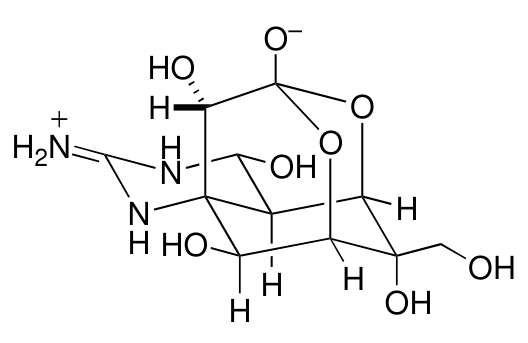
tetrodotoxin (1)
The guanidine group in tetrodotoxin exhibits strong basicity. The guanidinium ion resulting from protonation on the guanidine group is stabilized by the existence of the following resonance.
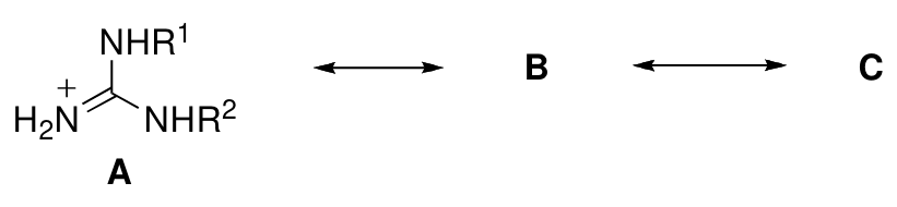
7.1 Draw two resonance structures B and C.
Many derivatization reactions were performed in structure studies of tetrodotoxin. Treatment of tetrodotoxin (1) (see reaction scheme 1) with ethanolic potassium hydroxide upon heating afforded quinazoline derivative 2, which provided an insight into the nature of the fundamental skeleton of tetrodotoxin. The reaction mechanism can be described as follows. First, tetrodotoxin is hydrolyzed into carboxylate 3. Then the hydroxyl group highlighted with a frame is eliminated by the base to give intermediate D. A retro-aldol reaction of D cleaves a carbon-carbon bond to provide intermediates E and F. Finally, dehydration and aromatization from E produce quinazoline derivative 2.
7.2 Draw structures of the postulated intermediates D, E, and F.
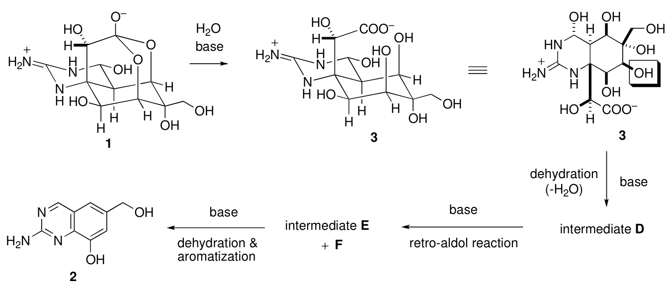
Reaction scheme 1
Although biosynthesis of tetrodotoxin still remains to be clarified, it is proposed that tetrodotoxin may be biologically synthesized from L-arginine and isopentenyl diphosphate.

7.3 Among the carbon atoms included in tetrodotoxin, circle all those that are expected to be of L-arginine origin.
In the 1990s, an alternative biosynthetic pathway of tetrodotoxin was proposed. Condensation between 2-deoxy-3-oxo-D-pentose and guanidine provides intermediate G with cyclic guanidine moiety (molecular formula \(\ce{C6H11N3O3}\)). Tetrodotoxin may be biologically synthesized from intermediate G and isopentenyl diphosphate.
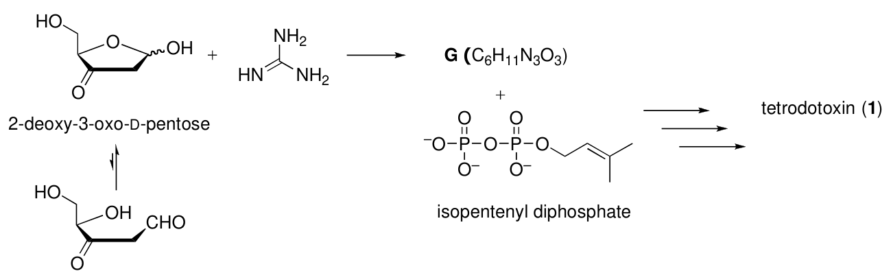
7.4 Draw a structure of the postulated intermediate G showing the stereochemistry.
Official English text of the problem.
Answers
- 7.1 The two remaining contributors of the disubstituted guanidinium ion are:
B: double bond and positive charge on the nitrogen bearing \(R^1\):
NC(=[NH+]([*:1]))N[*:2]
C: double bond and positive charge on the nitrogen bearing \(R^2\):
NC(N[*:1])=[NH+]([*:2])
Here [*:1] and [*:2] represent \(R^1\) and \(R^2\).
- 7.2 Using the atom numbering defined below:
D — dehydrated carboxylate, C8-ketone tautomer:
[NH2+]=C1N[C@H](O)[C@H]2[C@@H](O)[C@](CO)(O)CC(=O)[C@]2([C@H](O)C(=O)[O-])N1
E — retro-aldol product, C8-enol tautomer:
[NH2+]=C1N[C@H](O)[C@H]2[C@@H](O)[C@](CO)(O)CC(O)=C2N1
F — glyoxylate (2-oxoacetate), \(\ce{OHC-COO-}\):
O=CC(=O)[O-]
-
7.3 Circle six carbons: the guanidinium carbon C2, and C4, C4a, C8a, C9, and C10. These constitute the guanidinium carbon plus the five-carbon chain ending at the carboxylate carbon in 3.
-
7.4 G — 2-amino-6-[(1S)-1,2-dihydroxyethyl]-3,4-dihydropyrimidin-4-ol, a cyclic guanidine hemiaminal:
NC1=NC([C@H](O)CO)=CC(O)N1
The side-chain stereocenter retains the sugar’s configuration. The newly formed ring hemiaminal center is unspecified; draw its hydroxyl bond as wavy.
Solution
7.1 — Guanidinium resonance
In A, the positive charge is on the nitrogen of the exocyclic double bond. Either other nitrogen can donate its lone pair to the central carbon while the existing double-bond electrons move onto the initially positive nitrogen.
The resulting contributors are
\[ \mathbf B:\quad \ce{H2N-C(=N^{+}HR^{1})-NHR^{2}} \]
and
\[ \mathbf C:\quad \ce{H2N-C(NHR^{1})=N^{+}HR^{2}}. \]
Thus, each contributor has one carbon–nitrogen double bond, with the positive charge on that double-bonded nitrogen. All atoms retain octets. Hydrogen atoms and substituents remain attached to the same nitrogens: these are resonance contributors, so no proton transfer occurs.
7.2 — Dehydration, retro-aldol cleavage, and aromatization
To identify bonds precisely, use the quinazoline numbering inherited from product 2. The two fused rings follow these atom sequences:
\[ \mathrm{N1-C2-N3-C4-C4a-C8a-N1} \]
\[ \mathrm{C4a-C5-C6-C7-C8-C8a-C4a}. \]
In carboxylate 3, call the hydroxyl-bearing side-chain carbon attached to C8a C9, its carboxylate carbon C10, and the hydroxymethyl carbon attached to C6 C11.
The starting hydrolyzed tetrodotoxin carboxylate 3 is:
[NH2+]=C1N[C@H](O)[C@H]2[C@@H](O)[C@](CO)(O)[C@@H](O)[C@@H](O)[C@]2([C@H](O)C(=O)[O-])N1
Formation of D. The boxed hydroxyl is attached to C7. Of its neighboring carbons, C6 has no hydrogen, whereas C8 does. Elimination therefore removes the C7 hydroxyl and the C8 hydrogen, giving the C7=C8 enol.
This initial enol tautomer of D is:
[NH2+]=C1N[C@H](O)[C@H]2[C@@H](O)[C@](CO)(O)C=C(O)[C@]2([C@H](O)C(=O)[O-])N1
Keto–enol tautomerization gives the C8 ketone D listed in the Answers. To draw it from 3:
- Replace C7–OH by a methylene group at C7.
- Replace C8–OH and C8–H by a carbonyl at C8.
- Retain the remaining substituents and stereochemistry.
Formation of E and F. In the ketone tautomer of D, the relevant sequence is
\[ \mathrm{C8(=O)-C8a-C9(OH)-C10(=O)O^{-}}. \]
This is a β-hydroxy carbonyl arrangement. Base removes the C9 hydroxyl proton; the resulting oxygen lone pair forms the C9 carbonyl while the C8a–C9 bond breaks. The ring fragment becomes an enolate.
The detached two-carbon fragment is glyoxylate F, O=CC(=O)[O-]. Protonation of the ring enolate on oxygen gives the enol E listed above.
To draw E, remove the entire C9–C10 side chain from D, replace the C8 carbonyl by C8–OH, and draw a C8=C8a double bond. Its remaining stereocenters retain their configurations.
Formation of 2. The product is 2-amino-6-(hydroxymethyl)quinazolin-8-ol, Nc1ncc2cc(CO)cc(O)c2n1.
Starting from the enol representation of E, the three dehydrations are:
| Hydroxyl removed | Hydrogen removed | Double bond formed |
|---|---|---|
| C4–OH | N3–H | N3=C4 |
| C5–OH | C4a–H | C4a=C5 |
| C6–OH | C7–H | C6=C7 |
The existing C8=C8a bond completes the carbon-ring unsaturation. Deprotonation and redistribution of the guanidinium π bonds give the aromatic quinazoline.
For the protonation states shown, the atom- and charge-balanced equations are
\[ \underbrace{\ce{C11H17N3O8}}_{\mathbf 1} +\ce{H2O} \longrightarrow \underbrace{\ce{C11H19N3O9}}_{\mathbf 3}, \]
\[ \underbrace{\ce{C11H19N3O9}}_{\mathbf 3} \longrightarrow \underbrace{\ce{C11H17N3O8}}_{\mathbf D} +\ce{H2O}, \]
\[ \underbrace{\ce{C11H17N3O8}}_{\mathbf D} \longrightarrow \underbrace{\ce{C9H16N3O5+}}_{\mathbf E} + \underbrace{\ce{C2HO3-}}_{\mathbf F}, \]
\[ \underbrace{\ce{C9H16N3O5+}}_{\mathbf E} +\ce{OH-} \longrightarrow \underbrace{\ce{C9H9N3O2}}_{\mathbf 2} +4\ce{H2O}. \]
Here water is O, and the hydroxide ion is [OH-]. The last equation includes three dehydration waters and one water from neutralization.
The overall reaction is consequently
\[ \ce{C11H17N3O8 + OH- -> C9H9N3O2 + C2HO3- + 4H2O}. \]
7.3 — Carbon atoms originating from L-arginine
L-Arginine, in the protonation state drawn in the problem, has SMILES:
NC(=[NH2+])NCCC[C@H](N)C(=O)[O-]
Its guanidinium carbon and its uninterrupted five-carbon chain can be mapped onto tetrodotoxin without breaking that chain:
| Carbon in L-arginine | Corresponding carbon in tetrodotoxin |
|---|---|
| Guanidinium carbon | C2 |
| Side-chain carbon adjacent to the guanidino nitrogen, \(C_\delta\) | C4 |
| \(C_\gamma\) | C4a |
| \(C_\beta\) | C8a |
| Amino-acid \(C_\alpha\) | C9 |
| Carboxylate carbon | C10 |
The remaining five-carbon fragment is the branched chain
\[ \mathrm{C8-C7-C6(-C11)-C5}, \]
which matches the prenyl donor’s carbon connectivity.
On the original tetrodotoxin drawing, circle:
- The carbon bonded to all three nitrogens.
- The hydroxyl-bearing carbon directly bonded to the upper ring NH.
- Its adjacent methine carbon, C4a.
- The adjacent quaternary carbon bonded to the other ring NH, C8a.
- The hydroxyl-bearing methine above that quaternary carbon, C9.
- The carbon bearing the negatively charged oxygen, C10.
Hydrolysis from 1 to 3 changes oxygen connectivity but preserves these carbon–carbon connections, so the assignment is the same in both drawings.
7.4 — Formation and stereochemistry of G
The open-chain 2-deoxy-3-oxo-D-pentose is
\[ \ce{OHC-CH2-C(=O)-CH(OH)-CH2OH}, \]
with stereochemical SMILES O=CCC(=O)[C@H](O)CO.
Guanidine is N=C(N)N. The molecular formulas require
\[ \ce{C5H8O4 + CH5N3 -> C6H11N3O3 + H2O}. \]
Only one water molecule is lost. Complete condensation at both carbonyl groups would instead lose two waters and give \(\ce{C6H9N3O2}\), which is inconsistent with the stated formula.
The cyclic intermediate can form by condensation at the ketone, followed by intramolecular addition of another guanidine nitrogen to the aldehyde. This closes a six-membered ring while retaining the aldehyde oxygen as a hemiaminal hydroxyl. Proton transfers give the enamine tautomer of G specified in the Answers.
To draw that tautomer precisely, use the ring sequence
\[ \mathrm{N1=C2(-NH_2)-N3H-C4H(OH)-C5H=C6(-R)-N1}, \]
where the C6 substituent is the stereochemically defined dihydroxyethyl group encoded in
NC1=NC([C@H](O)CO)=CC(O)N1.
The original sugar stereocenter retains its spatial configuration. A Fischer projection of the side chain has:
- The heterocyclic ring above the stereocenter.
- The terminal hydroxymethyl group below it.
- The hydroxyl group on the right and hydrogen on the left.
Its descriptor is \(S\) in G, although the corresponding center is \(R\) in the starting sugar. This change results from a change in CIP priorities when the ketone carbon becomes part of the nitrogen-containing ring; it does not represent inversion.
The newly formed hemiaminal carbon has unspecified stereochemistry, consistent with showing its hydroxyl bond as wavy.
The answers were checked against the official answers published by the organisers.
IChO 2010 · Theorieaufgabe 7
Tetrodotoxin
Bestimmte Arten von Kugelfischen, auf Japanisch Fugu, sind in Japan als Speise sehr geschätzt. Da die Innereien (insbesondere Eierstöcke und Lebern) der Fische ein starkes Gift (Tetrodotoxin) enthalten, führt ihr Verzehr häufig zu Lebensmittelvergiftungen. Seit Beginn des 20. Jahrhunderts wird Tetrodotoxin (1) untersucht. Seine chemische Struktur wurde 1964 aufgeklärt.
Tetrodotoxin (1)
Die Guanidingruppe im Tetrodotoxin ist stark basisch. Das durch Protonierung der Guanidingruppe entstehende Guanidiniumion wird durch die folgende Mesomerie stabilisiert.
7.1 Zeichnen Sie zwei mesomere Grenzstrukturen B und C.
Bei den Untersuchungen zur Struktur von Tetrodotoxin wurden zahlreiche Derivatisierungsreaktionen durchgeführt. Die Behandlung von Tetrodotoxin (1) (siehe Reaktionsschema 1) mit ethanolischer Kaliumhydroxidlösung unter Erhitzen lieferte das Chinazolinderivat 2, das Aufschluss über das Grundgerüst von Tetrodotoxin gab. Der Reaktionsmechanismus lässt sich wie folgt beschreiben. Zunächst wird Tetrodotoxin zum Carboxylat 3 hydrolysiert. Anschließend wird die durch einen Rahmen hervorgehobene Hydroxygruppe durch die Base eliminiert, wobei das Zwischenprodukt D entsteht. Eine Retroaldolreaktion von D spaltet eine Kohlenstoff-Kohlenstoff-Bindung und liefert die Zwischenprodukte E und F. Schließlich entsteht aus E durch Dehydratisierung und Aromatisierung das Chinazolinderivat 2.
7.2 Zeichnen Sie die Strukturen der postulierten Zwischenprodukte D, E und F.
Reaktionsschema 1
Obwohl die Biosynthese von Tetrodotoxin noch nicht aufgeklärt ist, wird vorgeschlagen, dass Tetrodotoxin biosynthetisch aus L-Arginin und Isopentenyldiphosphat gebildet werden könnte.
7.3 Kreisen Sie unter den Kohlenstoffatomen des Tetrodotoxins alle diejenigen ein, die vermutlich aus L-Arginin stammen.
In den 1990er Jahren wurde ein alternativer Biosyntheseweg für Tetrodotoxin vorgeschlagen. Die Kondensation von 2-Desoxy-3-oxo-D-pentose mit Guanidin liefert das Zwischenprodukt G mit einer cyclischen Guanidineinheit (Summenformel \(\ce{C6H11N3O3}\)). Tetrodotoxin könnte biosynthetisch aus dem Zwischenprodukt G und Isopentenyldiphosphat gebildet werden.
7.4 Zeichnen Sie eine Struktur des postulierten Zwischenprodukts G unter Angabe der Stereochemie.
Aufgabentext aus dem offiziellen englischen Text übersetzt.
Antworten
- 7.1 Die beiden übrigen mesomeren Grenzstrukturen des disubstituierten Guanidiniumions sind:
B: Doppelbindung und positive Ladung am Stickstoffatom, das \(R^1\) trägt:
NC(=[NH+]([*:1]))N[*:2]
C: Doppelbindung und positive Ladung am Stickstoffatom, das \(R^2\) trägt:
NC(N[*:1])=[NH+]([*:2])
Dabei stehen [*:1] und [*:2] für \(R^1\) bzw. \(R^2\).
- 7.2 Unter Verwendung der unten definierten Atomnummerierung:
D — dehydratisiertes Carboxylat, C8-Ketotautomer:
[NH2+]=C1N[C@H](O)[C@H]2[C@@H](O)[C@](CO)(O)CC(=O)[C@]2([C@H](O)C(=O)[O-])N1
E — Retroaldolprodukt, C8-Enoltautomer:
[NH2+]=C1N[C@H](O)[C@H]2[C@@H](O)[C@](CO)(O)CC(O)=C2N1
F — Glyoxylat (2-Oxoacetat), \(\ce{OHC-COO-}\):
O=CC(=O)[O-]
-
7.3 Kreisen Sie sechs Kohlenstoffatome ein: das Guanidinium-Kohlenstoffatom C2 sowie C4, C4a, C8a, C9 und C10. Diese umfassen das Guanidinium-Kohlenstoffatom und die Kette aus fünf Kohlenstoffatomen, die in 3 am Carboxylat-Kohlenstoffatom endet.
-
7.4 G — 2-Amino-6-[(1S)-1,2-dihydroxyethyl]-3,4-dihydropyrimidin-4-ol, ein cyclisches Guanidin-Halbaminal:
NC1=NC([C@H](O)CO)=CC(O)N1
Das Stereozentrum der Seitenkette behält die Konfiguration des Zuckers bei. Die Konfiguration des neu gebildeten Halbaminalzentrums im Ring ist nicht festgelegt; zeichnen Sie seine Bindung zur Hydroxygruppe als Wellenlinie.
Lösung
7.1 — Mesomerie des Guanidiniumions
In A befindet sich die positive Ladung am Stickstoffatom der exocyclischen Doppelbindung. Jedes der beiden anderen Stickstoffatome kann sein freies Elektronenpaar dem zentralen Kohlenstoffatom zur Verfügung stellen, während die Elektronen der vorhandenen Doppelbindung auf das ursprünglich positiv geladene Stickstoffatom verschoben werden.
Die daraus resultierenden Grenzstrukturen sind
\[ \mathbf B:\quad \ce{H2N-C(=N^{+}HR^{1})-NHR^{2}} \]
und
\[ \mathbf C:\quad \ce{H2N-C(NHR^{1})=N^{+}HR^{2}}. \]
Jede Grenzstruktur besitzt somit eine Kohlenstoff–Stickstoff-Doppelbindung, wobei die positive Ladung an dem doppelt gebundenen Stickstoffatom liegt. Alle Atome behalten ihre Elektronenoktette bei. Wasserstoffatome und Substituenten bleiben an dieselben Stickstoffatome gebunden: Es handelt sich um mesomere Grenzstrukturen, daher findet keine Protonenübertragung statt.
7.2 — Dehydratisierung, Retroaldolspaltung und Aromatisierung
Um Bindungen eindeutig zu bezeichnen, wird die vom Produkt 2 übernommene Chinazolinnummerierung verwendet. Die beiden anellierten Ringe weisen folgende Atomfolgen auf:
\[ \mathrm{N1-C2-N3-C4-C4a-C8a-N1} \]
\[ \mathrm{C4a-C5-C6-C7-C8-C8a-C4a}. \]
Im Carboxylat 3 wird das an C8a gebundene, eine Hydroxygruppe tragende Kohlenstoffatom der Seitenkette als C9, sein Carboxylat-Kohlenstoffatom als C10 und das an C6 gebundene Hydroxymethyl-Kohlenstoffatom als C11 bezeichnet.
Das als Ausgangsstoff dienende Carboxylat 3 des hydrolysierten Tetrodotoxins ist:
[NH2+]=C1N[C@H](O)[C@H]2[C@@H](O)[C@](CO)(O)[C@@H](O)[C@@H](O)[C@]2([C@H](O)C(=O)[O-])N1
Bildung von D. Die eingerahmte Hydroxygruppe ist an C7 gebunden. Von den benachbarten Kohlenstoffatomen trägt C6 kein Wasserstoffatom, C8 dagegen schon. Bei der Eliminierung werden daher die Hydroxygruppe an C7 und das Wasserstoffatom an C8 abgespalten, wodurch das C7=C8-Enol entsteht.
Dieses zunächst gebildete Enoltautomer von D ist:
[NH2+]=C1N[C@H](O)[C@H]2[C@@H](O)[C@](CO)(O)C=C(O)[C@]2([C@H](O)C(=O)[O-])N1
Durch Keto-Enol-Tautomerisierung entsteht das unter Antworten aufgeführte C8-Keton D. Um es ausgehend von 3 zu zeichnen:
- Ersetzen Sie C7–OH durch eine Methylengruppe an C7.
- Ersetzen Sie C8–OH und C8–H durch eine Carbonylgruppe an C8.
- Behalten Sie die übrigen Substituenten und die Stereochemie bei.
Bildung von E und F. Im Ketotautomer von D lautet die relevante Atomfolge
\[ \mathrm{C8(=O)-C8a-C9(OH)-C10(=O)O^{-}}. \]
Dies ist eine β-Hydroxycarbonyl-Anordnung. Die Base spaltet das Proton der Hydroxygruppe an C9 ab; das dadurch entstehende freie Elektronenpaar am Sauerstoff bildet die Carbonylgruppe an C9, während die C8a–C9-Bindung gespalten wird. Das Ringfragment wird zu einem Enolat.
Das abgespaltene Fragment aus zwei Kohlenstoffatomen ist Glyoxylat F, O=CC(=O)[O-]. Die Protonierung des Ringenolats am Sauerstoff liefert das oben aufgeführte Enol E.
Um E zu zeichnen, entfernen Sie die gesamte C9–C10-Seitenkette aus D, ersetzen Sie die Carbonylgruppe an C8 durch C8–OH und zeichnen Sie eine C8=C8a-Doppelbindung. Die übrigen Stereozentren behalten ihre Konfigurationen bei.
Bildung von 2. Das Produkt ist 2-Amino-6-(hydroxymethyl)chinazolin-8-ol, Nc1ncc2cc(CO)cc(O)c2n1.
Ausgehend von der Enoldarstellung von E ergeben sich die drei Dehydratisierungen wie folgt:
| Abgespaltene Hydroxygruppe | Abgespaltenes Wasserstoffatom | Gebildete Doppelbindung |
|---|---|---|
| C4–OH | N3–H | N3=C4 |
| C5–OH | C4a–H | C4a=C5 |
| C6–OH | C7–H | C6=C7 |
Die bereits vorhandene C8=C8a-Bindung vervollständigt die Ungesättigtheit des Kohlenstoffrings. Deprotonierung und Umverteilung der π-Bindungen des Guanidiniumions ergeben das aromatische Chinazolin.
Für die dargestellten Protonierungszustände lauten die hinsichtlich Atomen und Ladungen ausgeglichenen Gleichungen
\[ \underbrace{\ce{C11H17N3O8}}_{\mathbf 1} +\ce{H2O} \longrightarrow \underbrace{\ce{C11H19N3O9}}_{\mathbf 3}, \]
\[ \underbrace{\ce{C11H19N3O9}}_{\mathbf 3} \longrightarrow \underbrace{\ce{C11H17N3O8}}_{\mathbf D} +\ce{H2O}, \]
\[ \underbrace{\ce{C11H17N3O8}}_{\mathbf D} \longrightarrow \underbrace{\ce{C9H16N3O5+}}_{\mathbf E} + \underbrace{\ce{C2HO3-}}_{\mathbf F}, \]
\[ \underbrace{\ce{C9H16N3O5+}}_{\mathbf E} +\ce{OH-} \longrightarrow \underbrace{\ce{C9H9N3O2}}_{\mathbf 2} +4\ce{H2O}. \]
Dabei ist Wasser O und das Hydroxidion [OH-]. Die letzte Gleichung umfasst drei Wassermoleküle aus den Dehydratisierungen und ein Wassermolekül aus der Neutralisation.
Die Gesamtreaktion lautet folglich
\[ \ce{C11H17N3O8 + OH- -> C9H9N3O2 + C2HO3- + 4H2O}. \]
7.3 — Aus L-Arginin stammende Kohlenstoffatome
L-Arginin hat in dem in der Aufgabenstellung gezeichneten Protonierungszustand die folgende SMILES-Darstellung:
NC(=[NH2+])NCCC[C@H](N)C(=O)[O-]
Sein Guanidinium-Kohlenstoffatom und seine durchgehende Kette aus fünf Kohlenstoffatomen lassen sich dem Tetrodotoxin zuordnen, ohne diese Kette zu spalten:
| Kohlenstoffatom in L-Arginin | Entsprechendes Kohlenstoffatom im Tetrodotoxin |
|---|---|
| Guanidinium-Kohlenstoffatom | C2 |
| Dem Guanidino-Stickstoffatom benachbartes Kohlenstoffatom der Seitenkette, \(C_\delta\) | C4 |
| \(C_\gamma\) | C4a |
| \(C_\beta\) | C8a |
| \(C_\alpha\) der Aminosäure | C9 |
| Carboxylat-Kohlenstoffatom | C10 |
Das verbleibende Fragment aus fünf Kohlenstoffatomen ist die verzweigte Kette
\[ \mathrm{C8-C7-C6(-C11)-C5}, \]
die der Verknüpfung der Kohlenstoffatome im Prenyldonor entspricht.
Kreisen Sie in der ursprünglichen Tetrodotoxinzeichnung Folgendes ein:
- Das Kohlenstoffatom, das an alle drei Stickstoffatome gebunden ist.
- Das eine Hydroxygruppe tragende Kohlenstoffatom, das direkt an die obere NH-Gruppe des Rings gebunden ist.
- Das benachbarte Methin-Kohlenstoffatom C4a.
- Das benachbarte quartäre Kohlenstoffatom C8a, das an die andere NH-Gruppe des Rings gebunden ist.
- Das eine Hydroxygruppe tragende Methin-Kohlenstoffatom C9 oberhalb dieses quartären Kohlenstoffatoms.
- Das Kohlenstoffatom C10, das das negativ geladene Sauerstoffatom trägt.
Die Hydrolyse von 1 zu 3 verändert die Verknüpfung der Sauerstoffatome, erhält jedoch diese Kohlenstoff–Kohlenstoff-Verknüpfungen, sodass die Zuordnung in beiden Zeichnungen gleich ist.
7.4 — Bildung und Stereochemie von G
Die offenkettige 2-Desoxy-3-oxo-D-pentose ist
\[ \ce{OHC-CH2-C(=O)-CH(OH)-CH2OH}, \]
mit der stereochemischen SMILES-Darstellung O=CCC(=O)[C@H](O)CO.
Guanidin ist N=C(N)N. Aus den Summenformeln ergibt sich zwingend
\[ \ce{C5H8O4 + CH5N3 -> C6H11N3O3 + H2O}. \]
Es wird nur ein Wassermolekül abgespalten. Bei vollständiger Kondensation an beiden Carbonylgruppen würden dagegen zwei Wassermoleküle abgespalten und \(\ce{C6H9N3O2}\) gebildet, was nicht mit der angegebenen Summenformel übereinstimmt.
Das cyclische Zwischenprodukt kann durch Kondensation an der Ketogruppe mit anschließender intramolekularer Addition eines weiteren Guanidin-Stickstoffatoms an die Aldehydgruppe entstehen. Dadurch wird ein sechsgliedriger Ring geschlossen, während der Aldehydsauerstoff als Hydroxygruppe eines Halbaminals erhalten bleibt. Protonenübertragungen ergeben das unter Antworten angegebene Enamintautomer von G.
Um dieses Tautomer genau zu zeichnen, verwenden Sie die folgende Atomfolge im Ring
\[ \mathrm{N1=C2(-NH_2)-N3H-C4H(OH)-C5H=C6(-R)-N1}, \]
wobei der Substituent an C6 die stereochemisch definierte Dihydroxyethylgruppe ist, die in der folgenden SMILES-Darstellung codiert ist:
NC1=NC([C@H](O)CO)=CC(O)N1.
Das ursprüngliche Stereozentrum des Zuckers behält seine räumliche Konfiguration bei. In einer Fischer-Projektion der Seitenkette befindet sich:
- Der heterocyclische Ring oberhalb des Stereozentrums.
- Die endständige Hydroxymethylgruppe unterhalb des Stereozentrums.
- Die Hydroxygruppe rechts und das Wasserstoffatom links.
Sein Deskriptor ist \(S\) in G, obwohl das entsprechende Zentrum im Ausgangszucker \(R\) ist. Diese Änderung ergibt sich aus einer Änderung der CIP-Prioritäten, wenn das Keton-Kohlenstoffatom Teil des stickstoffhaltigen Rings wird; sie bedeutet keine Inversion.
Die Stereochemie des neu gebildeten Halbaminal-Kohlenstoffatoms ist nicht festgelegt, weshalb seine Bindung zur Hydroxygruppe als Wellenlinie dargestellt wird.
Die Ergebnisse wurden mit den von den Veranstaltern veröffentlichten offiziellen Ergebnissen abgeglichen.
IChO 2010 · Teori sorusu 7
Tetrodotoksin
Japoncada Fugu olarak adlandırılan balon balığının bazı türleri, Japonya'da yiyecek olarak çok değerlidir. Balığın iç organları (özellikle yumurtalıkları ve karaciğeri) güçlü bir toksin (tetrodotoksin) içerdiğinden, tüketilmesi sıklıkla gıda zehirlenmesine yol açar. Tetrodotoksin (1) üzerine çalışmalar 20. yüzyılın başından beri yürütülmektedir. Kimyasal yapısı 1964 yılında aydınlatılmıştır.
tetrodotoksin (1)
Tetrodotoksindeki guanidin grubu güçlü bazlık gösterir. Guanidin grubunun protonlanmasıyla oluşan guanidinyum iyonu, aşağıdaki rezonans sayesinde kararlı hâle gelir.
7.1 İki rezonans yapısı olan B ve C'yi çiziniz.
Tetrodotoksinin yapı çalışmalarında çok sayıda türevlendirme tepkimesi gerçekleştirilmiştir. Tetrodotoksinin (1) (bkz. tepkime şeması 1) etanol içindeki potasyum hidroksit ile ısıtılarak tepkimeye sokulması, tetrodotoksinin temel iskeletinin niteliği hakkında bilgi sağlayan kinazolin türevi 2'yi vermiştir. Tepkime mekanizması aşağıdaki gibi açıklanabilir. Önce tetrodotoksin hidroliz edilerek karboksilat 3'e dönüşür. Ardından, çerçeve içinde gösterilen hidroksil grubu baz etkisiyle elimine edilerek ara ürün D oluşur. D'nin retro-aldol tepkimesi bir karbon-karbon bağını kopararak E ve F ara ürünlerini verir. Son olarak, E'nin dehidrasyonu ve aromatikleşmesiyle kinazolin türevi 2 oluşur.
7.2 Önerilen D, E ve F ara ürünlerinin yapılarını çiziniz.
Tepkime şeması 1
Tetrodotoksinin biyosentezi henüz aydınlatılmamış olmakla birlikte, tetrodotoksinin biyolojik olarak L-arjinin ve izopentenil difosfattan sentezlenebileceği öne sürülmektedir.
7.3 Tetrodotoksindeki karbon atomlarından L-arjinin kökenli olması beklenenlerin tümünü daire içine alınız.
1990'larda tetrodotoksin için alternatif bir biyosentez yolu önerilmiştir. 2-Deoksi-3-okso-D-pentoz ile guanidin arasındaki kondenzasyon, halkalı guanidin birimi içeren ara ürün G'yi (molekül formülü \(\ce{C6H11N3O3}\)) verir. Tetrodotoksin, ara ürün G ve izopentenil difosfattan biyolojik olarak sentezlenebilir.
7.4 Önerilen ara ürün G'nin yapısını stereokimyasını göstererek çiziniz.
Soru metni resmi İngilizce metinden çevrilmiştir.
Cevaplar
- 7.1 Disübstitüe guanidinyum iyonunun diğer iki rezonans katkı yapısı şunlardır:
B: çift bağ ve pozitif yük, \(R^1\) taşıyan azot üzerindedir:
NC(=[NH+]([*:1]))N[*:2]
C: çift bağ ve pozitif yük, \(R^2\) taşıyan azot üzerindedir:
NC(N[*:1])=[NH+]([*:2])
Burada [*:1] ve [*:2], \(R^1\) ve \(R^2\)'yi temsil eder.
- 7.2 Aşağıda tanımlanan atom numaralandırması kullanıldığında:
D — dehidrasyona uğramış karboksilat, C8-keton tautomeri:
[NH2+]=C1N[C@H](O)[C@H]2[C@@H](O)[C@](CO)(O)CC(=O)[C@]2([C@H](O)C(=O)[O-])N1
E — retro-aldol ürünü, C8-enol tautomeri:
[NH2+]=C1N[C@H](O)[C@H]2[C@@H](O)[C@](CO)(O)CC(O)=C2N1
F — glioksilat (2-oksoasetat), \(\ce{OHC-COO-}\):
O=CC(=O)[O-]
-
7.3 Altı karbonu daire içine alınız: guanidinyum karbonu C2 ile C4, C4a, C8a, C9 ve C10. Bunlar, guanidinyum karbonu ile 3'te karboksilat karbonunda sonlanan beş karbonlu zinciri oluşturur.
-
7.4 G — 2-amino-6-[(1S)-1,2-dihidroksietil]-3,4-dihidropirimidin-4-ol, halkalı bir guanidin hemiaminali:
NC1=NC([C@H](O)CO)=CC(O)N1
Yan zincirdeki stereomerkez, şekerin konfigürasyonunu korur. Halkada yeni oluşan hemiaminal merkezinin stereokimyası belirtilmemiştir; hidroksil bağını dalgalı çiziniz.
Çözüm
7.1 — Guanidinyum rezonansı
A'da pozitif yük, halka dışındaki çift bağın azotu üzerindedir. Diğer iki azottan herhangi biri, mevcut çift bağın elektronları başlangıçta pozitif yüklü olan azot üzerine geçerken, ortaklanmamış elektron çiftini merkezdeki karbona verebilir.
Oluşan katkı yapıları şunlardır:
\[ \mathbf B:\quad \ce{H2N-C(=N^{+}HR^{1})-NHR^{2}} \]
ve
\[ \mathbf C:\quad \ce{H2N-C(NHR^{1})=N^{+}HR^{2}}. \]
Böylece her katkı yapısında bir karbon–azot çift bağı bulunur ve pozitif yük bu çift bağdaki azot üzerindedir. Tüm atomlar oktetlerini korur. Hidrojen atomları ve sübstitüentler aynı azotlara bağlı kalır: bunlar rezonans katkı yapıları olduğundan proton aktarımı gerçekleşmez.
7.2 — Dehidrasyon, retro-aldol parçalanması ve aromatikleşme
Bağları kesin olarak tanımlamak için ürün 2'den alınan kinazolin numaralandırmasını kullanınız. İki kaynaşmış halka şu atom sıralarını izler:
\[ \mathrm{N1-C2-N3-C4-C4a-C8a-N1} \]
\[ \mathrm{C4a-C5-C6-C7-C8-C8a-C4a}. \]
Karboksilat 3'te, C8a'ya bağlı olan ve hidroksil taşıyan yan zincir karbonunu C9, bunun karboksilat karbonunu C10 ve C6'ya bağlı hidroksimetil karbonunu C11 olarak adlandırınız.
Başlangıçtaki hidroliz edilmiş tetrodotoksin karboksilatı 3 şöyledir:
[NH2+]=C1N[C@H](O)[C@H]2[C@@H](O)[C@](CO)(O)[C@@H](O)[C@@H](O)[C@]2([C@H](O)C(=O)[O-])N1
D'nin oluşumu. Çerçeve içindeki hidroksil C7'ye bağlıdır. Komşu karbonlarından C6'da hidrojen bulunmazken C8'de bulunur. Bu nedenle eliminasyonda C7 hidroksili ve C8 hidrojeni uzaklaşarak C7=C8 enolünü verir.
D'nin başlangıçta oluşan bu enol tautomeri şöyledir:
[NH2+]=C1N[C@H](O)[C@H]2[C@@H](O)[C@](CO)(O)C=C(O)[C@]2([C@H](O)C(=O)[O-])N1
Keto–enol tautomerleşmesi, Cevaplar bölümünde verilen C8 ketonu D'yi oluşturur. Bunu 3'ten hareketle çizmek için:
- C7–OH yerine C7'de bir metilen grubu oluşturunuz.
- C8–OH ve C8–H yerine C8'de bir karbonil grubu oluşturunuz.
- Diğer sübstitüentleri ve stereokimyayı koruyunuz.
E ve F'nin oluşumu. D'nin keton tautomerinde ilgili atom dizisi şöyledir:
\[ \mathrm{C8(=O)-C8a-C9(OH)-C10(=O)O^{-}}. \]
Bu, bir β-hidroksi karbonil düzenlenmesidir. Baz, C9 hidroksilinin protonunu uzaklaştırır; oluşan oksijen ortaklanmamış elektron çifti C9 karbonilini oluştururken C8a–C9 bağı kopar. Halka parçası bir enolata dönüşür.
Ayrılan iki karbonlu parça glioksilat F, O=CC(=O)[O-]'dir. Halka enolatının oksijen üzerinden protonlanması, yukarıda verilen enol E'yi oluşturur.
E'yi çizmek için C9–C10 yan zincirinin tamamını D'den çıkarınız, C8 karbonili yerine C8–OH koyunuz ve bir C8=C8a çift bağı çiziniz. Geriye kalan stereomerkezler konfigürasyonlarını korur.
2'nin oluşumu. Ürün, 2-amino-6-(hidroksimetil)kinazolin-8-ol, Nc1ncc2cc(CO)cc(O)c2n1'dir.
E'nin enol gösteriminden başlanarak gerçekleşen üç dehidrasyon şöyledir:
| Uzaklaştırılan hidroksil | Uzaklaştırılan hidrojen | Oluşan çift bağ |
|---|---|---|
| C4–OH | N3–H | N3=C4 |
| C5–OH | C4a–H | C4a=C5 |
| C6–OH | C7–H | C6=C7 |
Mevcut C8=C8a bağı, karbon halkasının doymamışlığını tamamlar. Deprotonlanma ve guanidinyum π bağlarının yeniden düzenlenmesi aromatik kinazolini verir.
Gösterilen protonlanma durumları için atom ve yük bakımından denkleştirilmiş denklemler şöyledir:
\[ \underbrace{\ce{C11H17N3O8}}_{\mathbf 1} +\ce{H2O} \longrightarrow \underbrace{\ce{C11H19N3O9}}_{\mathbf 3}, \]
\[ \underbrace{\ce{C11H19N3O9}}_{\mathbf 3} \longrightarrow \underbrace{\ce{C11H17N3O8}}_{\mathbf D} +\ce{H2O}, \]
\[ \underbrace{\ce{C11H17N3O8}}_{\mathbf D} \longrightarrow \underbrace{\ce{C9H16N3O5+}}_{\mathbf E} + \underbrace{\ce{C2HO3-}}_{\mathbf F}, \]
\[ \underbrace{\ce{C9H16N3O5+}}_{\mathbf E} +\ce{OH-} \longrightarrow \underbrace{\ce{C9H9N3O2}}_{\mathbf 2} +4\ce{H2O}. \]
Burada su O, hidroksit iyonu ise [OH-]'dir. Son denklem, dehidrasyondan gelen üç su molekülünü ve nötralleşmeden gelen bir su molekülünü içerir.
Dolayısıyla toplam tepkime şöyledir:
\[ \ce{C11H17N3O8 + OH- -> C9H9N3O2 + C2HO3- + 4H2O}. \]
7.3 — L-arjinin kökenli karbon atomları
L-Arjininin, soruda çizilen protonlanma durumundaki SMILES gösterimi şöyledir:
NC(=[NH2+])NCCC[C@H](N)C(=O)[O-]
Guanidinyum karbonu ve kesintisiz beş karbonlu zinciri, bu zincir kırılmadan tetrodotoksinle eşleştirilebilir:
| L-arjinindeki karbon | Tetrodotoksindeki karşılık gelen karbon |
|---|---|
| Guanidinyum karbonu | C2 |
| Guanidino azotuna komşu yan zincir karbonu, \(C_\delta\) | C4 |
| \(C_\gamma\) | C4a |
| \(C_\beta\) | C8a |
| Amino asidin \(C_\alpha\) karbonu | C9 |
| Karboksilat karbonu | C10 |
Geriye kalan beş karbonlu parça, şu dallanmış zincirdir:
\[ \mathrm{C8-C7-C6(-C11)-C5}, \]
Bu zincir, prenil vericisinin karbon bağlantı düzeniyle örtüşür.
Başlangıçta verilen tetrodotoksin çiziminde şunları daire içine alınız:
- Üç azotun tümüne bağlı olan karbon.
- Üstteki halka NH grubuna doğrudan bağlı, hidroksil taşıyan karbon.
- Buna komşu metin karbonu, C4a.
- Diğer halka NH grubuna bağlı olan komşu kuaterner karbon, C8a.
- Bu kuaterner karbonun üstündeki, hidroksil taşıyan metin karbonu, C9.
- Negatif yüklü oksijeni taşıyan karbon, C10.
1'den 3'e hidroliz, oksijenlerin bağlantı düzenini değiştirir ancak bu karbon–karbon bağlantılarını korur; dolayısıyla her iki çizimde de eşleştirme aynıdır.
7.4 — G'nin oluşumu ve stereokimyası
Açık zincirli 2-deoksi-3-okso-D-pentoz şöyledir:
\[ \ce{OHC-CH2-C(=O)-CH(OH)-CH2OH}, \]
Stereokimyasal SMILES gösterimi O=CCC(=O)[C@H](O)CO'dur.
Guanidin, N=C(N)N'dir. Molekül formülleri şu denklemi gerektirir:
\[ \ce{C5H8O4 + CH5N3 -> C6H11N3O3 + H2O}. \]
Yalnızca bir su molekülü kaybedilir. Her iki karbonil grubunda tam kondenzasyon gerçekleşseydi iki su molekülü kaybedilir ve verilen formülle uyuşmayan \(\ce{C6H9N3O2}\) elde edilirdi.
Halkalı ara ürün, ketonda kondenzasyonu takiben başka bir guanidin azotunun aldehide molekül içi katılmasıyla oluşabilir. Bu, aldehit oksijenini hemiaminal hidroksili olarak korurken altı üyeli bir halka oluşturur. Proton aktarımları, Cevaplar bölümünde belirtilen G'nin enamin tautomerini verir.
Bu tautomeri tam olarak çizmek için şu halka dizisini kullanınız:
\[ \mathrm{N1=C2(-NH_2)-N3H-C4H(OH)-C5H=C6(-R)-N1}, \]
Burada C6 sübstitüenti, aşağıdaki gösterimde kodlanan, stereokimyası tanımlı dihidroksietil grubudur:
NC1=NC([C@H](O)CO)=CC(O)N1.
Başlangıçtaki şekerin stereomerkezi uzaysal konfigürasyonunu korur. Yan zincirin Fischer izdüşümünde:
- Heterosiklik halka stereomerkezin üstündedir.
- Uçtaki hidroksimetil grubu stereomerkezin altındadır.
- Hidroksil grubu sağda, hidrojen soldadır.
Başlangıç şekerinde karşılık gelen merkez \(R\) olmasına rağmen, bu merkezin tanımlayıcısı G'de \(S\)'dir. Bu değişiklik, keton karbonu azot içeren halkanın bir parçası hâline geldiğinde CIP önceliklerinin değişmesinden kaynaklanır; inversiyonu ifade etmez.
Yeni oluşan hemiaminal karbonunun stereokimyası belirtilmemiştir; bu, hidroksil bağının dalgalı gösterilmesiyle uyumludur.
Sonuçlar, düzenleyicilerin yayımladığı resmi sonuçlarla karşılaştırılarak kontrol edildi.
IChO 2010 · Problema teórico 7
Tetrodotoxina
Ciertas variedades de pez globo, Fugu en japonés, son muy apreciadas como alimento en Japón. Dado que las vísceras (especialmente los ovarios y el hígado) del pez contienen una potente toxina (tetrodotoxina), su ingestión provoca a menudo intoxicaciones alimentarias. La tetrodotoxina (1) se estudia desde principios del siglo XX. Su estructura química se elucidó en 1964.
tetrodotoxina (1)
El grupo guanidina de la tetrodotoxina presenta una fuerte basicidad. El ion guanidinio resultante de la protonación del grupo guanidina se estabiliza por la existencia de la siguiente resonancia.
7.1 Dibuje dos estructuras de resonancia B y C.
En los estudios estructurales de la tetrodotoxina se llevaron a cabo muchas reacciones de derivatización. El tratamiento de la tetrodotoxina (1) (véase el esquema de reacción 1) con hidróxido de potasio en etanol y calentamiento dio el derivado de quinazolina 2, que permitió comprender la naturaleza del esqueleto fundamental de la tetrodotoxina. El mecanismo de reacción puede describirse como sigue. Primero, la tetrodotoxina se hidroliza para dar el carboxilato 3. A continuación, la base provoca la eliminación del grupo hidroxilo resaltado con un recuadro para dar el intermedio D. Una reacción retroaldólica de D rompe un enlace carbono-carbono para dar los intermedios E y F. Finalmente, la deshidratación y la aromatización de E producen el derivado de quinazolina 2.
7.2 Dibuje las estructuras de los intermedios propuestos D, E y F.
Esquema de reacción 1
Aunque la biosíntesis de la tetrodotoxina aún está por esclarecer, se propone que la tetrodotoxina podría sintetizarse biológicamente a partir de L-arginina y difosfato de isopentenilo.
7.3 De los átomos de carbono presentes en la tetrodotoxina, rodee con un círculo todos aquellos que se espera que procedan de la L-arginina.
En la década de 1990 se propuso una ruta biosintética alternativa para la tetrodotoxina. La condensación entre la 2-desoxi-3-oxo-D-pentosa y la guanidina da el intermedio G, con una unidad de guanidina cíclica (fórmula molecular \(\ce{C6H11N3O3}\)). La tetrodotoxina podría sintetizarse biológicamente a partir del intermedio G y difosfato de isopentenilo.
7.4 Dibuje una estructura del intermedio propuesto G que muestre la estereoquímica.
Enunciado traducido del texto oficial en inglés.
Respuestas
- 7.1 Las otras dos formas contribuyentes del ion guanidinio disustituido son:
B: doble enlace y carga positiva en el nitrógeno que lleva \(R^1\):
NC(=[NH+]([*:1]))N[*:2]
C: doble enlace y carga positiva en el nitrógeno que lleva \(R^2\):
NC(N[*:1])=[NH+]([*:2])
Aquí [*:1] y [*:2] representan \(R^1\) y \(R^2\).
- 7.2 Usando la numeración de átomos definida a continuación:
D — carboxilato deshidratado, tautómero cetónico en C8:
[NH2+]=C1N[C@H](O)[C@H]2[C@@H](O)[C@](CO)(O)CC(=O)[C@]2([C@H](O)C(=O)[O-])N1
E — producto retroaldólico, tautómero enólico en C8:
[NH2+]=C1N[C@H](O)[C@H]2[C@@H](O)[C@](CO)(O)CC(O)=C2N1
F — glioxilato (2-oxoacetato), \(\ce{OHC-COO-}\):
O=CC(=O)[O-]
-
7.3 Rodee con un círculo seis carbonos: el carbono del guanidinio C2, y C4, C4a, C8a, C9 y C10. Estos constituyen el carbono del guanidinio más la cadena de cinco carbonos que termina en el carbono del carboxilato en 3.
-
7.4 G — 2-amino-6-[(1S)-1,2-dihidroxietil]-3,4-dihidropirimidin-4-ol, un hemiaminal de guanidina cíclica:
NC1=NC([C@H](O)CO)=CC(O)N1
El estereocentro de la cadena lateral conserva la configuración del azúcar. La configuración del centro hemiaminal del anillo recién formado no está especificada; dibuje su enlace al hidroxilo como una línea ondulada.
Solución
7.1 — Resonancia del guanidinio
En A, la carga positiva se encuentra en el nitrógeno del doble enlace exocíclico. Cualquiera de los otros dos nitrógenos puede donar su par de electrones no compartido al carbono central mientras los electrones del doble enlace existente se desplazan hacia el nitrógeno inicialmente positivo.
Las formas contribuyentes resultantes son
\[ \mathbf B:\quad \ce{H2N-C(=N^{+}HR^{1})-NHR^{2}} \]
y
\[ \mathbf C:\quad \ce{H2N-C(NHR^{1})=N^{+}HR^{2}}. \]
Así, cada forma contribuyente tiene un doble enlace carbono–nitrógeno, con la carga positiva en el nitrógeno unido mediante ese doble enlace. Todos los átomos conservan sus octetos. Los átomos de hidrógeno y los sustituyentes permanecen unidos a los mismos nitrógenos: se trata de formas contribuyentes de resonancia, por lo que no se produce ninguna transferencia de protones.
7.2 — Deshidratación, ruptura retroaldólica y aromatización
Para identificar los enlaces con precisión, utilice la numeración de la quinazolina heredada del producto 2. Los dos anillos fusionados siguen estas secuencias de átomos:
\[ \mathrm{N1-C2-N3-C4-C4a-C8a-N1} \]
\[ \mathrm{C4a-C5-C6-C7-C8-C8a-C4a}. \]
En el carboxilato 3, denomine C9 al carbono de la cadena lateral que lleva el hidroxilo y está unido a C8a, C10 a su carbono del carboxilato y C11 al carbono del hidroximetilo unido a C6.
El carboxilato de tetrodotoxina hidrolizada 3 de partida es:
[NH2+]=C1N[C@H](O)[C@H]2[C@@H](O)[C@](CO)(O)[C@@H](O)[C@@H](O)[C@]2([C@H](O)C(=O)[O-])N1
Formación de D. El hidroxilo enmarcado está unido a C7. De sus carbonos vecinos, C6 no tiene hidrógeno, mientras que C8 sí lo tiene. Por tanto, la eliminación suprime el hidroxilo de C7 y el hidrógeno de C8, dando el enol C7=C8.
Este tautómero enólico inicial de D es:
[NH2+]=C1N[C@H](O)[C@H]2[C@@H](O)[C@](CO)(O)C=C(O)[C@]2([C@H](O)C(=O)[O-])N1
La tautomerización ceto–enólica da la cetona en C8 D indicada en las Respuestas. Para dibujarla a partir de 3:
- Sustituya C7–OH por un grupo metileno en C7.
- Sustituya C8–OH y C8–H por un carbonilo en C8.
- Conserve los demás sustituyentes y la estereoquímica.
Formación de E y F. En el tautómero cetónico de D, la secuencia pertinente es
\[ \mathrm{C8(=O)-C8a-C9(OH)-C10(=O)O^{-}}. \]
Esta disposición corresponde a un compuesto β-hidroxicarbonílico. La base elimina el protón del hidroxilo de C9; el par de electrones no compartido del oxígeno resultante forma el carbonilo de C9 mientras se rompe el enlace C8a–C9. El fragmento cíclico se convierte en un enolato.
El fragmento de dos carbonos que se separa es el glioxilato F, O=CC(=O)[O-]. La protonación del enolato cíclico en el oxígeno da el enol E indicado anteriormente.
Para dibujar E, elimine toda la cadena lateral C9–C10 de D, sustituya el carbonilo de C8 por C8–OH y dibuje un doble enlace C8=C8a. Sus estereocentros restantes conservan sus configuraciones.
Formación de 2. El producto es 2-amino-6-(hidroximetil)quinazolin-8-ol, Nc1ncc2cc(CO)cc(O)c2n1.
Partiendo de la representación enólica de E, las tres deshidrataciones son:
| Hidroxilo eliminado | Hidrógeno eliminado | Doble enlace formado |
|---|---|---|
| C4–OH | N3–H | N3=C4 |
| C5–OH | C4a–H | C4a=C5 |
| C6–OH | C7–H | C6=C7 |
El enlace C8=C8a existente completa la insaturación del anillo carbonado. La desprotonación y la redistribución de los enlaces π del guanidinio dan la quinazolina aromática.
Para los estados de protonación mostrados, las ecuaciones ajustadas en átomos y carga son
\[ \underbrace{\ce{C11H17N3O8}}_{\mathbf 1} +\ce{H2O} \longrightarrow \underbrace{\ce{C11H19N3O9}}_{\mathbf 3}, \]
\[ \underbrace{\ce{C11H19N3O9}}_{\mathbf 3} \longrightarrow \underbrace{\ce{C11H17N3O8}}_{\mathbf D} +\ce{H2O}, \]
\[ \underbrace{\ce{C11H17N3O8}}_{\mathbf D} \longrightarrow \underbrace{\ce{C9H16N3O5+}}_{\mathbf E} + \underbrace{\ce{C2HO3-}}_{\mathbf F}, \]
\[ \underbrace{\ce{C9H16N3O5+}}_{\mathbf E} +\ce{OH-} \longrightarrow \underbrace{\ce{C9H9N3O2}}_{\mathbf 2} +4\ce{H2O}. \]
Aquí el agua es O, y el ion hidróxido es [OH-]. La última ecuación incluye tres moléculas de agua de deshidratación y una de neutralización.
Por consiguiente, la reacción global es
\[ \ce{C11H17N3O8 + OH- -> C9H9N3O2 + C2HO3- + 4H2O}. \]
7.3 — Átomos de carbono procedentes de la L-arginina
La L-arginina, en el estado de protonación dibujado en el problema, tiene el SMILES:
NC(=[NH2+])NCCC[C@H](N)C(=O)[O-]
Su carbono del guanidinio y su cadena ininterrumpida de cinco carbonos pueden hacerse corresponder con los de la tetrodotoxina sin romper dicha cadena:
| Carbono en la L-arginina | Carbono correspondiente en la tetrodotoxina |
|---|---|
| Carbono del guanidinio | C2 |
| Carbono de la cadena lateral adyacente al nitrógeno del grupo guanidino, \(C_\delta\) | C4 |
| \(C_\gamma\) | C4a |
| \(C_\beta\) | C8a |
| \(C_\alpha\) del aminoácido | C9 |
| Carbono del carboxilato | C10 |
El fragmento restante de cinco carbonos es la cadena ramificada
\[ \mathrm{C8-C7-C6(-C11)-C5}, \]
que coincide con la conectividad de los carbonos del donante de prenilo.
En el dibujo original de la tetrodotoxina, rodee con un círculo:
- El carbono unido a los tres nitrógenos.
- El carbono que lleva el hidroxilo y está unido directamente al NH superior del anillo.
- Su carbono metínico adyacente, C4a.
- El carbono cuaternario adyacente unido al otro NH del anillo, C8a.
- El carbono metínico que lleva el hidroxilo situado por encima de ese carbono cuaternario, C9.
- El carbono que lleva el oxígeno con carga negativa, C10.
La hidrólisis de 1 a 3 cambia la conectividad de los oxígenos, pero conserva estas conexiones carbono–carbono, por lo que la asignación es la misma en ambos dibujos.
7.4 — Formación y estereoquímica de G
La 2-desoxi-3-oxo-D-pentosa de cadena abierta es
\[ \ce{OHC-CH2-C(=O)-CH(OH)-CH2OH}, \]
con el SMILES estereoquímico O=CCC(=O)[C@H](O)CO.
La guanidina es N=C(N)N. Las fórmulas moleculares exigen
\[ \ce{C5H8O4 + CH5N3 -> C6H11N3O3 + H2O}. \]
Solo se pierde una molécula de agua. En cambio, la condensación completa en ambos grupos carbonilo perdería dos moléculas de agua y daría \(\ce{C6H9N3O2}\), lo cual es incompatible con la fórmula indicada.
El intermedio cíclico puede formarse por condensación en la cetona, seguida de la adición intramolecular de otro nitrógeno de la guanidina al aldehído. Así se cierra un anillo de seis miembros, conservando el oxígeno del aldehído como hidroxilo hemiaminal. Las transferencias de protones dan el tautómero enamínico de G especificado en las Respuestas.
Para dibujar ese tautómero con precisión, utilice la secuencia del anillo
\[ \mathrm{N1=C2(-NH_2)-N3H-C4H(OH)-C5H=C6(-R)-N1}, \]
donde el sustituyente de C6 es el grupo dihidroxietilo estereoquímicamente definido codificado en
NC1=NC([C@H](O)CO)=CC(O)N1.
El estereocentro original del azúcar conserva su configuración espacial. Una proyección de Fischer de la cadena lateral tiene:
- El anillo heterocíclico por encima del estereocentro.
- El grupo hidroximetilo terminal por debajo de él.
- El grupo hidroxilo a la derecha y el hidrógeno a la izquierda.
Su descriptor es \(S\) en G, aunque el centro correspondiente es \(R\) en el azúcar de partida. Este cambio se debe a un cambio en las prioridades CIP cuando el carbono de la cetona pasa a formar parte del anillo que contiene nitrógeno; no representa una inversión.
El carbono hemiaminal recién formado tiene una estereoquímica no especificada, lo cual es coherente con representar su enlace al hidroxilo mediante una línea ondulada.
Los resultados se comprobaron con los resultados oficiales publicados por los organizadores.
IChO 2010 · 理论题 7
河豚毒素
某些种类的河豚在日语中称为 Fugu,在日本被视为珍贵的食材。由于河豚的内脏(尤其是卵巢和肝脏)含有一种强效毒素(河豚毒素),食用后常会引起食物中毒。对河豚毒素(1)的研究始于20世纪初。其化学结构于1964年得到阐明。
河豚毒素 (1)
河豚毒素中的胍基具有强碱性。胍基质子化后形成的胍鎓离子因存在以下共振而得到稳定。
7.1 画出两个共振结构 B 和 C。
在河豚毒素的结构研究中,人们进行了许多衍生化反应。将河豚毒素(1)(见反应方案1)与氢氧化钾的乙醇溶液一起加热,得到喹唑啉衍生物 2,这为认识河豚毒素的基本骨架提供了线索。该反应的机理可描述如下。首先,河豚毒素水解生成羧酸盐 3。随后,方框标出的羟基在碱的作用下被消去,生成中间体 D。D 发生逆羟醛反应,使一个碳—碳键断裂,生成中间体 E 和 F。最后,E 经脱水和芳构化生成喹唑啉衍生物 2。
7.2 画出所推测的中间体 D、E 和 F 的结构。
反应方案1
虽然河豚毒素的生物合成途径仍有待阐明,但有人提出,河豚毒素可能由 L-精氨酸和异戊烯基二磷酸经生物合成得到。
7.3 在河豚毒素所含的碳原子中,圈出所有预计来源于 L-精氨酸的碳原子。
20世纪90年代,人们提出了河豚毒素的另一条生物合成途径。2-脱氧-3-氧代-D-戊糖与胍缩合,生成含有环状胍结构单元的中间体 G(分子式为 \(\ce{C6H11N3O3}\))。河豚毒素可能由中间体 G 和异戊烯基二磷酸经生物合成得到。
7.4 画出所推测的中间体 G 的结构,并标明其立体化学。
题目译自官方英文原文。
答案
- 7.1 二取代胍鎓离子的另外两个共振贡献式为:
B:双键和正电荷位于连接 \(R^1\) 的氮原子上:
NC(=[NH+]([*:1]))N[*:2]
C:双键和正电荷位于连接 \(R^2\) 的氮原子上:
NC(N[*:1])=[NH+]([*:2])
其中,[*:1] 和 [*:2] 分别代表 \(R^1\) 和 \(R^2\)。
- 7.2 采用下文定义的原子编号:
D — 脱水后的羧酸盐,C8 酮式互变异构体:
[NH2+]=C1N[C@H](O)[C@H]2[C@@H](O)[C@](CO)(O)CC(=O)[C@]2([C@H](O)C(=O)[O-])N1
E — 逆羟醛反应产物,C8 烯醇式互变异构体:
[NH2+]=C1N[C@H](O)[C@H]2[C@@H](O)[C@](CO)(O)CC(O)=C2N1
F — 乙醛酸根(2-氧代乙酸根),\(\ce{OHC-COO-}\):
O=CC(=O)[O-]
-
7.3 圈出六个碳原子:胍鎓碳 C2,以及 C4、C4a、C8a、C9 和 C10。它们由胍鎓碳和一条五碳链组成,该碳链以 3 中的羧酸根碳为末端。
-
7.4 G — 2-氨基-6-[(1S)-1,2-二羟基乙基]-3,4-二氢嘧啶-4-醇,一种环状胍半缩醛胺:
NC1=NC([C@H](O)CO)=CC(O)N1
侧链上的立体中心保留糖的构型。新形成的环上半缩醛胺中心的构型未作指定;将连接其羟基的键画成波浪线。
解答
7.1 — 胍鎓离子的共振
在 A 中,正电荷位于环外双键的氮原子上。其余两个氮原子中的任何一个都可以将其孤电子对提供给中心碳原子,同时原有双键的电子移向最初带正电荷的氮原子。
由此得到的共振贡献式为
\[ \mathbf B:\quad \ce{H2N-C(=N^{+}HR^{1})-NHR^{2}} \]
和
\[ \mathbf C:\quad \ce{H2N-C(NHR^{1})=N^{+}HR^{2}}. \]
因此,每个共振贡献式都含有一个碳—氮双键,正电荷位于形成该双键的氮原子上。所有原子均保持八隅体结构。氢原子和取代基仍连接在原来的氮原子上:这些是共振贡献式,因此不发生质子转移。
7.2 — 脱水、逆羟醛断裂和芳构化
为准确标识各化学键,采用产物 2 的喹唑啉编号。两个稠合环的原子排列顺序如下:
\[ \mathrm{N1-C2-N3-C4-C4a-C8a-N1} \]
\[ \mathrm{C4a-C5-C6-C7-C8-C8a-C4a}. \]
在羧酸盐 3 中,将连接在 C8a 上、带有羟基的侧链碳记为 C9,将其羧酸根碳记为 C10,将连接在 C6 上的羟甲基碳记为 C11。
起始的河豚毒素水解产物羧酸盐 3 为:
[NH2+]=C1N[C@H](O)[C@H]2[C@@H](O)[C@](CO)(O)[C@@H](O)[C@@H](O)[C@]2([C@H](O)C(=O)[O-])N1
D 的形成。方框标出的羟基连接在 C7 上。在其相邻的碳原子中,C6 上没有氢,而 C8 上有氢。因此,消去反应除去 C7 上的羟基和 C8 上的氢,生成含 C7=C8 双键的烯醇。
这一最初形成的 D 的烯醇式互变异构体为:
[NH2+]=C1N[C@H](O)[C@H]2[C@@H](O)[C@](CO)(O)C=C(O)[C@]2([C@H](O)C(=O)[O-])N1
酮—烯醇互变异构化生成答案中列出的 C8 酮式 D。从 3 出发画出该结构时:
- 将 C7–OH 替换为 C7 处的亚甲基。
- 将 C8–OH 和 C8–H 替换为 C8 处的羰基。
- 保留其余取代基及立体化学。
E 和 F 的形成。在 D 的酮式互变异构体中,相关的原子排列顺序为
\[ \mathrm{C8(=O)-C8a-C9(OH)-C10(=O)O^{-}}. \]
这是一个 β-羟基羰基结构。碱夺取 C9 羟基上的质子;随后,氧原子上的孤电子对形成 C9 羰基,同时 C8a–C9 键断裂。环状片段转变为烯醇负离子。
脱离的二碳片段为乙醛酸根 F,O=CC(=O)[O-]。环状烯醇负离子在氧原子上质子化,生成上文列出的烯醇 E。
画 E 时,从 D 中移去整个 C9–C10 侧链,将 C8 羰基替换为 C8–OH,并画出 C8=C8a 双键。其余立体中心均保留原有构型。
2 的形成。产物为 2-氨基-6-(羟甲基)喹唑啉-8-醇,Nc1ncc2cc(CO)cc(O)c2n1。
从 E 的烯醇式结构出发,三次脱水分别为:
| 脱去的羟基 | 脱去的氢 | 形成的双键 |
|---|---|---|
| C4–OH | N3–H | N3=C4 |
| C5–OH | C4a–H | C4a=C5 |
| C6–OH | C7–H | C6=C7 |
原有的 C8=C8a 键使碳环的不饱和键体系完整形成。经去质子化和胍鎓部分 π 键的重新分布,得到芳香性的喹唑啉。
对于所示的质子化状态,原子数和电荷均配平的方程式为
\[ \underbrace{\ce{C11H17N3O8}}_{\mathbf 1} +\ce{H2O} \longrightarrow \underbrace{\ce{C11H19N3O9}}_{\mathbf 3}, \]
\[ \underbrace{\ce{C11H19N3O9}}_{\mathbf 3} \longrightarrow \underbrace{\ce{C11H17N3O8}}_{\mathbf D} +\ce{H2O}, \]
\[ \underbrace{\ce{C11H17N3O8}}_{\mathbf D} \longrightarrow \underbrace{\ce{C9H16N3O5+}}_{\mathbf E} + \underbrace{\ce{C2HO3-}}_{\mathbf F}, \]
\[ \underbrace{\ce{C9H16N3O5+}}_{\mathbf E} +\ce{OH-} \longrightarrow \underbrace{\ce{C9H9N3O2}}_{\mathbf 2} +4\ce{H2O}. \]
这里,水表示为 O,氢氧根离子表示为 [OH-]。最后一个方程式中的水包括脱水产生的三个水分子和中和产生的一个水分子。
因此,总反应为
\[ \ce{C11H17N3O8 + OH- -> C9H9N3O2 + C2HO3- + 4H2O}. \]
7.3 — 来源于 L-精氨酸的碳原子
L-精氨酸在题目所画质子化状态下的 SMILES 为:
NC(=[NH2+])NCCC[C@H](N)C(=O)[O-]
其胍鎓碳和连续的五碳链可在不使该碳链断裂的情况下,与河豚毒素中的碳原子对应:
| L-精氨酸中的碳原子 | 河豚毒素中对应的碳原子 |
|---|---|
| 胍鎓碳 | C2 |
| 与胍基氮相邻的侧链碳,\(C_\delta\) | C4 |
| \(C_\gamma\) | C4a |
| \(C_\beta\) | C8a |
| 氨基酸的 \(C_\alpha\) | C9 |
| 羧酸根碳 | C10 |
其余五碳片段为支链
\[ \mathrm{C8-C7-C6(-C11)-C5}, \]
其碳原子的连接方式与异戊烯基供体相符。
在原始的河豚毒素结构图中,圈出:
- 与三个氮原子均相连的碳原子。
- 与上方环内 NH 直接相连、带有羟基的碳原子。
- 与其相邻的次甲基碳 C4a。
- 与 C4a 相邻、并与另一个环内 NH 相连的季碳 C8a。
- 该季碳上方带有羟基的次甲基碳 C9。
- 连接带负电荷氧原子的碳 C10。
从 1 到 3 的水解改变了氧原子的连接方式,但保留了这些碳—碳连接,因此两幅结构图中的归属相同。
7.4 — G 的形成及立体化学
开链形式的 2-脱氧-3-氧代-D-戊糖为
\[ \ce{OHC-CH2-C(=O)-CH(OH)-CH2OH}, \]
其立体化学 SMILES 为 O=CCC(=O)[C@H](O)CO。
胍为 N=C(N)N。根据分子式,反应必须满足
\[ \ce{C5H8O4 + CH5N3 -> C6H11N3O3 + H2O}. \]
反应仅失去一个水分子。若两个羰基均完全缩合,则会失去两个水分子并生成 \(\ce{C6H9N3O2}\),这与给定的分子式不符。
该环状中间体可先通过酮羰基处的缩合形成,随后胍中的另一个氮原子对醛基发生分子内加成。这样形成一个六元环,同时醛基的氧原子保留下来,成为半缩醛胺的羟基。经质子转移,得到答案中给出的 G 的烯胺式互变异构体。
为准确画出该互变异构体,采用以下环原子顺序
\[ \mathrm{N1=C2(-NH_2)-N3H-C4H(OH)-C5H=C6(-R)-N1}, \]
其中,C6 上的取代基是具有确定立体化学的二羟基乙基,其编码见
NC1=NC([C@H](O)CO)=CC(O)N1。
原糖的立体中心保留其空间构型。侧链的费歇尔投影式具有以下特征:
- 杂环位于立体中心上方。
- 末端羟甲基位于其下方。
- 羟基位于右侧,氢位于左侧。
该中心在 G 中的构型描述符为 \(S\),而在起始糖中对应中心的构型描述符为 \(R\)。这一变化源于酮羰基碳成为含氮环的一部分后,CIP 优先顺序发生改变;它并不表示构型反转。
新形成的半缩醛胺碳的立体化学未作指定,因此将连接其羟基的键画为波浪线与此相符。
结果已与主办方公布的官方答案核对。
IChO 2010 · Теоретическая задача 7
Тетродотоксин
Некоторые виды рыб-иглобрюхов, по-японски Fugu, высоко ценятся в Японии как пищевой продукт. Поскольку внутренние органы этих рыб (особенно яичники и печень) содержат сильнодействующий токсин (тетродотоксин), их употребление в пищу нередко приводит к пищевому отравлению. Исследования тетродотоксина (1) проводятся с начала XX века. Его химическая структура была установлена в 1964 году.
тетродотоксин (1)
Гуанидиновая группа в тетродотоксине проявляет сильные основные свойства. Ион гуанидиния, образующийся при протонировании гуанидиновой группы, стабилизируется за счёт следующего резонанса.
7.1 Изобразите две резонансные структуры B и C.
В ходе исследований структуры тетродотоксина было проведено множество реакций получения его производных. Обработка тетродотоксина (1) (см. схему реакции 1) спиртовым раствором гидроксида калия при нагревании приводила к производному хиназолина 2, что позволило получить представление об основном скелете тетродотоксина. Механизм реакции можно описать следующим образом. Сначала тетродотоксин гидролизуется с образованием карбоксилата 3. Затем под действием основания отщепляется выделенная рамкой гидроксильная группа, образуя промежуточное соединение D. В результате ретроальдольной реакции D происходит разрыв связи углерод–углерод с образованием промежуточных соединений E и F. Наконец, дегидратация и ароматизация E приводят к производному хиназолина 2.
7.2 Изобразите структуры предполагаемых промежуточных соединений D, E и F.
Схема реакции 1
Хотя биосинтез тетродотоксина ещё предстоит выяснить, предполагается, что тетродотоксин может синтезироваться в живых организмах из L-аргинина и изопентенилдифосфата.
7.3 Среди атомов углерода тетродотоксина обведите все те, которые, предположительно, происходят из L-аргинина.
В 1990-х годах был предложен альтернативный путь биосинтеза тетродотоксина. Конденсация 2-дезокси-3-оксо-D-пентозы с гуанидином приводит к промежуточному соединению G, содержащему циклический гуанидиновый фрагмент (молекулярная формула \(\ce{C6H11N3O3}\)). Тетродотоксин может синтезироваться в живых организмах из промежуточного соединения G и изопентенилдифосфата.
7.4 Изобразите структуру предполагаемого промежуточного соединения G, указав стереохимию.
Условие переведено с официального английского текста.
Ответы
- 7.1 Две оставшиеся резонансные структуры двузамещённого иона гуанидиния:
B: двойная связь и положительный заряд на атоме азота, несущем \(R^1\):
NC(=[NH+]([*:1]))N[*:2]
C: двойная связь и положительный заряд на атоме азота, несущем \(R^2\):
NC(N[*:1])=[NH+]([*:2])
Здесь [*:1] и [*:2] обозначают \(R^1\) и \(R^2\).
- 7.2 При использовании приведённой ниже нумерации атомов:
D — дегидратированный карбоксилат, таутомер с кетогруппой при C8:
[NH2+]=C1N[C@H](O)[C@H]2[C@@H](O)[C@](CO)(O)CC(=O)[C@]2([C@H](O)C(=O)[O-])N1
E — продукт ретроальдольной реакции, таутомер с енольной группой при C8:
[NH2+]=C1N[C@H](O)[C@H]2[C@@H](O)[C@](CO)(O)CC(O)=C2N1
F — глиоксилат (2-оксоацетат), \(\ce{OHC-COO-}\):
O=CC(=O)[O-]
-
7.3 Обведите шесть атомов углерода: гуанидиниевый атом углерода C2, а также C4, C4a, C8a, C9 и C10. Это гуанидиниевый атом углерода и пятиуглеродная цепь, заканчивающаяся карбоксилатным атомом углерода в 3.
-
7.4 G — 2-амино-6-[(1S)-1,2-дигидроксиэтил]-3,4-дигидропиримидин-4-ол, циклический гуанидиновый полуаминаль:
NC1=NC([C@H](O)CO)=CC(O)N1
Стереоцентр боковой цепи сохраняет конфигурацию исходного сахара. Конфигурация вновь образовавшегося полуаминального центра в кольце не определена; изобразите его связь с гидроксильной группой волнистой линией.
Решение
7.1 — Резонанс в ионе гуанидиния
В A положительный заряд находится на атоме азота экзоциклической двойной связи. Любой из двух других атомов азота может предоставить свою неподелённую электронную пару центральному атому углерода, при этом электроны существующей двойной связи переходят к первоначально положительно заряженному атому азота.
Получаются следующие резонансные структуры:
\[ \mathbf B:\quad \ce{H2N-C(=N^{+}HR^{1})-NHR^{2}} \]
и
\[ \mathbf C:\quad \ce{H2N-C(NHR^{1})=N^{+}HR^{2}}. \]
Таким образом, каждая резонансная структура содержит одну двойную связь углерод–азот, а положительный заряд находится на атоме азота, участвующем в этой двойной связи. Все атомы сохраняют октеты. Атомы водорода и заместители остаются связанными с теми же атомами азота: это резонансные структуры, поэтому переноса протона не происходит.
7.2 — Дегидратация, ретроальдольное расщепление и ароматизация
Для точного обозначения связей используем нумерацию хиназолина, соответствующую продукту 2. Два конденсированных кольца образованы следующими последовательностями атомов:
\[ \mathrm{N1-C2-N3-C4-C4a-C8a-N1} \]
\[ \mathrm{C4a-C5-C6-C7-C8-C8a-C4a}. \]
В карбоксилате 3 обозначим несущий гидроксильную группу атом углерода боковой цепи, связанный с C8a, как C9, карбоксилатный атом углерода этой цепи — как C10, а атом углерода гидроксиметильной группы, связанной с C6, — как C11.
Исходный карбоксилат 3, полученный гидролизом тетродотоксина, имеет вид:
[NH2+]=C1N[C@H](O)[C@H]2[C@@H](O)[C@](CO)(O)[C@@H](O)[C@@H](O)[C@]2([C@H](O)C(=O)[O-])N1
Образование D. Выделенная рамкой гидроксильная группа связана с C7. Из соседних атомов углерода C6 не имеет атома водорода, тогда как C8 имеет. Поэтому при элиминировании удаляются гидроксильная группа при C7 и атом водорода при C8, что приводит к енолу C7=C8.
Этот первоначальный енольный таутомер D имеет вид:
[NH2+]=C1N[C@H](O)[C@H]2[C@@H](O)[C@](CO)(O)C=C(O)[C@]2([C@H](O)C(=O)[O-])N1
Кето-енольная таутомеризация приводит к кетону D с карбонильной группой при C8, приведённому в разделе «Ответы». Чтобы изобразить его на основе 3:
- Замените C7–OH метиленовой группой в положении C7.
- Замените C8–OH и C8–H карбонильной группой при C8.
- Сохраните остальные заместители и стереохимию.
Образование E и F. В кетонном таутомере D соответствующая последовательность атомов имеет вид
\[ \mathrm{C8(=O)-C8a-C9(OH)-C10(=O)O^{-}}. \]
Это β-гидроксикарбонильный фрагмент. Основание отщепляет протон гидроксильной группы при C9; образовавшаяся неподелённая электронная пара кислорода формирует карбонильную группу при C9, при этом связь C8a–C9 разрывается. Циклический фрагмент превращается в енолят.
Отщепившийся двухуглеродный фрагмент — глиоксилат F, O=CC(=O)[O-]. Протонирование циклического енолята по кислороду даёт приведённый выше енол E.
Чтобы изобразить E, удалите из D всю боковую цепь C9–C10, замените карбонильную группу при C8 на C8–OH и изобразите двойную связь C8=C8a. Остальные стереоцентры сохраняют свои конфигурации.
Образование 2. Продукт представляет собой 2-амино-6-(гидроксиметил)хиназолин-8-ол, Nc1ncc2cc(CO)cc(O)c2n1.
Для енольной формы E три стадии дегидратации имеют следующий вид:
| Удаляемая гидроксильная группа | Удаляемый атом водорода | Образующаяся двойная связь |
|---|---|---|
| C4–OH | N3–H | N3=C4 |
| C5–OH | C4a–H | C4a=C5 |
| C6–OH | C7–H | C6=C7 |
Уже имеющаяся связь C8=C8a дополняет систему ненасыщенных связей углеродного кольца. Депротонирование и перераспределение π-связей гуанидиниевого фрагмента приводят к ароматическому хиназолину.
Для показанных состояний протонирования уравнения, сбалансированные по атомам и зарядам, имеют вид
\[ \underbrace{\ce{C11H17N3O8}}_{\mathbf 1} +\ce{H2O} \longrightarrow \underbrace{\ce{C11H19N3O9}}_{\mathbf 3}, \]
\[ \underbrace{\ce{C11H19N3O9}}_{\mathbf 3} \longrightarrow \underbrace{\ce{C11H17N3O8}}_{\mathbf D} +\ce{H2O}, \]
\[ \underbrace{\ce{C11H17N3O8}}_{\mathbf D} \longrightarrow \underbrace{\ce{C9H16N3O5+}}_{\mathbf E} + \underbrace{\ce{C2HO3-}}_{\mathbf F}, \]
\[ \underbrace{\ce{C9H16N3O5+}}_{\mathbf E} +\ce{OH-} \longrightarrow \underbrace{\ce{C9H9N3O2}}_{\mathbf 2} +4\ce{H2O}. \]
Здесь вода обозначена как O, а гидроксид-ион — как [OH-]. Последнее уравнение включает три молекулы воды, образующиеся при дегидратации, и одну молекулу воды, образующуюся при нейтрализации.
Следовательно, суммарная реакция имеет вид
\[ \ce{C11H17N3O8 + OH- -> C9H9N3O2 + C2HO3- + 4H2O}. \]
7.3 — Атомы углерода, происходящие из L-аргинина
L-Аргинин в состоянии протонирования, изображённом в условии задачи, имеет следующую запись SMILES:
NC(=[NH2+])NCCC[C@H](N)C(=O)[O-]
Его гуанидиниевому атому углерода и непрерывной пятиуглеродной цепи можно сопоставить атомы тетродотоксина без разрыва этой цепи:
| Атом углерода в L-аргинине | Соответствующий атом углерода в тетродотоксине |
|---|---|
| Гуанидиниевый атом углерода | C2 |
| Атом углерода боковой цепи, соседний с атомом азота гуанидиновой группы, \(C_\delta\) | C4 |
| \(C_\gamma\) | C4a |
| \(C_\beta\) | C8a |
| \(C_\alpha\) аминокислоты | C9 |
| Карбоксилатный атом углерода | C10 |
Оставшийся пятиуглеродный фрагмент представляет собой разветвлённую цепь
\[ \mathrm{C8-C7-C6(-C11)-C5}, \]
что соответствует порядку соединения атомов углерода в доноре пренильной группы.
На исходном рисунке тетродотоксина обведите:
- Атом углерода, связанный со всеми тремя атомами азота.
- Несущий гидроксильную группу атом углерода, непосредственно связанный с верхней кольцевой группой NH.
- Соседний с ним метиновый атом углерода C4a.
- Соседний четвертичный атом углерода C8a, связанный с другой кольцевой группой NH.
- Несущий гидроксильную группу метиновый атом углерода C9, расположенный над этим четвертичным атомом углерода.
- Атом углерода C10, несущий отрицательно заряженный атом кислорода.
Гидролиз 1 с образованием 3 изменяет порядок соединения атомов кислорода, но сохраняет эти связи углерод–углерод, поэтому отнесение атомов одинаково на обоих рисунках.
7.4 — Образование и стереохимия G
2-Дезокси-3-оксо-D-пентоза с открытой цепью имеет вид
\[ \ce{OHC-CH2-C(=O)-CH(OH)-CH2OH}, \]
со стереохимической записью SMILES O=CCC(=O)[C@H](O)CO.
Гуанидин имеет запись N=C(N)N. Из молекулярных формул следует
\[ \ce{C5H8O4 + CH5N3 -> C6H11N3O3 + H2O}. \]
Отщепляется только одна молекула воды. При полной конденсации по обеим карбонильным группам вместо этого отщепились бы две молекулы воды и образовалось бы соединение \(\ce{C6H9N3O2}\), что не соответствует указанной формуле.
Циклическое промежуточное соединение может образоваться в результате конденсации по кетонной группе с последующим внутримолекулярным присоединением другого атома азота гуанидина к альдегидной группе. При этом замыкается шестичленное кольцо, а кислород альдегидной группы сохраняется в виде полуаминальной гидроксильной группы. Переносы протонов приводят к енаминному таутомеру G, указанному в разделе «Ответы».
Чтобы точно изобразить этот таутомер, используйте следующую последовательность атомов кольца:
\[ \mathrm{N1=C2(-NH_2)-N3H-C4H(OH)-C5H=C6(-R)-N1}, \]
где заместителем при C6 является дигидроксиэтильная группа с определённой стереохимией, заданной в записи
NC1=NC([C@H](O)CO)=CC(O)N1.
Стереоцентр исходного сахара сохраняет свою пространственную конфигурацию. В проекции Фишера боковой цепи:
- Гетероциклическое кольцо расположено над стереоцентром.
- Концевая гидроксиметильная группа расположена под ним.
- Гидроксильная группа находится справа, а атом водорода — слева.
Его дескриптор — \(S\) в G, хотя соответствующий центр в исходном сахаре имеет дескриптор \(R\). Это изменение обусловлено изменением приоритетов по правилам Кана–Ингольда–Прелога (CIP), когда кетонный атом углерода становится частью азотсодержащего кольца; оно не означает обращения конфигурации.
Стереохимия вновь образовавшегося полуаминального атома углерода не определена, что соответствует изображению его связи с гидроксильной группой волнистой линией.
Ответы проверены по официальным ответам, опубликованным организаторами.
IChO 2010 · مسألة نظرية 7
التيترودوتوكسين
تحظى بعض أنواع الأسماك المنتفخة، التي تُسمّى فوغو باليابانية، بتقدير كبير بوصفها طعامًا في اليابان. ولأن أحشاء هذه الأسماك (ولا سيما المبايض والأكباد) تحتوي على سمّ قوي (التيترودوتوكسين)، فإن تناولها كثيرًا ما يؤدي إلى التسمم الغذائي. وقد أُجريت دراسات على التيترودوتوكسين (1) منذ بداية القرن العشرين. وكُشف عن بنيته الكيميائية في عام 1964.
التيترودوتوكسين (1)
تُظهر مجموعة الغوانيدين في التيترودوتوكسين قاعدية قوية. ويستقر أيون الغوانيدينيوم الناتج عن بروتنة مجموعة الغوانيدين بفعل الرنين الآتي.
7.1 ارسم بنيتي الرنين B وC.
أُجريت تفاعلات عديدة لتكوين مشتقات في أثناء الدراسات البنيوية للتيترودوتوكسين. وقد أدت معالجة التيترودوتوكسين (1) (انظر مخطط التفاعل 1) بهيدروكسيد البوتاسيوم في الإيثانول مع التسخين إلى الحصول على مشتق الكينازولين 2، الذي أتاح فهمًا لطبيعة الهيكل الأساسي للتيترودوتوكسين. ويمكن وصف آلية التفاعل كما يأتي. أولًا، يتحلل التيترودوتوكسين مائيًا إلى الكربوكسيلات 3. ثم تُحذف مجموعة الهيدروكسيل المحددة بإطار بفعل القاعدة لتعطي الوسيط D. ويشطر تفاعل ألدول عكسي للوسيط D رابطة كربون–كربون ليعطي الوسيطين E وF. وأخيرًا، يؤدي نزع الماء والتحول إلى بنية عطرية انطلاقًا من E إلى تكوين مشتق الكينازولين 2.
7.2 ارسم بُنى الوسائط المفترضة D وE وF.
مخطط التفاعل 1
على الرغم من أن التخليق الحيوي للتيترودوتوكسين لا يزال بحاجة إلى توضيح، فقد اقتُرح أن التيترودوتوكسين قد يُخلَّق حيويًا من L-أرجينين وثنائي فوسفات الإيزوبنتينيل.
7.3 من بين ذرات الكربون الموجودة في التيترودوتوكسين، ضع دائرة حول كل ذرة يُتوقَّع أن يكون منشؤها L-أرجينين.
في تسعينيات القرن العشرين، اقتُرح مسار بديل للتخليق الحيوي للتيترودوتوكسين. ويعطي التكاثف بين 2-ديوكسي-3-أوكسو-D-بنتوز والغوانيدين الوسيط G، الذي يحتوي على جزء غوانيدين حلقي (صيغته الجزيئية \(\ce{C6H11N3O3}\)). وقد يُخلَّق التيترودوتوكسين حيويًا من الوسيط G وثنائي فوسفات الإيزوبنتينيل.
7.4 ارسم بنية الوسيط المفترض G مع إظهار الكيمياء الفراغية.
نص المسألة مترجم عن النص الإنجليزي الرسمي.
الإجابات
- 7.1 البنيتان الرنينيتان المساهمتان المتبقيتان لأيون الغوانيدينيوم ثنائي الاستبدال هما:
B: الرابطة المزدوجة والشحنة الموجبة على ذرة النيتروجين الحاملة لـ \(R^1\):
NC(=[NH+]([*:1]))N[*:2]
C: الرابطة المزدوجة والشحنة الموجبة على ذرة النيتروجين الحاملة لـ \(R^2\):
NC(N[*:1])=[NH+]([*:2])
يمثّل [*:1] و[*:2] هنا \(R^1\) و\(R^2\).
- 7.2 باستخدام ترقيم الذرات المحدد أدناه:
D — الكربوكسيلات المنزوع منها الماء، المتماكب التوتومري الكيتوني عند C8:
[NH2+]=C1N[C@H](O)[C@H]2[C@@H](O)[C@](CO)(O)CC(=O)[C@]2([C@H](O)C(=O)[O-])N1
E — ناتج تفاعل الألدول العكسي، المتماكب التوتومري الإينولي عند C8:
[NH2+]=C1N[C@H](O)[C@H]2[C@@H](O)[C@](CO)(O)CC(O)=C2N1
F — الغليوكسيلات (2-أوكسوأسيتات)، \(\ce{OHC-COO-}\):
O=CC(=O)[O-]
-
7.3 ضع دوائر حول ست ذرات كربون: كربون الغوانيدينيوم C2، وC4 وC4a وC8a وC9 وC10. وهي تمثّل كربون الغوانيدينيوم، إضافة إلى السلسلة المكوَّنة من خمس ذرات كربون التي تنتهي بكربون الكربوكسيلات في 3.
-
7.4 G — 2-أمينو-6-[(1S)-1،2-ثنائي هيدروكسي إيثيل]-3،4-ثنائي هيدروبيريميدين-4-أول، وهو هيمي أمينال غوانيديني حلقي:
NC1=NC([C@H](O)CO)=CC(O)N1
يحتفظ المركز الفراغي في السلسلة الجانبية بالتكوين الفراغي للسكر. أما مركز الهيمي أمينال الحلقي المتكوّن حديثًا فتكوينه الفراغي غير محدد؛ ارسم رابطته مع مجموعة الهيدروكسيل بخط متموج.
الحل
7.1 — رنين الغوانيدينيوم
في A، تقع الشحنة الموجبة على ذرة النيتروجين في الرابطة المزدوجة خارج الحلقة. ويمكن لأيٍّ من ذرتي النيتروجين الأخريين أن تمنح زوجها الإلكتروني الحر إلى ذرة الكربون المركزية، بينما تنتقل إلكترونات الرابطة المزدوجة الموجودة إلى ذرة النيتروجين التي كانت موجبة الشحنة في البداية.
البنيتان الرنينيتان المساهمتان الناتجتان هما
\[ \mathbf B:\quad \ce{H2N-C(=N^{+}HR^{1})-NHR^{2}} \]
و
\[ \mathbf C:\quad \ce{H2N-C(NHR^{1})=N^{+}HR^{2}}. \]
وهكذا، تحتوي كل بنية مساهمة على رابطة مزدوجة واحدة بين الكربون والنيتروجين، وتقع الشحنة الموجبة على ذرة النيتروجين المرتبطة بهذه الرابطة المزدوجة. وتحتفظ جميع الذرات بثمانياتها الإلكترونية. وتبقى ذرات الهيدروجين والمستبدلات متصلة بذرات النيتروجين نفسها: فهذه بُنى رنينية مساهمة، ولذلك لا يحدث انتقال للبروتونات.
7.2 — نزع الماء، والشطر بتفاعل الألدول العكسي، والتحول إلى بنية عطرية
لتحديد الروابط بدقة، استخدم ترقيم الكينازولين الموروث من الناتج 2. وتتبع الحلقتان المندمجتان تسلسلي الذرات الآتيين:
\[ \mathrm{N1-C2-N3-C4-C4a-C8a-N1} \]
\[ \mathrm{C4a-C5-C6-C7-C8-C8a-C4a}. \]
في الكربوكسيلات 3، سمِّ ذرة كربون السلسلة الجانبية الحاملة للهيدروكسيل والمتصلة بـ C8a باسم C9، وذرة كربون الكربوكسيلات فيها باسم C10، وذرة كربون الهيدروكسي ميثيل المتصلة بـ C6 باسم C11.
كربوكسيلات التيترودوتوكسين المتحلل مائيًا 3 التي يبدأ منها التفاعل هي:
[NH2+]=C1N[C@H](O)[C@H]2[C@@H](O)[C@](CO)(O)[C@@H](O)[C@@H](O)[C@]2([C@H](O)C(=O)[O-])N1
تكوُّن D. تتصل مجموعة الهيدروكسيل المحاطة بإطار بـ C7. ومن بين ذرتي الكربون المجاورتين لها، لا تحمل C6 هيدروجينًا، في حين تحمل C8 هيدروجينًا. ولذلك يزيل الحذف مجموعة الهيدروكسيل عند C7 والهيدروجين عند C8، فيعطي الإينول ذا الرابطة C7=C8.
المتماكب التوتومري الإينولي الأولي هذا للوسيط D هو:
[NH2+]=C1N[C@H](O)[C@H]2[C@@H](O)[C@](CO)(O)C=C(O)[C@]2([C@H](O)C(=O)[O-])N1
يعطي التحول التوتومري الكيتوني–الإينولي الكيتون D عند C8 الوارد في الإجابات. ولرسمه انطلاقًا من 3:
- استبدل بـ C7–OH مجموعة ميثيلين عند C7.
- استبدل بـ C8–OH وC8–H مجموعة كربونيل عند C8.
- احتفظ ببقية المستبدلات والكيمياء الفراغية.
تكوُّن E وF. في المتماكب التوتومري الكيتوني للوسيط D، يكون التسلسل المعني هو
\[ \mathrm{C8(=O)-C8a-C9(OH)-C10(=O)O^{-}}. \]
وهذا ترتيب كربونيلي يحمل مجموعة هيدروكسيل في الموضع β. تنتزع القاعدة بروتون مجموعة الهيدروكسيل عند C9؛ ويكوّن الزوج الإلكتروني الحر الناتج على الأكسجين مجموعة الكربونيل عند C9، بينما تنكسر الرابطة C8a–C9. ويتحول الجزء الحلقي إلى إينولات.
الجزء المنفصل المكوَّن من ذرتي كربون هو الغليوكسيلات F، O=CC(=O)[O-]. وتعطي بروتنة إينولات الحلقة عند الأكسجين الإينول E الوارد أعلاه.
لرسم E، أزل السلسلة الجانبية C9–C10 بأكملها من D، واستبدل بمجموعة الكربونيل عند C8 مجموعة C8–OH، وارسم رابطة مزدوجة C8=C8a. وتحتفظ المراكز الفراغية المتبقية بتكويناتها الفراغية.
تكوُّن 2. الناتج هو 2-أمينو-6-(هيدروكسي ميثيل)كينازولين-8-أول، Nc1ncc2cc(CO)cc(O)c2n1.
انطلاقًا من التمثيل الإينولي للوسيط E، تكون عمليات نزع الماء الثلاث كما يأتي:
| مجموعة الهيدروكسيل المنزوعة | الهيدروجين المنزوع | الرابطة المزدوجة المتكوّنة |
|---|---|---|
| C4–OH | N3–H | N3=C4 |
| C5–OH | C4a–H | C4a=C5 |
| C6–OH | C7–H | C6=C7 |
تُكمل الرابطة C8=C8a الموجودة عدم إشباع الحلقة الكربونية. ويعطي نزع البروتون وإعادة توزيع روابط π للغوانيدينيوم الكينازولين العطري.
لحالات البروتنة الموضحة، تكون المعادلات الموزونة من حيث الذرات والشحنات كما يأتي
\[ \underbrace{\ce{C11H17N3O8}}_{\mathbf 1} +\ce{H2O} \longrightarrow \underbrace{\ce{C11H19N3O9}}_{\mathbf 3}, \]
\[ \underbrace{\ce{C11H19N3O9}}_{\mathbf 3} \longrightarrow \underbrace{\ce{C11H17N3O8}}_{\mathbf D} +\ce{H2O}, \]
\[ \underbrace{\ce{C11H17N3O8}}_{\mathbf D} \longrightarrow \underbrace{\ce{C9H16N3O5+}}_{\mathbf E} + \underbrace{\ce{C2HO3-}}_{\mathbf F}, \]
\[ \underbrace{\ce{C9H16N3O5+}}_{\mathbf E} +\ce{OH-} \longrightarrow \underbrace{\ce{C9H9N3O2}}_{\mathbf 2} +4\ce{H2O}. \]
يُمثَّل الماء هنا بـ O، وأيون الهيدروكسيد بـ [OH-]. وتشمل المعادلة الأخيرة ثلاثة جزيئات ماء ناتجة عن نزع الماء وجزيء ماء واحدًا ناتجًا عن التعادل.
وبالتالي يكون التفاعل الكلي
\[ \ce{C11H17N3O8 + OH- -> C9H9N3O2 + C2HO3- + 4H2O}. \]
7.3 — ذرات الكربون ذات المنشأ من L-أرجينين
تمثيل SMILES لـ L-أرجينين، في حالة البروتنة المرسومة في المسألة، هو:
NC(=[NH2+])NCCC[C@H](N)C(=O)[O-]
يمكن مطابقة كربون الغوانيدينيوم وسلسلته المتصلة المكوَّنة من خمس ذرات كربون مع ذرات التيترودوتوكسين دون قطع تلك السلسلة:
| الكربون في L-أرجينين | الكربون المقابل في التيترودوتوكسين |
|---|---|
| كربون الغوانيدينيوم | C2 |
| كربون السلسلة الجانبية المجاور لنيتروجين مجموعة الغوانيدينو، \(C_\delta\) | C4 |
| \(C_\gamma\) | C4a |
| \(C_\beta\) | C8a |
| \(C_\alpha\) للحمض الأميني | C9 |
| كربون الكربوكسيلات | C10 |
الجزء المتبقي المكوَّن من خمس ذرات كربون هو السلسلة المتفرعة
\[ \mathrm{C8-C7-C6(-C11)-C5}, \]
وهي تطابق نمط اتصال ذرات الكربون في مانح البرينيل.
على الرسم الأصلي للتيترودوتوكسين، ضع دوائر حول:
- ذرة الكربون المرتبطة بذرات النيتروجين الثلاث جميعها.
- ذرة الكربون الحاملة للهيدروكسيل والمرتبطة مباشرةً بمجموعة NH العلوية في الحلقة.
- ذرة كربون الميثين المجاورة لها، C4a.
- ذرة الكربون الرباعية المجاورة المرتبطة بمجموعة NH الأخرى في الحلقة، C8a.
- ذرة كربون الميثين الحاملة للهيدروكسيل فوق ذرة الكربون الرباعية تلك، C9.
- ذرة الكربون الحاملة للأكسجين ذي الشحنة السالبة، C10.
يغيّر التحلل المائي من 1 إلى 3 نمط اتصال الأكسجين، لكنه يحافظ على اتصالات الكربون–الكربون هذه، ولذلك يكون التعيين نفسه في كلا الرسمين.
7.4 — تكوُّن G وكيمياؤه الفراغية
الصيغة مفتوحة السلسلة لـ 2-ديوكسي-3-أوكسو-D-بنتوز هي
\[ \ce{OHC-CH2-C(=O)-CH(OH)-CH2OH}, \]
وتمثيل SMILES المحدِّد للكيمياء الفراغية لها هو O=CCC(=O)[C@H](O)CO.
الغوانيدين هو N=C(N)N. وتقتضي الصيغ الجزيئية المعادلة
\[ \ce{C5H8O4 + CH5N3 -> C6H11N3O3 + H2O}. \]
يُفقَد جزيء ماء واحد فقط. أما التكاثف الكامل عند مجموعتي الكربونيل كلتيهما فيؤدي بدلًا من ذلك إلى فقدان جزيئَي ماء وإعطاء \(\ce{C6H9N3O2}\)، وهو ما لا يتفق مع الصيغة المذكورة.
يمكن أن يتكوّن الوسيط الحلقي بتكاثف عند مجموعة الكيتون، تتبعه إضافة داخل الجزيء لذرة نيتروجين أخرى من الغوانيدين إلى مجموعة الألدهيد. ويؤدي ذلك إلى إغلاق حلقة سداسية الأعضاء مع الاحتفاظ بأكسجين الألدهيد في صورة مجموعة هيدروكسيل هيمي أمينالية. وتعطي انتقالات البروتونات المتماكب التوتومري الإيناميني للوسيط G المحدد في الإجابات.
لرسم ذلك المتماكب التوتومري بدقة، استخدم تسلسل الحلقة
\[ \mathrm{N1=C2(-NH_2)-N3H-C4H(OH)-C5H=C6(-R)-N1}, \]
حيث يكون المستبدل عند C6 هو مجموعة ثنائي هيدروكسي إيثيل المحددة فراغيًا والممثلة في
NC1=NC([C@H](O)CO)=CC(O)N1.
يحتفظ المركز الفراغي الأصلي للسكر بتكوينه المكاني. وفي إسقاط فيشر للسلسلة الجانبية تكون:
- الحلقة غير المتجانسة فوق المركز الفراغي.
- مجموعة الهيدروكسي ميثيل الطرفية تحته.
- مجموعة الهيدروكسيل إلى اليمين والهيدروجين إلى اليسار.
واصفه هو \(S\) في G، على الرغم من أن المركز المقابل هو \(R\) في السكر الابتدائي. وينتج هذا التغير عن تغير أولويات CIP عندما تصبح ذرة كربون الكيتون جزءًا من الحلقة المحتوية على النيتروجين؛ وهو لا يمثّل انقلابًا في التكوين الفراغي.
ذرة كربون الهيمي أمينال المتكوّنة حديثًا ذات كيمياء فراغية غير محددة، وهو ما يتفق مع إظهار رابطتها مع مجموعة الهيدروكسيل بخط متموج.
تمت مراجعة النتائج بمقارنتها بالنتائج الرسمية التي نشرها المنظمون.