IChO 2006 · Theory problem 4
The Chemistry of DNA
4.1 In 1944 Oswald Avery isolated a genetic material and showed by elemental analysis that it was a sodium salt of deoxyribonucleic acid. A segment of DNA with formula mass of 1323.72 is shown.
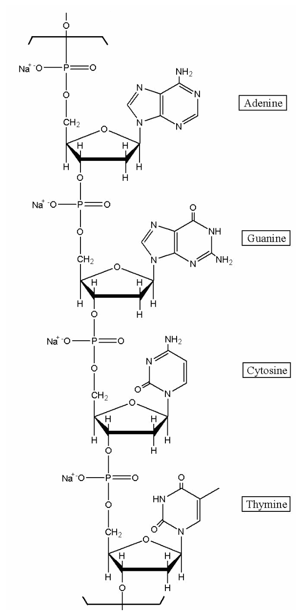
Assuming that equimolar amounts of the four bases are present in DNA, write the number of \(\ce{H}\) atoms per \(\ce{P}\) atom. Calculate (to 3 significant figures) the theoretical weight percentage of \(\ce{H}\) expected upon elemental analysis of DNA.
4.2 Chargaff extracted the separated bases and determined their concentrations by measuring UV absorbance. The Beer-Lambert law was used to obtain the molar concentration. Chargaff discovered the following molar ratio for bases in DNA:
| adenine to guanine = 1.43 | thymine to cytosine = 1.43 |
| adenine to thymine = 1.02 | guanine to cytosine = 1.02 |
Chargaff’s discovery suggested that the bases might exist as pairs in DNA. Watson and Crick mentioned in their celebrated 1953 paper in Nature: "It has not escaped our notice that the specific pairing we have postulated immediately suggests a possible copying mechanism for the genetic material."
Draw structures of the specific pairing found in DNA. Indicate hydrogen bonds. Omit the sugar-phosphate backbone.
4.3 Mutation can occur through base pairings different from the above. Draw structures of any three alternative base pairs.
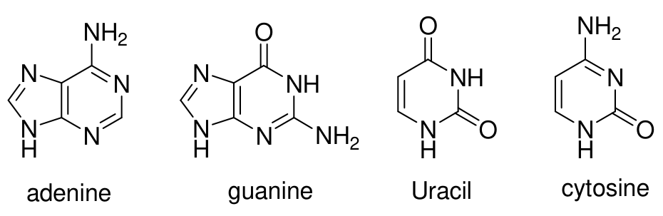
4.4 The plausibility of the formation of purine and pyrimidine bases in the prebiotic atmosphere of the Earth from \(\ce{HCN}\), \(\ce{NH3}\), and \(\ce{H2O}\) has been demonstrated in the laboratory. Write the minimum number of \(\ce{HCN}\) and \(\ce{H2O}\) molecules required for formation of the following compounds.
Official English text of the problem.
Answers
- 4.1: 11.25 hydrogen atoms per phosphorus atom; theoretical hydrogen content 3.43% by mass.
- 4.2: Adenine–thymine, with two hydrogen bonds, and guanine–cytosine, with three hydrogen bonds. Structures and precise hydrogen-bond assignments are given below.
- 4.3: Three examples are guanine–thymine wobble, imino-adenine–cytosine, and guanine–adenine pairs, each with the two hydrogen bonds specified below.
- 4.4: Minimum numbers of molecules per molecule of base:
| Base | \(\ce{HCN}\) | \(\ce{H2O}\) |
|---|---|---|
| Adenine | 5 | 0 |
| Guanine | 5 | 1 |
| Uracil | 4 | 2 |
| Cytosine | 4 | 1 |
Solution
4.1 — Hydrogen content of sodium DNA
The pictured structure is an internal sodium DNA segment with sequence \(5'\)-AGCT-\(3'\). Its chain-fragment SMILES, including the sugar stereochemistry and sodium counterions, is:
*P(=O)([O-])OC[C@H]1O[C@@H](n2cnc3c(N)ncnc32)C[C@@H]1OP(=O)([O-])OC[C@H]1O[C@@H](n2cnc3c(=O)[nH]c(N)nc32)C[C@@H]1OP(=O)([O-])OC[C@H]1O[C@@H](n2ccc(N)nc2=O)C[C@@H]1OP(=O)([O-])OC[C@H]1O[C@@H](n2cc(C)c(=O)[nH]c2=O)C[C@@H]1O*.[Na+].[Na+].[Na+].[Na+]
The first * represents the preceding residue’s \(3'\) oxygen, and the last represents the following phosphate phosphorus. These placeholders lie outside the counted segment.
Each sugar residue has seven carbon-bound hydrogen atoms: one, two, one, one, and two on \(C1'\), \(C2'\), \(C3'\), \(C4'\), and \(C5'\), respectively. The internal sodium phosphodiester groups contribute no additional hydrogen atoms.
Each attached base has one fewer hydrogen than its corresponding free base because its glycosidic attachment replaces an N–H hydrogen.
| Attached base | Hydrogen atoms on base | Hydrogen atoms on sugar | Total |
|---|---|---|---|
| Adenine | 4 | 7 | 11 |
| Guanine | 4 | 7 | 11 |
| Cytosine | 4 | 7 | 11 |
| Thymine | 5 | 7 | 12 |
| Total | 17 | 28 | 45 |
Thus the segment contains 45 hydrogen atoms and four phosphorus atoms:
\[ \boxed{\frac{N_{\ce{H}}}{N_{\ce{P}}}=\frac{45}{4}=11.25}. \]
The counted segment has composition \(\ce{C39H45N15Na4O24P4}\), consistent with the supplied formula mass.
Using \(M(\ce{H})=1.008\ \mathrm{g\,mol^{-1}}\),
\[ \text{Hydrogen mass percentage} = \frac{45(1.008\ \mathrm{g\,mol^{-1}})} {1323.72\ \mathrm{g\,mol^{-1}}}\times100\% = 3.4267\%. \]
To three significant figures,
\[ \boxed{3.43\%\text{ hydrogen by mass}}. \]
4.2 — Normal complementary base pairs
The ratios adenine/thymine and guanine/cytosine are both approximately unity. These suggest the complementary stoichiometries A:T \(=1:1\) and G:C \(=1:1\). The hydrogen-bond donors and acceptors of the depicted bases support these pairings.
The required covalent structures are:
| Base | SMILES |
|---|---|
| Adenine (A) | Nc1ncnc2[nH]cnc12 |
| Thymine (T) | Cc1c[nH]c(=O)[nH]c1=O |
| Guanine (G) | Nc1nc2[nH]cnc2c(=O)[nH]1 |
| Cytosine (C) | Nc1cc[nH]c(=O)n1 |
These are free-base SMILES. In DNA, the hydrogen at N9 of a purine or N1 of a pyrimidine is replaced by the sugar attachment. Those attachment sites do not participate in the hydrogen bonds listed here.
For the requested drawings, place the paired bases approximately coplanar, with their Watson–Crick edges facing each other. Draw a dashed line from each indicated donor hydrogen to its acceptor:
| Pair | Hydrogen-bond donor | Acceptor |
|---|---|---|
| A–T | Adenine N6–H | Thymine O4 |
| A–T | Thymine N3–H | Adenine N1 |
| G–C | Cytosine N4–H | Guanine O6 |
| G–C | Guanine N1–H | Cytosine N3 |
| G–C | Guanine N2–H | Cytosine O2 |
Here N6 of adenine, N4 of cytosine, and N2 of guanine are the exocyclic amino nitrogens; the numbered oxygen atoms are carbonyl oxygens.
Therefore, A–T has two hydrogen bonds, while G–C has three. The SMILES specify covalent connectivity; the table specifies the intermolecular hydrogen bonds and the arrangement required for the drawings.
4.3 — Three alternative base pairs
The following are three possible mismatched pairs.
- Guanine–thymine wobble pair
Use the ordinary guanine and thymine structures given in 4.2. Shift the bases sideways relative to a Watson–Crick arrangement so that:
- Guanine N1–H donates a hydrogen bond to thymine O2.
- Thymine N3–H donates a hydrogen bond to guanine O6.
Draw these two dashed hydrogen bonds. Guanine O6 and thymine O2 are the acceptors.
- Imino-adenine–cytosine pair
Use the rare neutral imino tautomer of adenine, N=c1[nH]cnc2[nH]cnc12, together with ordinary cytosine.
Relative to ordinary adenine, this tautomer has a hydrogen transferred from the exocyclic N6 amino group to ring N1, with the accompanying double-bond rearrangement. Consequently, N1–H is a donor and the exocyclic imino N6 is an acceptor.
Place the bases with their corresponding pairing edges facing each other and draw:
- Cytosine N4–H donating to imino-adenine N6.
- Imino-adenine N1–H donating to cytosine N3.
This gives two hydrogen bonds.
- Guanine–adenine pair
Use the ordinary guanine and adenine structures from 4.2, with their Watson–Crick edges facing each other. Draw:
- Adenine N6–H donating to guanine O6.
- Guanine N1–H donating to adenine N1.
This gives two hydrogen bonds. Because both bases are purines, this arrangement is wider than a normal purine–pyrimidine pair and requires distortion of the local DNA geometry.
4.4 — Minimum precursor requirements
The starting substances are hydrogen cyanide, C#N; ammonia, N; and water, O.
Hydrogen cyanide is the only carbon source, so at least one \(\ce{HCN}\) molecule is required for every carbon atom in the product. Water is the only oxygen source, so at least one \(\ce{H2O}\) molecule is required for every oxygen atom. Ammonia contains neither carbon nor oxygen and cannot reduce these lower bounds.
Adenine, guanine, and cytosine have the structures listed in 4.2. Uracil has SMILES O=c1cc[nH]c(=O)[nH]1.
| Product | Molecular formula | Minimum \(\ce{HCN}\) | Minimum \(\ce{H2O}\) |
|---|---|---|---|
| Adenine | \(\ce{C5H5N5}\) | 5 | 0 |
| Guanine | \(\ce{C5H5N5O}\) | 5 | 1 |
| Uracil | \(\ce{C4H4N2O2}\) | 4 | 2 |
| Cytosine | \(\ce{C4H5N3O}\) | 4 | 1 |
These minima are compatible with complete atom conservation. For example, the following formal overall balances achieve them:
\[ \ce{5HCN -> C5H5N5} \qquad\text{(adenine)} \]
\[ \ce{5HCN + H2O -> C5H5N5O + H2} \qquad\text{(guanine)} \]
\[ \ce{4HCN + 2H2O -> C4H4N2O2 + N2 + 2H2} \qquad\text{(uracil)} \]
\[ \ce{8HCN + 2H2O -> 2C4H5N3O + N2 + H2} \qquad\text{(cytosine)} \]
The additional products in these formal balances are dinitrogen, N#N, and dihydrogen, [H][H]. The cytosine equation produces two molecules, so its requirement per molecule is four \(\ce{HCN}\) and one \(\ce{H2O}\).
These equations establish stoichiometric consistency; they do not assert particular experimental pathways or byproducts, which the problem does not specify.
The solution was checked against the official solution.
IChO 2006 · Theorieaufgabe 4
Die Chemie der DNA
4.1 Im Jahr 1944 isolierte Oswald Avery genetisches Material und zeigte durch Elementaranalyse, dass es sich um ein Natriumsalz der Desoxyribonukleinsäure handelte. Ein DNA-Abschnitt mit der Formelmasse 1323.72 ist dargestellt.
Geben Sie unter der Annahme, dass die vier Basen in DNA in äquimolaren Mengen vorliegen, die Anzahl der \(\ce{H}\)-Atome pro \(\ce{P}\)-Atom an. Berechnen Sie (auf 3 signifikante Stellen) den theoretischen Massenanteil von \(\ce{H}\) in Prozent, der bei der Elementaranalyse von DNA zu erwarten ist.
4.2 Chargaff extrahierte die getrennten Basen und bestimmte ihre Konzentrationen durch Messung der UV-Absorbanz. Zur Ermittlung der Stoffmengenkonzentration wurde das Lambert-Beer-Gesetz verwendet. Chargaff entdeckte die folgenden Stoffmengenverhältnisse der Basen in DNA:
| Adenin zu Guanin = 1.43 | Thymin zu Cytosin = 1.43 |
| Adenin zu Thymin = 1.02 | Guanin zu Cytosin = 1.02 |
Chargaffs Entdeckung legte nahe, dass die Basen in DNA als Paare vorliegen könnten. Watson und Crick bemerkten in ihrer berühmten Veröffentlichung von 1953 in Nature: „Es ist uns nicht entgangen, dass die von uns postulierte spezifische Paarung unmittelbar einen möglichen Kopiermechanismus für das genetische Material nahelegt.“
Zeichnen Sie die Strukturen der spezifischen Basenpaarungen, die in DNA vorkommen. Kennzeichnen Sie die Wasserstoffbrückenbindungen. Lassen Sie das Zucker-Phosphat-Rückgrat weg.
4.3 Mutationen können durch andere als die oben genannten Basenpaarungen auftreten. Zeichnen Sie die Strukturen von drei beliebigen alternativen Basenpaaren.
4.4 Im Labor wurde gezeigt, dass die Bildung von Purin- und Pyrimidinbasen aus \(\ce{HCN}\), \(\ce{NH3}\) und \(\ce{H2O}\) in der präbiotischen Atmosphäre der Erde plausibel ist. Geben Sie die Mindestanzahl an \(\ce{HCN}\)- und \(\ce{H2O}\)-Molekülen an, die zur Bildung der folgenden Verbindungen erforderlich ist.
Aufgabentext aus dem offiziellen englischen Text übersetzt.
Antworten
- 4.1: 11.25 Wasserstoffatome pro Phosphoratom; theoretischer Wasserstoffgehalt 3.43 Massen-%.
- 4.2: Adenin–Thymin mit zwei Wasserstoffbrückenbindungen und Guanin–Cytosin mit drei Wasserstoffbrückenbindungen. Die Strukturen und die genauen Zuordnungen der Wasserstoffbrückenbindungen sind unten angegeben.
- 4.3: Drei Beispiele sind das Guanin–Thymin-Wobble-Paar, das Imino-Adenin–Cytosin-Paar und das Guanin–Adenin-Paar, jeweils mit den beiden unten angegebenen Wasserstoffbrückenbindungen.
- 4.4: Mindestanzahl der Moleküle pro Basenmolekül:
| Base | \(\ce{HCN}\) | \(\ce{H2O}\) |
|---|---|---|
| Adenin | 5 | 0 |
| Guanin | 5 | 1 |
| Uracil | 4 | 2 |
| Cytosin | 4 | 1 |
Lösung
4.1 — Wasserstoffgehalt des Natriumsalzes der DNA
Die abgebildete Struktur ist ein innerer Abschnitt des Natriumsalzes der DNA mit der Sequenz \(5'\)-AGCT-\(3'\). Die SMILES-Darstellung dieses Kettenfragments einschließlich der Stereochemie der Zucker und der Natriumgegenionen lautet:
*P(=O)([O-])OC[C@H]1O[C@@H](n2cnc3c(N)ncnc32)C[C@@H]1OP(=O)([O-])OC[C@H]1O[C@@H](n2cnc3c(=O)[nH]c(N)nc32)C[C@@H]1OP(=O)([O-])OC[C@H]1O[C@@H](n2ccc(N)nc2=O)C[C@@H]1OP(=O)([O-])OC[C@H]1O[C@@H](n2cc(C)c(=O)[nH]c2=O)C[C@@H]1O*.[Na+].[Na+].[Na+].[Na+]
Das erste * steht für das \(3'\)-Sauerstoffatom des vorhergehenden Restes, das letzte für das Phosphoratom der nachfolgenden Phosphatgruppe. Diese Platzhalter liegen außerhalb des bei der Zählung berücksichtigten Abschnitts.
Jeder Zuckerrest besitzt sieben an Kohlenstoff gebundene Wasserstoffatome: eines, zwei, eines, eines und zwei an \(C1'\), \(C2'\), \(C3'\), \(C4'\) beziehungsweise \(C5'\). Die inneren Phosphodiestergruppen in ihrer Natriumsalzform tragen keine zusätzlichen Wasserstoffatome bei.
Jede gebundene Base besitzt ein Wasserstoffatom weniger als die entsprechende freie Base, da bei der glykosidischen Verknüpfung ein N–H-Wasserstoffatom ersetzt wird.
| Gebundene Base | Wasserstoffatome an der Base | Wasserstoffatome am Zucker | Summe |
|---|---|---|---|
| Adenin | 4 | 7 | 11 |
| Guanin | 4 | 7 | 11 |
| Cytosin | 4 | 7 | 11 |
| Thymin | 5 | 7 | 12 |
| Summe | 17 | 28 | 45 |
Der Abschnitt enthält somit 45 Wasserstoffatome und vier Phosphoratome:
\[ \boxed{\frac{N_{\ce{H}}}{N_{\ce{P}}}=\frac{45}{4}=11.25}. \]
Der bei der Zählung berücksichtigte Abschnitt hat die Zusammensetzung \(\ce{C39H45N15Na4O24P4}\), die mit der angegebenen Formelmasse übereinstimmt.
Mit \(M(\ce{H})=1.008\ \mathrm{g\,mol^{-1}}\) ergibt sich:
\[ \text{Wasserstoffmassenanteil in Prozent} = \frac{45(1.008\ \mathrm{g\,mol^{-1}})} {1323.72\ \mathrm{g\,mol^{-1}}}\times100\% = 3.4267\%. \]
Auf drei signifikante Stellen gerundet:
\[ \boxed{3.43\%\text{ Wasserstoff nach Masse}}. \]
4.2 — Normale komplementäre Basenpaare
Die Verhältnisse Adenin/Thymin und Guanin/Cytosin betragen beide ungefähr eins. Dies legt die komplementären Stöchiometrien A:T \(=1:1\) und G:C \(=1:1\) nahe. Die Wasserstoffbrückendonoren und -akzeptoren der dargestellten Basen stützen diese Paarungen.
Die erforderlichen kovalenten Strukturen sind:
| Base | SMILES |
|---|---|
| Adenin (A) | Nc1ncnc2[nH]cnc12 |
| Thymin (T) | Cc1c[nH]c(=O)[nH]c1=O |
| Guanin (G) | Nc1nc2[nH]cnc2c(=O)[nH]1 |
| Cytosin (C) | Nc1cc[nH]c(=O)n1 |
Dies sind SMILES-Darstellungen der freien Basen. In DNA wird das Wasserstoffatom an N9 eines Purins beziehungsweise an N1 eines Pyrimidins durch die Bindung zum Zucker ersetzt. Diese Verknüpfungsstellen sind nicht an den hier aufgeführten Wasserstoffbrückenbindungen beteiligt.
Ordnen Sie für die geforderten Zeichnungen die gepaarten Basen annähernd koplanar an, wobei ihre Watson–Crick-Kanten einander zugewandt sind. Zeichnen Sie von jedem angegebenen Donorwasserstoffatom eine gestrichelte Linie zu seinem Akzeptor:
| Paar | Wasserstoffbrückendonor | Akzeptor |
|---|---|---|
| A–T | Adenin N6–H | Thymin O4 |
| A–T | Thymin N3–H | Adenin N1 |
| G–C | Cytosin N4–H | Guanin O6 |
| G–C | Guanin N1–H | Cytosin N3 |
| G–C | Guanin N2–H | Cytosin O2 |
Dabei sind N6 von Adenin, N4 von Cytosin und N2 von Guanin die exocyclischen Aminostickstoffatome; die nummerierten Sauerstoffatome sind Carbonylsauerstoffatome.
Somit besitzt A–T zwei Wasserstoffbrückenbindungen, während G–C drei besitzt. Die SMILES-Darstellungen geben die kovalente Verknüpfung der Atome an; die Tabelle legt die intermolekularen Wasserstoffbrückenbindungen und die für die Zeichnungen erforderliche Anordnung fest.
4.3 — Drei alternative Basenpaare
Die folgenden drei Fehlpaarungen sind möglich.
- Guanin–Thymin-Wobble-Paar
Verwenden Sie die in 4.2 angegebenen gewöhnlichen Strukturen von Guanin und Thymin. Verschieben Sie die Basen gegenüber einer Watson–Crick-Anordnung seitlich, sodass:
- Guanin N1–H als Donor eine Wasserstoffbrückenbindung zu Thymin O2 bildet.
- Thymin N3–H als Donor eine Wasserstoffbrückenbindung zu Guanin O6 bildet.
Zeichnen Sie diese beiden Wasserstoffbrückenbindungen als gestrichelte Linien. Guanin O6 und Thymin O2 sind die Akzeptoren.
- Imino-Adenin–Cytosin-Paar
Verwenden Sie das seltene neutrale Imino-Tautomer von Adenin, N=c1[nH]cnc2[nH]cnc12, zusammen mit gewöhnlichem Cytosin.
Gegenüber gewöhnlichem Adenin ist bei diesem Tautomer ein Wasserstoffatom von der exocyclischen N6-Aminogruppe auf das Ringatom N1 übertragen, begleitet von einer Umlagerung der Doppelbindungen. Daher ist N1–H ein Donor und das exocyclische Imino-N6 ein Akzeptor.
Ordnen Sie die Basen so an, dass ihre entsprechenden Paarungskanten einander zugewandt sind, und zeichnen Sie:
- Cytosin N4–H als Donor einer Wasserstoffbrückenbindung zu Imino-Adenin N6.
- Imino-Adenin N1–H als Donor einer Wasserstoffbrückenbindung zu Cytosin N3.
Dadurch entstehen zwei Wasserstoffbrückenbindungen.
- Guanin–Adenin-Paar
Verwenden Sie die gewöhnlichen Strukturen von Guanin und Adenin aus 4.2, wobei ihre Watson–Crick-Kanten einander zugewandt sind. Zeichnen Sie:
- Adenin N6–H als Donor einer Wasserstoffbrückenbindung zu Guanin O6.
- Guanin N1–H als Donor einer Wasserstoffbrückenbindung zu Adenin N1.
Dadurch entstehen zwei Wasserstoffbrückenbindungen. Da beide Basen Purine sind, ist diese Anordnung breiter als ein normales Purin–Pyrimidin-Paar und erfordert eine Verzerrung der lokalen DNA-Geometrie.
4.4 — Mindestbedarf an Ausgangsstoffen
Die Ausgangsstoffe sind Cyanwasserstoff, C#N, Ammoniak, N, und Wasser, O.
Cyanwasserstoff ist die einzige Kohlenstoffquelle, daher wird für jedes Kohlenstoffatom im Produkt mindestens ein \(\ce{HCN}\)-Molekül benötigt. Wasser ist die einzige Sauerstoffquelle, daher wird für jedes Sauerstoffatom mindestens ein \(\ce{H2O}\)-Molekül benötigt. Ammoniak enthält weder Kohlenstoff noch Sauerstoff und kann diese Untergrenzen nicht herabsetzen.
Adenin, Guanin und Cytosin besitzen die in 4.2 aufgeführten Strukturen. Uracil hat die SMILES-Darstellung O=c1cc[nH]c(=O)[nH]1.
| Produkt | Summenformel | Mindestanzahl \(\ce{HCN}\) | Mindestanzahl \(\ce{H2O}\) |
|---|---|---|---|
| Adenin | \(\ce{C5H5N5}\) | 5 | 0 |
| Guanin | \(\ce{C5H5N5O}\) | 5 | 1 |
| Uracil | \(\ce{C4H4N2O2}\) | 4 | 2 |
| Cytosin | \(\ce{C4H5N3O}\) | 4 | 1 |
Diese Mindestwerte sind mit einer vollständigen Erhaltung aller Atome vereinbar. Beispielsweise werden sie durch die folgenden formalen Gesamtbilanzen erreicht:
\[ \ce{5HCN -> C5H5N5} \qquad\text{(Adenin)} \]
\[ \ce{5HCN + H2O -> C5H5N5O + H2} \qquad\text{(Guanin)} \]
\[ \ce{4HCN + 2H2O -> C4H4N2O2 + N2 + 2H2} \qquad\text{(Uracil)} \]
\[ \ce{8HCN + 2H2O -> 2C4H5N3O + N2 + H2} \qquad\text{(Cytosin)} \]
Die zusätzlichen Produkte in diesen formalen Bilanzen sind Distickstoff, N#N, und Diwasserstoff, [H][H]. Die Cytosin-Gleichung liefert zwei Moleküle, sodass pro Molekül vier \(\ce{HCN}\) und ein \(\ce{H2O}\) benötigt werden.
Diese Gleichungen belegen die stöchiometrische Konsistenz; sie machen keine Aussage über bestimmte experimentelle Reaktionswege oder Nebenprodukte, die in der Aufgabe nicht angegeben sind.
Die Lösung wurde mit der offiziellen Lösung abgeglichen.
IChO 2006 · Teori sorusu 4
DNA'nın Kimyası
4.1 1944 yılında Oswald Avery bir genetik materyali izole etti ve element analiziyle bunun deoksiribonükleik asidin sodyum tuzu olduğunu gösterdi. Formül kütlesi 1323.72 olan bir DNA parçası gösterilmiştir.
DNA'da dört bazın eşmolar miktarlarda bulunduğunu varsayarak, her bir \(\ce{P}\) atomu başına düşen \(\ce{H}\) atomu sayısını yazınız. DNA'nın element analizinde beklenen teorik \(\ce{H}\) kütle yüzdesini (3 anlamlı rakamla) hesaplayınız.
4.2 Chargaff, ayrılmış bazları ekstrakte etti ve UV absorbansını ölçerek derişimlerini belirledi. Molar derişimi elde etmek için Beer–Lambert yasası kullanıldı. Chargaff, DNA'daki bazlar için aşağıdaki molar oranları buldu:
| adeninin guanine oranı = 1.43 | timinin sitozine oranı = 1.43 |
| adeninin timine oranı = 1.02 | guaninin sitozine oranı = 1.02 |
Chargaff'ın keşfi, bazların DNA'da çiftler hâlinde bulunabileceğini düşündürdü. Watson ve Crick, 1953 yılında Nature dergisinde yayımlanan ünlü makalelerinde şöyle belirttiler: "Öne sürdüğümüz özgül eşleşmenin, genetik materyal için olası bir kopyalama mekanizmasını doğrudan akla getirdiği dikkatimizden kaçmamıştır."
DNA'da bulunan özgül eşleşmelerin yapılarını çiziniz. Hidrojen bağlarını gösteriniz. Şeker–fosfat omurgasını çizime dâhil etmeyiniz.
4.3 Yukarıdakilerden farklı baz eşleşmeleri yoluyla mutasyon meydana gelebilir. Herhangi üç alternatif baz çiftinin yapılarını çiziniz.
4.4 Dünya'nın prebiyotik atmosferinde \(\ce{HCN}\), \(\ce{NH3}\) ve \(\ce{H2O}\)'dan pürin ve pirimidin bazlarının oluşmasının mümkün olduğu laboratuvarda gösterilmiştir. Aşağıdaki bileşiklerin oluşumu için gereken en az \(\ce{HCN}\) ve \(\ce{H2O}\) molekülü sayılarını yazınız.
Soru metni resmi İngilizce metinden çevrilmiştir.
Cevaplar
- 4.1: Fosfor atomu başına 11.25 hidrojen atomu; teorik hidrojen içeriği kütlece %3.43.
- 4.2: İki hidrojen bağı içeren adenin–timin ve üç hidrojen bağı içeren guanin–sitozin. Yapılar ve hidrojen bağlarının hangi atomlar arasında kurulduğu aşağıda ayrıntılı olarak verilmiştir.
- 4.3: Üç örnek, her biri aşağıda belirtilen iki hidrojen bağını içeren guanin–timin yalpalama, imino-adenin–sitozin ve guanin–adenin çiftleridir.
- 4.4: Baz molekülü başına gereken en az molekül sayıları:
| Baz | \(\ce{HCN}\) | \(\ce{H2O}\) |
|---|---|---|
| Adenin | 5 | 0 |
| Guanin | 5 | 1 |
| Urasil | 4 | 2 |
| Sitozin | 4 | 1 |
Çözüm
4.1 — Sodyum DNA'nın hidrojen içeriği
Şekildeki yapı, dizisi \(5'\)-AGCT-\(3'\) olan, sodyum DNA zincirinin iç kısmından bir parçadır. Şekerin stereokimyasını ve sodyum karşı iyonlarını da içeren zincir parçasının SMILES gösterimi şöyledir:
*P(=O)([O-])OC[C@H]1O[C@@H](n2cnc3c(N)ncnc32)C[C@@H]1OP(=O)([O-])OC[C@H]1O[C@@H](n2cnc3c(=O)[nH]c(N)nc32)C[C@@H]1OP(=O)([O-])OC[C@H]1O[C@@H](n2ccc(N)nc2=O)C[C@@H]1OP(=O)([O-])OC[C@H]1O[C@@H](n2cc(C)c(=O)[nH]c2=O)C[C@@H]1O*.[Na+].[Na+].[Na+].[Na+]
İlk *, önceki kalıntının \(3'\) oksijenini, sonuncusu ise sonraki fosfatın fosforunu temsil eder. Bu yer tutucular, atom sayımına dâhil edilen parçanın dışında kalır.
Her şeker kalıntısında karbona bağlı yedi hidrojen atomu vardır: \(C1'\), \(C2'\), \(C3'\), \(C4'\) ve \(C5'\) üzerinde sırasıyla bir, iki, bir, bir ve iki. Zincirin iç kısmındaki sodyum fosfodiester grupları ek hidrojen atomu sağlamaz.
Bağlı her baz, glikozidik bağlanma sırasında bir N–H hidrojeninin yerini bağ aldığı için, karşılık gelen serbest bazdan bir hidrojen daha az içerir.
| Bağlı baz | Bazdaki hidrojen atomları | Şekerdeki hidrojen atomları | Toplam |
|---|---|---|---|
| Adenin | 4 | 7 | 11 |
| Guanin | 4 | 7 | 11 |
| Sitozin | 4 | 7 | 11 |
| Timin | 5 | 7 | 12 |
| Toplam | 17 | 28 | 45 |
Dolayısıyla bu parça 45 hidrojen atomu ve dört fosfor atomu içerir:
\[ \boxed{\frac{N_{\ce{H}}}{N_{\ce{P}}}=\frac{45}{4}=11.25}. \]
Atomları sayılan parçanın bileşimi \(\ce{C39H45N15Na4O24P4}\) olup verilen formül kütlesiyle uyumludur.
\(M(\ce{H})=1.008\ \mathrm{g\,mol^{-1}}\) kullanılarak,
\[ \text{Hidrojenin kütle yüzdesi} = \frac{45(1.008\ \mathrm{g\,mol^{-1}})} {1323.72\ \mathrm{g\,mol^{-1}}}\times100\% = 3.4267\%. \]
Üç anlamlı rakamla,
\[ \boxed{3.43\%\text{ kütlece hidrojen}}. \]
4.2 — Normal tamamlayıcı baz çiftleri
Adenin/timin ve guanin/sitozin oranlarının ikisi de yaklaşık olarak birdir. Bunlar, A:T \(=1:1\) ve G:C \(=1:1\) tamamlayıcı stokiyometrilerini düşündürür. Gösterilen bazların hidrojen bağı vericileri ve alıcıları bu eşleşmeleri destekler.
İstenen kovalent yapılar şöyledir:
| Baz | SMILES |
|---|---|
| Adenin (A) | Nc1ncnc2[nH]cnc12 |
| Timin (T) | Cc1c[nH]c(=O)[nH]c1=O |
| Guanin (G) | Nc1nc2[nH]cnc2c(=O)[nH]1 |
| Sitozin (C) | Nc1cc[nH]c(=O)n1 |
Bunlar serbest bazların SMILES gösterimleridir. DNA'da, bir pürinin N9 konumundaki veya bir pirimidinin N1 konumundaki hidrojenin yerini şekere bağlanma alır. Bu bağlanma noktaları, burada listelenen hidrojen bağlarına katılmaz.
İstenen çizimler için eşleşen bazları, Watson–Crick kenarları birbirine bakacak şekilde yaklaşık olarak aynı düzleme yerleştiriniz. Belirtilen her verici hidrojenden alıcısına doğru kesikli bir çizgi çiziniz:
| Çift | Hidrojen bağı vericisi | Alıcı |
|---|---|---|
| A–T | Adenin N6–H | Timin O4 |
| A–T | Timin N3–H | Adenin N1 |
| G–C | Sitozin N4–H | Guanin O6 |
| G–C | Guanin N1–H | Sitozin N3 |
| G–C | Guanin N2–H | Sitozin O2 |
Burada adeninin N6, sitozinin N4 ve guaninin N2 atomları halka dışı amino azotlarıdır; numaralandırılmış oksijen atomları ise karbonil oksijenleridir.
Dolayısıyla A–T iki hidrojen bağı içerirken, G–C üç hidrojen bağı içerir. SMILES gösterimleri kovalent bağlanma düzenini belirtir; tablo ise moleküller arası hidrojen bağlarını ve çizimler için gereken yerleşimi belirtir.
4.3 — Üç alternatif baz çifti
Aşağıdakiler, mümkün olan üç hatalı eşleşmiş baz çiftidir.
- Guanin–timin yalpalama çifti
4.2'de verilen olağan guanin ve timin yapılarını kullanınız. Bazları Watson–Crick düzenine göre yana kaydırarak şu bağların kurulmasını sağlayınız:
- Guanin N1–H, timin O2'ye hidrojen bağı verir.
- Timin N3–H, guanin O6'ya hidrojen bağı verir.
Bu iki hidrojen bağını kesikli çizgilerle çiziniz. Guanin O6 ve timin O2 alıcılardır.
- İmino-adenin–sitozin çifti
Adeninin nadir görülen nötr imino tautomerini, N=c1[nH]cnc2[nH]cnc12, olağan sitozinle birlikte kullanınız.
Olağan adenine göre bu tautomerde, halka dışı N6 amino grubundan halka N1 atomuna bir hidrojen aktarılmış ve buna eşlik eden bir çift bağ yeniden düzenlenmesi gerçekleşmiştir. Sonuç olarak N1–H bir verici, halka dışı imino N6 ise bir alıcıdır.
Bazları karşılık gelen eşleşme kenarları birbirine bakacak şekilde yerleştiriniz ve şu bağları çiziniz:
- Sitozin N4–H'den imino-adenin N6'ya verilen hidrojen bağı.
- İmino-adenin N1–H'den sitozin N3'e verilen hidrojen bağı.
Böylece iki hidrojen bağı oluşur.
- Guanin–adenin çifti
4.2'deki olağan guanin ve adenin yapılarını, Watson–Crick kenarları birbirine bakacak şekilde kullanınız. Şu bağları çiziniz:
- Adenin N6–H'den guanin O6'ya verilen hidrojen bağı.
- Guanin N1–H'den adenin N1'e verilen hidrojen bağı.
Böylece iki hidrojen bağı oluşur. Her iki baz da pürin olduğundan, bu düzen normal bir pürin–pirimidin çiftinden daha geniştir ve DNA geometrisinin yerel olarak bozulmasını gerektirir.
4.4 — Gereken en az öncül miktarları
Başlangıç maddeleri hidrojen siyanür, C#N; amonyak, N; ve su, O'dur.
Hidrojen siyanür tek karbon kaynağı olduğundan, üründeki her karbon atomu için en az bir \(\ce{HCN}\) molekülü gerekir. Su tek oksijen kaynağı olduğundan, her oksijen atomu için en az bir \(\ce{H2O}\) molekülü gerekir. Amonyak ne karbon ne de oksijen içerir ve bu alt sınırları düşüremez.
Adenin, guanin ve sitozinin yapıları 4.2'de listelenmiştir. Urasilin SMILES gösterimi O=c1cc[nH]c(=O)[nH]1'dir.
| Ürün | Molekül formülü | En az \(\ce{HCN}\) | En az \(\ce{H2O}\) |
|---|---|---|---|
| Adenin | \(\ce{C5H5N5}\) | 5 | 0 |
| Guanin | \(\ce{C5H5N5O}\) | 5 | 1 |
| Urasil | \(\ce{C4H4N2O2}\) | 4 | 2 |
| Sitozin | \(\ce{C4H5N3O}\) | 4 | 1 |
Bu en küçük değerler, tüm atomların korunmasıyla uyumludur. Örneğin, aşağıdaki biçimsel toplam denkleştirmeler bu değerleri sağlar:
\[ \ce{5HCN -> C5H5N5} \qquad\text{(adenin)} \]
\[ \ce{5HCN + H2O -> C5H5N5O + H2} \qquad\text{(guanin)} \]
\[ \ce{4HCN + 2H2O -> C4H4N2O2 + N2 + 2H2} \qquad\text{(urasil)} \]
\[ \ce{8HCN + 2H2O -> 2C4H5N3O + N2 + H2} \qquad\text{(sitozin)} \]
Bu biçimsel denkleştirmelerdeki ek ürünler diazot, N#N, ve dihidrojen, [H][H]'dir. Sitozin denklemi iki molekül üretir; dolayısıyla molekül başına gereken miktarlar dört \(\ce{HCN}\) ve bir \(\ce{H2O}\)'dur.
Bu denklemler stokiyometrik tutarlılığı ortaya koyar; soruda belirtilmeyen belirli deneysel tepkime yolları veya yan ürünler hakkında bir iddiada bulunmaz.
Çözüm, resmi çözümle karşılaştırılarak kontrol edildi.
IChO 2006 · Problema teórico 4
La química del ADN
4.1 En 1944, Oswald Avery aisló un material genético y demostró mediante análisis elemental que se trataba de una sal sódica del ácido desoxirribonucleico. Se muestra un segmento de ADN con una masa fórmula de 1323.72.
Suponiendo que el ADN contiene cantidades equimolares de las cuatro bases, indique el número de átomos de \(\ce{H}\) por átomo de \(\ce{P}\). Calcule (con 3 cifras significativas) el porcentaje en masa teórico de \(\ce{H}\) esperado en el análisis elemental del ADN.
4.2 Chargaff extrajo las bases separadas y determinó sus concentraciones midiendo la absorbancia UV. Se utilizó la ley de Beer-Lambert para obtener la concentración molar. Chargaff descubrió las siguientes relaciones molares entre las bases del ADN:
| adenina/guanina = 1.43 | timina/citosina = 1.43 |
| adenina/timina = 1.02 | guanina/citosina = 1.02 |
El descubrimiento de Chargaff sugirió que las bases podrían formar pares en el ADN. Watson y Crick señalaron en su célebre artículo de 1953 en Nature: «No ha escapado a nuestra atención que el apareamiento específico que hemos postulado sugiere de inmediato un posible mecanismo de copia del material genético».
Dibuje las estructuras del apareamiento específico que se encuentra en el ADN. Indique los enlaces de hidrógeno. Omita el esqueleto de azúcar-fosfato.
4.3 Pueden producirse mutaciones mediante apareamientos de bases distintos de los anteriores. Dibuje las estructuras de tres pares de bases alternativos cualesquiera.
4.4 Se ha demostrado en el laboratorio la plausibilidad de la formación de bases púricas y pirimidínicas en la atmósfera prebiótica de la Tierra a partir de \(\ce{HCN}\), \(\ce{NH3}\) y \(\ce{H2O}\). Indique el número mínimo de moléculas de \(\ce{HCN}\) y \(\ce{H2O}\) necesarias para formar los siguientes compuestos.
Enunciado traducido del texto oficial en inglés.
Respuestas
- 4.1: 11.25 átomos de hidrógeno por átomo de fósforo; contenido teórico de hidrógeno del 3.43% en masa.
- 4.2: Adenina–timina, con dos enlaces de hidrógeno, y guanina–citosina, con tres enlaces de hidrógeno. A continuación se presentan las estructuras y las asignaciones precisas de los enlaces de hidrógeno.
- 4.3: Tres ejemplos son los pares guanina–timina con apareamiento oscilante, iminoadenina–citosina y guanina–adenina, cada uno con los dos enlaces de hidrógeno especificados a continuación.
- 4.4: Números mínimos de moléculas por molécula de base:
| Base | \(\ce{HCN}\) | \(\ce{H2O}\) |
|---|---|---|
| Adenina | 5 | 0 |
| Guanina | 5 | 1 |
| Uracilo | 4 | 2 |
| Citosina | 4 | 1 |
Solución
4.1 — Contenido de hidrógeno del ADN sódico
La estructura representada es un segmento interno de ADN sódico con la secuencia \(5'\)-AGCT-\(3'\). Su SMILES como fragmento de cadena, que incluye la estereoquímica del azúcar y los contraiones de sodio, es:
*P(=O)([O-])OC[C@H]1O[C@@H](n2cnc3c(N)ncnc32)C[C@@H]1OP(=O)([O-])OC[C@H]1O[C@@H](n2cnc3c(=O)[nH]c(N)nc32)C[C@@H]1OP(=O)([O-])OC[C@H]1O[C@@H](n2ccc(N)nc2=O)C[C@@H]1OP(=O)([O-])OC[C@H]1O[C@@H](n2cc(C)c(=O)[nH]c2=O)C[C@@H]1O*.[Na+].[Na+].[Na+].[Na+]
El primer * representa el oxígeno \(3'\) del residuo precedente y el último representa el fósforo del fosfato siguiente. Estos marcadores se encuentran fuera del segmento considerado en el recuento.
Cada residuo de azúcar tiene siete átomos de hidrógeno unidos a átomos de carbono: uno, dos, uno, uno y dos en \(C1'\), \(C2'\), \(C3'\), \(C4'\) y \(C5'\), respectivamente. Los grupos fosfodiéster sódicos internos no aportan átomos de hidrógeno adicionales.
Cada base unida tiene un hidrógeno menos que la base libre correspondiente, porque su enlace glucosídico sustituye un hidrógeno N–H.
| Base unida | Átomos de hidrógeno en la base | Átomos de hidrógeno en el azúcar | Total |
|---|---|---|---|
| Adenina | 4 | 7 | 11 |
| Guanina | 4 | 7 | 11 |
| Citosina | 4 | 7 | 11 |
| Timina | 5 | 7 | 12 |
| Total | 17 | 28 | 45 |
Por tanto, el segmento contiene 45 átomos de hidrógeno y cuatro átomos de fósforo:
\[ \boxed{\frac{N_{\ce{H}}}{N_{\ce{P}}}=\frac{45}{4}=11.25}. \]
El segmento considerado en el recuento tiene la composición \(\ce{C39H45N15Na4O24P4}\), coherente con la masa fórmula proporcionada.
Utilizando \(M(\ce{H})=1.008\ \mathrm{g\,mol^{-1}}\),
\[ \text{Porcentaje en masa de hidrógeno} = \frac{45(1.008\ \mathrm{g\,mol^{-1}})} {1323.72\ \mathrm{g\,mol^{-1}}}\times100\% = 3.4267\%. \]
Con tres cifras significativas,
\[ \boxed{3.43\%\text{ de hidrógeno en masa}}. \]
4.2 — Pares de bases complementarios normales
Las relaciones adenina/timina y guanina/citosina son ambas aproximadamente iguales a la unidad. Estas sugieren las estequiometrías complementarias A:T \(=1:1\) y G:C \(=1:1\). Los donadores y aceptores de enlaces de hidrógeno de las bases representadas respaldan estos apareamientos.
Las estructuras covalentes requeridas son:
| Base | SMILES |
|---|---|
| Adenina (A) | Nc1ncnc2[nH]cnc12 |
| Timina (T) | Cc1c[nH]c(=O)[nH]c1=O |
| Guanina (G) | Nc1nc2[nH]cnc2c(=O)[nH]1 |
| Citosina (C) | Nc1cc[nH]c(=O)n1 |
Estos son los SMILES de las bases libres. En el ADN, el hidrógeno en N9 de una purina o en N1 de una pirimidina se sustituye por la unión al azúcar. Esos sitios de unión no participan en los enlaces de hidrógeno enumerados aquí.
Para los dibujos solicitados, coloque las bases apareadas aproximadamente en el mismo plano, con sus bordes de Watson–Crick enfrentados. Trace una línea discontinua desde cada hidrógeno donador indicado hasta su aceptor:
| Par | Donador del enlace de hidrógeno | Aceptor |
|---|---|---|
| A–T | N6–H de la adenina | O4 de la timina |
| A–T | N3–H de la timina | N1 de la adenina |
| G–C | N4–H de la citosina | O6 de la guanina |
| G–C | N1–H de la guanina | N3 de la citosina |
| G–C | N2–H de la guanina | O2 de la citosina |
Aquí, N6 de la adenina, N4 de la citosina y N2 de la guanina son los nitrógenos de los grupos amino exocíclicos; los átomos de oxígeno numerados son oxígenos carbonílicos.
Por tanto, A–T tiene dos enlaces de hidrógeno, mientras que G–C tiene tres. Los SMILES especifican la conectividad covalente; la tabla especifica los enlaces de hidrógeno intermoleculares y la disposición requerida para los dibujos.
4.3 — Tres pares de bases alternativos
Los siguientes son tres posibles pares de bases mal apareadas.
- Par guanina–timina con apareamiento oscilante
Utilice las estructuras habituales de la guanina y la timina indicadas en 4.2. Desplace lateralmente las bases con respecto a una disposición de Watson–Crick de modo que:
- N1–H de la guanina done un enlace de hidrógeno a O2 de la timina.
- N3–H de la timina done un enlace de hidrógeno a O6 de la guanina.
Dibuje estos dos enlaces de hidrógeno con líneas discontinuas. O6 de la guanina y O2 de la timina son los aceptores.
- Par iminoadenina–citosina
Utilice el tautómero imino de la adenina, neutro y poco frecuente, N=c1[nH]cnc2[nH]cnc12, junto con la citosina habitual.
Con respecto a la adenina habitual, este tautómero presenta un hidrógeno transferido del grupo amino exocíclico N6 al N1 del anillo, con el consiguiente reordenamiento de los dobles enlaces. En consecuencia, N1–H es un donador y el N6 imínico exocíclico es un aceptor.
Coloque las bases con sus correspondientes bordes de apareamiento enfrentados y dibuje:
- N4–H de la citosina donando un enlace de hidrógeno a N6 de la iminoadenina.
- N1–H de la iminoadenina donando un enlace de hidrógeno a N3 de la citosina.
Así se forman dos enlaces de hidrógeno.
- Par guanina–adenina
Utilice las estructuras habituales de la guanina y la adenina de 4.2, con sus bordes de Watson–Crick enfrentados. Dibuje:
- N6–H de la adenina donando un enlace de hidrógeno a O6 de la guanina.
- N1–H de la guanina donando un enlace de hidrógeno a N1 de la adenina.
Así se forman dos enlaces de hidrógeno. Dado que ambas bases son purinas, esta disposición es más ancha que un par purina–pirimidina normal y requiere una distorsión de la geometría local del ADN.
4.4 — Cantidades mínimas de precursores necesarias
Las sustancias de partida son cianuro de hidrógeno, C#N; amoníaco, N; y agua, O.
El cianuro de hidrógeno es la única fuente de carbono, por lo que se necesita al menos una molécula de \(\ce{HCN}\) por cada átomo de carbono del producto. El agua es la única fuente de oxígeno, por lo que se necesita al menos una molécula de \(\ce{H2O}\) por cada átomo de oxígeno. El amoníaco no contiene ni carbono ni oxígeno y no puede reducir estos límites inferiores.
La adenina, la guanina y la citosina tienen las estructuras indicadas en 4.2. El uracilo tiene el SMILES O=c1cc[nH]c(=O)[nH]1.
| Producto | Fórmula molecular | Mínimo de \(\ce{HCN}\) | Mínimo de \(\ce{H2O}\) |
|---|---|---|---|
| Adenina | \(\ce{C5H5N5}\) | 5 | 0 |
| Guanina | \(\ce{C5H5N5O}\) | 5 | 1 |
| Uracilo | \(\ce{C4H4N2O2}\) | 4 | 2 |
| Citosina | \(\ce{C4H5N3O}\) | 4 | 1 |
Estos mínimos son compatibles con la conservación de todos los átomos. Por ejemplo, los siguientes balances globales formales permiten alcanzarlos:
\[ \ce{5HCN -> C5H5N5} \qquad\text{(adenina)} \]
\[ \ce{5HCN + H2O -> C5H5N5O + H2} \qquad\text{(guanina)} \]
\[ \ce{4HCN + 2H2O -> C4H4N2O2 + N2 + 2H2} \qquad\text{(uracilo)} \]
\[ \ce{8HCN + 2H2O -> 2C4H5N3O + N2 + H2} \qquad\text{(citosina)} \]
Los productos adicionales de estos balances formales son dinitrógeno, N#N, y dihidrógeno, [H][H]. La ecuación de la citosina produce dos moléculas, por lo que se necesitan cuatro \(\ce{HCN}\) y una \(\ce{H2O}\) por molécula.
Estas ecuaciones establecen la coherencia estequiométrica; no afirman que existan vías experimentales ni subproductos concretos, que el problema no especifica.
La solución se comprobó con la solución oficial.
IChO 2006 · 理论题 4
DNA 的化学
4.1 1944 年,Oswald Avery 分离出一种遗传物质,并通过元素分析证明它是脱氧核糖核酸的钠盐。图中所示为一段式量为 1323.72 的 DNA 片段。
假设 DNA 中四种碱基的物质的量相等,写出每个 \(\ce{P}\) 原子所对应的 \(\ce{H}\) 原子数。计算 DNA 元素分析中预期得到的 \(\ce{H}\) 的理论质量百分含量(保留 3 位有效数字)。
4.2 Chargaff 提取了分离后的碱基,并通过测量紫外吸光度确定其浓度。利用比尔–朗伯定律求得摩尔浓度。Chargaff 发现 DNA 中的碱基具有如下摩尔比:
| 腺嘌呤与鸟嘌呤之比 = 1.43 | 胸腺嘧啶与胞嘧啶之比 = 1.43 |
| 腺嘌呤与胸腺嘧啶之比 = 1.02 | 鸟嘌呤与胞嘧啶之比 = 1.02 |
Chargaff 的发现表明,碱基在 DNA 中可能以配对形式存在。Watson 和 Crick 在 1953 年发表于 Nature 的著名论文中提到:“我们注意到,我们所提出的特异性配对方式直接提示了遗传物质的一种可能的复制机制。”
画出 DNA 中特异性碱基配对的结构。标出氢键。省略糖–磷酸骨架。
4.3 不同于上述方式的碱基配对可能引起突变。画出任意三种其他碱基对的结构。
4.4 实验室研究已证实,在地球生命起源前的大气中,由 \(\ce{HCN}\)、\(\ce{NH3}\) 和 \(\ce{H2O}\) 形成嘌呤和嘧啶碱基是可能的。写出形成下列化合物所需的 \(\ce{HCN}\) 和 \(\ce{H2O}\) 分子的最少数目。
题目译自官方英文原文。
答案
- 4.1: 每个磷原子对应 11.25 个氢原子;理论氢含量为质量分数 3.43%。
- 4.2: 腺嘌呤–胸腺嘧啶之间有两条氢键,鸟嘌呤–胞嘧啶之间有三条氢键。结构及氢键的确切对应关系见下文。
- 4.3: 三个例子为鸟嘌呤–胸腺嘧啶摆动配对、亚氨基式腺嘌呤–胞嘧啶配对和鸟嘌呤–腺嘌呤配对,各自均含有下文所指定的两条氢键。
- 4.4: 形成每个碱基分子所需的最少分子数:
| 碱基 | \(\ce{HCN}\) | \(\ce{H2O}\) |
|---|---|---|
| 腺嘌呤 | 5 | 0 |
| 鸟嘌呤 | 5 | 1 |
| 尿嘧啶 | 4 | 2 |
| 胞嘧啶 | 4 | 1 |
解答
4.1 — DNA 钠盐的氢含量
图示结构为 DNA 钠盐链内部的一段片段,其序列为 \(5'\)-AGCT-\(3'\)。该链片段的 SMILES 如下,其中包含糖的立体化学信息和钠反离子:
*P(=O)([O-])OC[C@H]1O[C@@H](n2cnc3c(N)ncnc32)C[C@@H]1OP(=O)([O-])OC[C@H]1O[C@@H](n2cnc3c(=O)[nH]c(N)nc32)C[C@@H]1OP(=O)([O-])OC[C@H]1O[C@@H](n2ccc(N)nc2=O)C[C@@H]1OP(=O)([O-])OC[C@H]1O[C@@H](n2cc(C)c(=O)[nH]c2=O)C[C@@H]1O*.[Na+].[Na+].[Na+].[Na+]
第一个 * 表示前一个残基的 \(3'\) 位氧原子,最后一个则表示后一个磷酸基团的磷原子。这些占位符位于所计数片段之外。
每个糖残基含有七个与碳相连的氢原子:\(C1'\)、\(C2'\)、\(C3'\)、\(C4'\) 和 \(C5'\) 上分别有一个、两个、一个、一个和两个。链内部的磷酸二酯钠盐基团不再贡献任何氢原子。
每个与糖相连的碱基都比相应的游离碱基少一个氢原子,因为形成糖苷键时取代了一个 N–H 上的氢原子。
| 与糖相连的碱基 | 碱基上的氢原子数 | 糖上的氢原子数 | 合计 |
|---|---|---|---|
| 腺嘌呤 | 4 | 7 | 11 |
| 鸟嘌呤 | 4 | 7 | 11 |
| 胞嘧啶 | 4 | 7 | 11 |
| 胸腺嘧啶 | 5 | 7 | 12 |
| 总计 | 17 | 28 | 45 |
因此,该片段含有 45 个氢原子和四个磷原子:
\[ \boxed{\frac{N_{\ce{H}}}{N_{\ce{P}}}=\frac{45}{4}=11.25}. \]
所计数片段的组成为 \(\ce{C39H45N15Na4O24P4}\),与所给式量一致。
取 \(M(\ce{H})=1.008\ \mathrm{g\,mol^{-1}}\),
\[ \text{氢的质量百分含量} = \frac{45(1.008\ \mathrm{g\,mol^{-1}})} {1323.72\ \mathrm{g\,mol^{-1}}}\times100\% = 3.4267\%. \]
保留三位有效数字,
\[ \boxed{3.43\%\text{(氢的质量分数)}}. \]
4.2 — 正常的互补碱基对
腺嘌呤/胸腺嘧啶和鸟嘌呤/胞嘧啶的比值均约为 1。这提示互补配对的化学计量比为 A:T \(=1:1\) 和 G:C \(=1:1\)。图示碱基的氢键供体和受体支持这些配对方式。
所需的共价结构如下:
| 碱基 | SMILES |
|---|---|
| 腺嘌呤 (A) | Nc1ncnc2[nH]cnc12 |
| 胸腺嘧啶 (T) | Cc1c[nH]c(=O)[nH]c1=O |
| 鸟嘌呤 (G) | Nc1nc2[nH]cnc2c(=O)[nH]1 |
| 胞嘧啶 (C) | Nc1cc[nH]c(=O)n1 |
这些是游离碱基的 SMILES。在 DNA 中,嘌呤 N9 或嘧啶 N1 上的氢被与糖的连接所取代。这些连接位点不参与此处列出的氢键。
作图时,将配对的碱基置于大致共面的状态,使它们的 Watson–Crick 配对边缘相对。从每个指定的供体氢原子向其受体画一条虚线:
| 碱基对 | 氢键供体 | 受体 |
|---|---|---|
| A–T | 腺嘌呤 N6–H | 胸腺嘧啶 O4 |
| A–T | 胸腺嘧啶 N3–H | 腺嘌呤 N1 |
| G–C | 胞嘧啶 N4–H | 鸟嘌呤 O6 |
| G–C | 鸟嘌呤 N1–H | 胞嘧啶 N3 |
| G–C | 鸟嘌呤 N2–H | 胞嘧啶 O2 |
这里,腺嘌呤的 N6、胞嘧啶的 N4 和鸟嘌呤的 N2 均为环外氨基氮原子;带编号的氧原子均为羰基氧原子。
因此,A–T 之间有两条氢键,而 G–C 之间有三条氢键。SMILES 指定了共价连接关系;表格则指定了分子间氢键以及作图所需的排列方式。
4.3 — 三种其他碱基对
以下为三种可能的错配碱基对。
- 鸟嘌呤–胸腺嘧啶摆动碱基对
使用 4.2 中给出的普通鸟嘌呤和胸腺嘧啶结构。相对于 Watson–Crick 排列,将碱基横向错开,使得:
- 鸟嘌呤 N1–H 作为氢键供体,与胸腺嘧啶 O2 形成氢键。
- 胸腺嘧啶 N3–H 作为氢键供体,与鸟嘌呤 O6 形成氢键。
用虚线画出这两条氢键。鸟嘌呤 O6 和胸腺嘧啶 O2 是受体。
- 亚氨基式腺嘌呤–胞嘧啶碱基对
使用罕见的中性腺嘌呤亚氨基互变异构体 N=c1[nH]cnc2[nH]cnc12,与普通胞嘧啶配对。
相对于普通腺嘌呤,该互变异构体中的一个氢从环外 N6 氨基转移到环内 N1,同时伴随双键重排。因此,N1–H 是供体,环外亚氨基 N6 是受体。
将碱基的相应配对边缘相对放置,并画出:
- 胞嘧啶 N4–H 作为供体,与亚氨基式腺嘌呤 N6 形成氢键。
- 亚氨基式腺嘌呤 N1–H 作为供体,与胞嘧啶 N3 形成氢键。
这样形成两条氢键。
- 鸟嘌呤–腺嘌呤碱基对
使用 4.2 中的普通鸟嘌呤和腺嘌呤结构,使它们的 Watson–Crick 配对边缘相对。画出:
- 腺嘌呤 N6–H 作为供体,与鸟嘌呤 O6 形成氢键。
- 鸟嘌呤 N1–H 作为供体,与腺嘌呤 N1 形成氢键。
这样形成两条氢键。由于两个碱基都是嘌呤,这种排列比正常的嘌呤–嘧啶碱基对更宽,需要 DNA 的局部几何结构发生畸变。
4.4 — 前体的最少需求量
起始物质为氰化氢 C#N、氨 N 和水 O。
氰化氢是唯一的碳源,因此产物中的每个碳原子至少需要一个 \(\ce{HCN}\) 分子。水是唯一的氧源,因此每个氧原子至少需要一个 \(\ce{H2O}\) 分子。氨既不含碳也不含氧,不能降低这些下限。
腺嘌呤、鸟嘌呤和胞嘧啶的结构已列于 4.2。尿嘧啶的 SMILES 为 O=c1cc[nH]c(=O)[nH]1。
| 产物 | 分子式 | \(\ce{HCN}\) 的最少分子数 | \(\ce{H2O}\) 的最少分子数 |
|---|---|---|---|
| 腺嘌呤 | \(\ce{C5H5N5}\) | 5 | 0 |
| 鸟嘌呤 | \(\ce{C5H5N5O}\) | 5 | 1 |
| 尿嘧啶 | \(\ce{C4H4N2O2}\) | 4 | 2 |
| 胞嘧啶 | \(\ce{C4H5N3O}\) | 4 | 1 |
这些最小值符合全部原子的守恒要求。例如,以下形式上的总反应配平方程式可达到这些最小值:
\[ \ce{5HCN -> C5H5N5} \qquad\text{(腺嘌呤)} \]
\[ \ce{5HCN + H2O -> C5H5N5O + H2} \qquad\text{(鸟嘌呤)} \]
\[ \ce{4HCN + 2H2O -> C4H4N2O2 + N2 + 2H2} \qquad\text{(尿嘧啶)} \]
\[ \ce{8HCN + 2H2O -> 2C4H5N3O + N2 + H2} \qquad\text{(胞嘧啶)} \]
这些形式上的配平方程式中的其他产物为氮气 N#N 和氢气 [H][H]。胞嘧啶的方程式生成两个分子,因此每个分子需要四个 \(\ce{HCN}\) 和一个 \(\ce{H2O}\)。
这些方程式确立了化学计量关系的一致性;它们并不表示特定的实验途径或副产物,题目也未对此作出规定。
解答已与官方解答核对。
IChO 2006 · Теоретическая задача 4
Химия ДНК
4.1 В 1944 году Освальд Эвери выделил генетический материал и с помощью элементного анализа показал, что он представляет собой натриевую соль дезоксирибонуклеиновой кислоты. На рисунке показан фрагмент ДНК с формульной массой 1323.72.
Предполагая, что четыре основания присутствуют в ДНК в эквимолярных количествах, укажите число атомов \(\ce{H}\), приходящихся на один атом \(\ce{P}\). Рассчитайте (до 3 значащих цифр) теоретическую массовую долю \(\ce{H}\) в процентах, ожидаемую при элементном анализе ДНК.
4.2 Чаргафф экстрагировал разделённые основания и определил их концентрации, измеряя поглощение УФ-излучения. Для определения молярной концентрации использовался закон Бугера — Ламберта — Бера. Чаргафф обнаружил следующие молярные соотношения оснований в ДНК:
| аденин к гуанину = 1.43 | тимин к цитозину = 1.43 |
| аденин к тимину = 1.02 | гуанин к цитозину = 1.02 |
Открытие Чаргаффа позволило предположить, что основания в ДНК могут существовать в виде пар. Уотсон и Крик отметили в своей знаменитой статье 1953 года в журнале Nature: «От нашего внимания не ускользнуло, что постулированное нами специфическое спаривание непосредственно указывает на возможный механизм копирования генетического материала».
Изобразите структуры специфических пар оснований, встречающихся в ДНК. Укажите водородные связи. Сахарофосфатный остов не изображайте.
4.3 Мутация может возникать вследствие спаривания оснований, отличного от описанного выше. Изобразите структуры любых трёх альтернативных пар оснований.
4.4 Возможность образования пуриновых и пиримидиновых оснований в пребиотической атмосфере Земли из \(\ce{HCN}\), \(\ce{NH3}\) и \(\ce{H2O}\) была продемонстрирована в лаборатории. Укажите минимальное число молекул \(\ce{HCN}\) и \(\ce{H2O}\), необходимое для образования следующих соединений.
Условие переведено с официального английского текста.
Ответы
- 4.1: 11.25 атома водорода на один атом фосфора; теоретическая массовая доля водорода 3.43%.
- 4.2: Аденин–тимин с двумя водородными связями и гуанин–цитозин с тремя водородными связями. Структуры и точное указание атомов, участвующих в водородных связях, приведены ниже.
- 4.3: Три примера: неканоническая (wobble) пара гуанин–тимин, пара иминоаденин–цитозин и пара гуанин–аденин, каждая с двумя водородными связями, указанными ниже.
- 4.4: Минимальные числа молекул на одну молекулу основания:
| Основание | \(\ce{HCN}\) | \(\ce{H2O}\) |
|---|---|---|
| Аденин | 5 | 0 |
| Гуанин | 5 | 1 |
| Урацил | 4 | 2 |
| Цитозин | 4 | 1 |
Решение
4.1 — Содержание водорода в натриевой соли ДНК
Изображённая структура представляет собой внутренний фрагмент натриевой соли ДНК с последовательностью \(5'\)-AGCT-\(3'\). Его запись SMILES для фрагмента цепи, включающая стереохимию сахарных остатков и противоионы натрия:
*P(=O)([O-])OC[C@H]1O[C@@H](n2cnc3c(N)ncnc32)C[C@@H]1OP(=O)([O-])OC[C@H]1O[C@@H](n2cnc3c(=O)[nH]c(N)nc32)C[C@@H]1OP(=O)([O-])OC[C@H]1O[C@@H](n2ccc(N)nc2=O)C[C@@H]1OP(=O)([O-])OC[C@H]1O[C@@H](n2cc(C)c(=O)[nH]c2=O)C[C@@H]1O*.[Na+].[Na+].[Na+].[Na+]
Первый символ * обозначает атом кислорода в положении \(3'\) предыдущего остатка, а последний — атом фосфора следующей фосфатной группы. Эти условно обозначенные атомы находятся за пределами учитываемого фрагмента.
Каждый сахарный остаток содержит семь атомов водорода, связанных с атомами углерода: один, два, один, один и два при \(C1'\), \(C2'\), \(C3'\), \(C4'\) и \(C5'\) соответственно. Внутренние фосфодиэфирные группы в форме натриевых солей не содержат дополнительных атомов водорода.
Каждое присоединённое основание содержит на один атом водорода меньше, чем соответствующее свободное основание, поскольку при образовании гликозидной связи замещается водород связи N–H.
| Присоединённое основание | Атомы водорода в основании | Атомы водорода в сахарном остатке | Всего |
|---|---|---|---|
| Аденин | 4 | 7 | 11 |
| Гуанин | 4 | 7 | 11 |
| Цитозин | 4 | 7 | 11 |
| Тимин | 5 | 7 | 12 |
| Всего | 17 | 28 | 45 |
Таким образом, фрагмент содержит 45 атомов водорода и четыре атома фосфора:
\[ \boxed{\frac{N_{\ce{H}}}{N_{\ce{P}}}=\frac{45}{4}=11.25}. \]
Учитываемый фрагмент имеет состав \(\ce{C39H45N15Na4O24P4}\), согласующийся с приведённой формульной массой.
Используя \(M(\ce{H})=1.008\ \mathrm{g\,mol^{-1}}\), получаем
\[ \text{Массовая доля водорода в процентах} = \frac{45(1.008\ \mathrm{g\,mol^{-1}})} {1323.72\ \mathrm{g\,mol^{-1}}}\times100\% = 3.4267\%. \]
С точностью до трёх значащих цифр:
\[ \boxed{3.43\%\text{ водорода по массе}}. \]
4.2 — Обычные комплементарные пары оснований
Отношения аденин/тимин и гуанин/цитозин оба приблизительно равны единице. Это указывает на комплементарные стехиометрические соотношения A:T \(=1:1\) и G:C \(=1:1\). Расположение доноров и акцепторов водородных связей в изображённых основаниях позволяет образовывать такие пары.
Необходимые структуры с указанием ковалентных связей:
| Основание | SMILES |
|---|---|
| Аденин (A) | Nc1ncnc2[nH]cnc12 |
| Тимин (T) | Cc1c[nH]c(=O)[nH]c1=O |
| Гуанин (G) | Nc1nc2[nH]cnc2c(=O)[nH]1 |
| Цитозин (C) | Nc1cc[nH]c(=O)n1 |
Это записи SMILES свободных оснований. В ДНК водород при N9 пурина или N1 пиримидина замещён связью с сахарным остатком. Эти места присоединения не участвуют в перечисленных здесь водородных связях.
Чтобы выполнить требуемые рисунки, расположите основания каждой пары приблизительно в одной плоскости, обращёнными друг к другу сторонами, участвующими в уотсон-криковском спаривании. Проведите пунктирную линию от каждого указанного донорного атома водорода к его акцептору:
| Пара | Донор водородной связи | Акцептор |
|---|---|---|
| A–T | N6–H аденина | O4 тимина |
| A–T | N3–H тимина | N1 аденина |
| G–C | N4–H цитозина | O6 гуанина |
| G–C | N1–H гуанина | N3 цитозина |
| G–C | N2–H гуанина | O2 цитозина |
Здесь N6 аденина, N4 цитозина и N2 гуанина — атомы азота экзоциклических аминогрупп; пронумерованные атомы кислорода — атомы кислорода карбонильных групп.
Следовательно, в паре A–T образуются две водородные связи, а в паре G–C — три. Записи SMILES задают соединение атомов ковалентными связями; таблица указывает межмолекулярные водородные связи и взаимное расположение, необходимое для рисунков.
4.3 — Три альтернативные пары оснований
Ниже приведены три возможные некомплементарные пары.
- Неканоническая (wobble) пара гуанин–тимин
Используйте обычные структуры гуанина и тимина, приведённые в 4.2. Сместите основания вбок относительно уотсон-криковского расположения так, чтобы:
- Группа N1–H гуанина выступала донором водородной связи с O2 тимина.
- Группа N3–H тимина выступала донором водородной связи с O6 гуанина.
Изобразите эти две водородные связи пунктирными линиями. Акцепторами служат O6 гуанина и O2 тимина.
- Пара иминоаденин–цитозин
Используйте редкий нейтральный иминотаутомер аденина, N=c1[nH]cnc2[nH]cnc12, вместе с обычным цитозином.
По сравнению с обычным аденином в этом таутомере атом водорода перенесён с экзоциклической аминогруппы N6 на атом N1 кольца, что сопровождается перераспределением двойных связей. Поэтому N1–H служит донором, а экзоциклический иминный атом N6 — акцептором.
Расположите основания так, чтобы их соответствующие стороны спаривания были обращены друг к другу, и изобразите:
- Группу N4–H цитозина, выступающую донором водородной связи с N6 иминоаденина.
- Группу N1–H иминоаденина, выступающую донором водородной связи с N3 цитозина.
При этом образуются две водородные связи.
- Пара гуанин–аденин
Используйте обычные структуры гуанина и аденина из 4.2, расположив их сторонами уотсон-криковского спаривания друг к другу. Изобразите:
- Группу N6–H аденина, выступающую донором водородной связи с O6 гуанина.
- Группу N1–H гуанина, выступающую донором водородной связи с N1 аденина.
При этом образуются две водородные связи. Поскольку оба основания являются пуринами, такая пара шире обычной пурин-пиримидиновой пары и требует локального искажения геометрии ДНК.
4.4 — Минимальные количества исходных веществ
Исходными веществами являются циановодород, C#N; аммиак, N; и вода, O.
Циановодород — единственный источник углерода, поэтому на каждый атом углерода в продукте требуется как минимум одна молекула \(\ce{HCN}\). Вода — единственный источник кислорода, поэтому на каждый атом кислорода требуется как минимум одна молекула \(\ce{H2O}\). Аммиак не содержит ни углерода, ни кислорода и не может уменьшить эти нижние пределы.
Структуры аденина, гуанина и цитозина приведены в 4.2. Урацил имеет запись SMILES O=c1cc[nH]c(=O)[nH]1.
| Продукт | Молекулярная формула | Минимум \(\ce{HCN}\) | Минимум \(\ce{H2O}\) |
|---|---|---|---|
| Аденин | \(\ce{C5H5N5}\) | 5 | 0 |
| Гуанин | \(\ce{C5H5N5O}\) | 5 | 1 |
| Урацил | \(\ce{C4H4N2O2}\) | 4 | 2 |
| Цитозин | \(\ce{C4H5N3O}\) | 4 | 1 |
Эти минимальные количества совместимы с полным сохранением числа атомов. Например, они достигаются в следующих формальных суммарных уравнениях:
\[ \ce{5HCN -> C5H5N5} \qquad\text{(аденин)} \]
\[ \ce{5HCN + H2O -> C5H5N5O + H2} \qquad\text{(гуанин)} \]
\[ \ce{4HCN + 2H2O -> C4H4N2O2 + N2 + 2H2} \qquad\text{(урацил)} \]
\[ \ce{8HCN + 2H2O -> 2C4H5N3O + N2 + H2} \qquad\text{(цитозин)} \]
Дополнительными продуктами в этих формальных уравнениях являются молекулярный азот, N#N, и молекулярный водород, [H][H]. В уравнении образования цитозина получаются две молекулы, поэтому на одну его молекулу требуются четыре молекулы \(\ce{HCN}\) и одна молекула \(\ce{H2O}\).
Эти уравнения устанавливают стехиометрическую согласованность; они не утверждают существования конкретных экспериментальных путей реакций или побочных продуктов, которые в условии задачи не указаны.
Решение проверено по официальному решению.
IChO 2006 · مسألة نظرية 4
كيمياء الحمض النووي DNA
4.1 في عام 1944، عزل أوزوالد أفري مادة وراثية وأظهر بالتحليل العنصري أنها ملح الصوديوم للحمض النووي الريبي منقوص الأكسجين. يظهر في الشكل مقطع من DNA كتلته الصيغية 1323.72.
بافتراض وجود كميات متساوية بالمولات من القواعد الأربع في DNA، اكتب عدد ذرات \(\ce{H}\) لكل ذرة \(\ce{P}\). احسب (إلى 3 أرقام معنوية) النسبة المئوية الكتلية النظرية لـ \(\ce{H}\) المتوقعة عند إجراء التحليل العنصري لـ DNA.
4.2 استخلص تشارغاف القواعد المفصولة وحدد تراكيزها بقياس امتصاصية الأشعة فوق البنفسجية. واستُخدم قانون بير–لامبرت للحصول على التركيز المولي. اكتشف تشارغاف النسب المولية التالية للقواعد في DNA:
| الأدينين إلى الغوانين = 1.43 | الثايمين إلى السيتوسين = 1.43 |
| الأدينين إلى الثايمين = 1.02 | الغوانين إلى السيتوسين = 1.02 |
أوحى اكتشاف تشارغاف بأن القواعد قد توجد على هيئة أزواج في DNA. وذكر واتسون وكريك في بحثهما الشهير المنشور عام 1953 في مجلة Nature: «لم يغب عن ملاحظتنا أن الاقتران النوعي الذي افترضناه يوحي مباشرة بآلية محتملة لنسخ المادة الوراثية».
ارسم تراكيب أزواج القواعد النوعية الموجودة في DNA. بيّن الروابط الهيدروجينية. احذف العمود الفقري السكري الفوسفاتي.
4.3 يمكن أن تحدث طفرة من خلال اقتران القواعد على نحو مختلف عما سبق. ارسم تراكيب أي ثلاثة أزواج بديلة من القواعد.
4.4 أُثبتت في المختبر إمكانية تكوّن قواعد البيورين والبيريميدين في الغلاف الجوي للأرض قبل نشوء الحياة، انطلاقًا من \(\ce{HCN}\) و\(\ce{NH3}\) و\(\ce{H2O}\). اكتب الحد الأدنى لعدد جزيئات \(\ce{HCN}\) و\(\ce{H2O}\) اللازمة لتكوين المركبات التالية.
نص المسألة مترجم عن النص الإنجليزي الرسمي.
الإجابات
- 4.1: 11.25 ذرة هيدروجين لكل ذرة فوسفور؛ محتوى الهيدروجين النظري 3.43% بالكتلة.
- 4.2: الأدينين–الثايمين، برابطتين هيدروجينيتين، والغوانين–السيتوسين، بثلاث روابط هيدروجينية. ترد أدناه التراكيب والتعيينات الدقيقة للروابط الهيدروجينية.
- 4.3: ثلاثة أمثلة هي أزواج الغوانين–الثايمين المتمايلة، والأدينين الإيميني–السيتوسين، والغوانين–الأدينين، ولكل منها الرابطتان الهيدروجينيتان المحددتان أدناه.
- 4.4: الحد الأدنى لعدد الجزيئات لكل جزيء من القاعدة:
| القاعدة | \(\ce{HCN}\) | \(\ce{H2O}\) |
|---|---|---|
| الأدينين | 5 | 0 |
| الغوانين | 5 | 1 |
| اليوراسيل | 4 | 2 |
| السيتوسين | 4 | 1 |
الحل
4.1 — محتوى الهيدروجين في ملح الصوديوم لـ DNA
التركيب المصوَّر هو مقطع داخلي من ملح الصوديوم لـ DNA ذو التسلسل \(5'\)-AGCT-\(3'\). وتمثيل SMILES لهذا الجزء من السلسلة، بما يشمل الكيمياء الفراغية للسكر وأيونات الصوديوم المقابلة، هو:
*P(=O)([O-])OC[C@H]1O[C@@H](n2cnc3c(N)ncnc32)C[C@@H]1OP(=O)([O-])OC[C@H]1O[C@@H](n2cnc3c(=O)[nH]c(N)nc32)C[C@@H]1OP(=O)([O-])OC[C@H]1O[C@@H](n2ccc(N)nc2=O)C[C@@H]1OP(=O)([O-])OC[C@H]1O[C@@H](n2cc(C)c(=O)[nH]c2=O)C[C@@H]1O*.[Na+].[Na+].[Na+].[Na+]
تمثل العلامة * الأولى ذرة الأكسجين عند الموضع \(3'\) من الوحدة السابقة، وتمثل الأخيرة ذرة فوسفور الفوسفات التالية. تقع هذه الرموز النائبة خارج المقطع المشمول في العدّ.
تحتوي كل وحدة سكرية على سبع ذرات هيدروجين مرتبطة بالكربون: واحدة، واثنتان، وواحدة، وواحدة، واثنتان عند \(C1'\) و\(C2'\) و\(C3'\) و\(C4'\) و\(C5'\)، على الترتيب. ولا تسهم مجموعات فوسفوديستر الصوديوم الداخلية بأي ذرات هيدروجين إضافية.
تحتوي كل قاعدة مرتبطة على ذرة هيدروجين أقل من القاعدة الحرة المناظرة لها، لأن ارتباطها الغليكوسيدي يحل محل ذرة هيدروجين في رابطة N–H.
| القاعدة المرتبطة | ذرات الهيدروجين على القاعدة | ذرات الهيدروجين على السكر | المجموع |
|---|---|---|---|
| الأدينين | 4 | 7 | 11 |
| الغوانين | 4 | 7 | 11 |
| السيتوسين | 4 | 7 | 11 |
| الثايمين | 5 | 7 | 12 |
| المجموع | 17 | 28 | 45 |
إذن يحتوي المقطع على 45 ذرة هيدروجين وأربع ذرات فوسفور:
\[ \boxed{\frac{N_{\ce{H}}}{N_{\ce{P}}}=\frac{45}{4}=11.25}. \]
تركيب المقطع المشمول في العدّ هو \(\ce{C39H45N15Na4O24P4}\)، وهو متوافق مع الكتلة الصيغية المعطاة.
باستخدام \(M(\ce{H})=1.008\ \mathrm{g\,mol^{-1}}\)،
\[ \text{النسبة المئوية الكتلية للهيدروجين} = \frac{45(1.008\ \mathrm{g\,mol^{-1}})} {1323.72\ \mathrm{g\,mol^{-1}}}\times100\% = 3.4267\%. \]
إلى ثلاثة أرقام معنوية،
\[ \boxed{3.43\%\text{ هيدروجين بالكتلة}}. \]
4.2 — أزواج القواعد المتكاملة المعتادة
كلٌّ من نسبتي الأدينين/الثايمين والغوانين/السيتوسين يساوي الواحد تقريبًا. وتشير هاتان النسبتان إلى النسب الستوكيومترية التكاملية A:T \(=1:1\) وG:C \(=1:1\). وتدعم المجموعات المانحة والمستقبلة للروابط الهيدروجينية في القواعد المبيَّنة هذه الاقترانات.
التراكيب التساهمية المطلوبة هي:
| القاعدة | SMILES |
|---|---|
| الأدينين (A) | Nc1ncnc2[nH]cnc12 |
| الثايمين (T) | Cc1c[nH]c(=O)[nH]c1=O |
| الغوانين (G) | Nc1nc2[nH]cnc2c(=O)[nH]1 |
| السيتوسين (C) | Nc1cc[nH]c(=O)n1 |
هذه تمثيلات SMILES للقواعد الحرة. في DNA، يحل الارتباط بالسكر محل الهيدروجين عند N9 في البيورين أو N1 في البيريميدين. ولا تشارك مواضع الارتباط هذه في الروابط الهيدروجينية المدرجة هنا.
لإعداد الرسوم المطلوبة، ضع القاعدتين المقترنتين في مستوى واحد تقريبًا، بحيث تتقابل حافتاهما الخاصتان باقتران واتسون–كريك. ارسم خطًا متقطعًا من كل ذرة هيدروجين مانحة محددة إلى الذرة المستقبلة لها:
| الزوج | مانح الرابطة الهيدروجينية | المستقبِل |
|---|---|---|
| A–T | N6–H في الأدينين | O4 في الثايمين |
| A–T | N3–H في الثايمين | N1 في الأدينين |
| G–C | N4–H في السيتوسين | O6 في الغوانين |
| G–C | N1–H في الغوانين | N3 في السيتوسين |
| G–C | N2–H في الغوانين | O2 في السيتوسين |
هنا، N6 في الأدينين وN4 في السيتوسين وN2 في الغوانين هي ذرات نيتروجين مجموعات الأمينو خارج الحلقة؛ أما ذرات الأكسجين المرقَّمة فهي ذرات أكسجين الكربونيل.
وعليه، يحتوي A–T على رابطتين هيدروجينيتين، بينما يحتوي G–C على ثلاث. تحدد تمثيلات SMILES كيفية اتصال الذرات بروابط تساهمية؛ ويحدد الجدول الروابط الهيدروجينية بين الجزيئات والترتيب اللازم للرسوم.
4.3 — ثلاثة أزواج بديلة من القواعد
فيما يلي ثلاثة أزواج ممكنة غير متطابقة.
- زوج الغوانين–الثايمين المتمايل
استخدم تركيبي الغوانين والثايمين المعتادين الواردين في 4.2. أزح القاعدتين جانبيًا بالنسبة إلى ترتيب واتسون–كريك بحيث:
- تمنح N1–H في الغوانين رابطة هيدروجينية إلى O2 في الثايمين.
- تمنح N3–H في الثايمين رابطة هيدروجينية إلى O6 في الغوانين.
ارسم هاتين الرابطتين الهيدروجينيتين بخطين متقطعين. وتكون O6 في الغوانين وO2 في الثايمين هما المستقبِلتين.
- زوج الأدينين الإيميني–السيتوسين
استخدم التوتومر الإيميني للأدينين النادر والمتعادل، N=c1[nH]cnc2[nH]cnc12، مع السيتوسين المعتاد.
بالمقارنة مع الأدينين المعتاد، تنتقل في هذا التوتومر ذرة هيدروجين من مجموعة الأمينو N6 خارج الحلقة إلى N1 في الحلقة، مع ما يصاحب ذلك من إعادة ترتيب للروابط المزدوجة. وبناءً عليه، تكون N1–H مانحة، وتكون N6 الإيمينية خارج الحلقة مستقبِلة.
ضع القاعدتين بحيث تتقابل حافتا الاقتران المتناظرتان فيهما، وارسم:
- N4–H في السيتوسين مانحةً إلى N6 في الأدينين الإيميني.
- N1–H في الأدينين الإيميني مانحةً إلى N3 في السيتوسين.
ينتج عن ذلك رابطتان هيدروجينيتان.
- زوج الغوانين–الأدينين
استخدم تركيبي الغوانين والأدينين المعتادين الواردين في 4.2، بحيث تتقابل حافتاهما الخاصتان باقتران واتسون–كريك. ارسم:
- N6–H في الأدينين مانحةً إلى O6 في الغوانين.
- N1–H في الغوانين مانحةً إلى N1 في الأدينين.
ينتج عن ذلك رابطتان هيدروجينيتان. ولأن كلتا القاعدتين من البيورينات، فإن هذا الترتيب أعرض من زوج البيورين–البيريميدين المعتاد، ويتطلب تشوّهًا في البنية الهندسية الموضعية لـ DNA.
4.4 — الحد الأدنى من المواد الأولية اللازمة
المواد الابتدائية هي سيانيد الهيدروجين، C#N؛ والأمونيا، N؛ والماء، O.
سيانيد الهيدروجين هو المصدر الوحيد للكربون، لذا يلزم جزيء واحد على الأقل من \(\ce{HCN}\) لكل ذرة كربون في الناتج. والماء هو المصدر الوحيد للأكسجين، لذا يلزم جزيء واحد على الأقل من \(\ce{H2O}\) لكل ذرة أكسجين. ولا تحتوي الأمونيا على الكربون أو الأكسجين، ولا يمكنها خفض هذه الحدود الدنيا.
للأدينين والغوانين والسيتوسين التراكيب المدرجة في 4.2. أما اليوراسيل فتمثيله بصيغة SMILES هو O=c1cc[nH]c(=O)[nH]1.
| الناتج | الصيغة الجزيئية | الحد الأدنى من \(\ce{HCN}\) | الحد الأدنى من \(\ce{H2O}\) |
|---|---|---|---|
| الأدينين | \(\ce{C5H5N5}\) | 5 | 0 |
| الغوانين | \(\ce{C5H5N5O}\) | 5 | 1 |
| اليوراسيل | \(\ce{C4H4N2O2}\) | 4 | 2 |
| السيتوسين | \(\ce{C4H5N3O}\) | 4 | 1 |
تتوافق هذه الحدود الدنيا مع الانحفاظ الكامل للذرات. فعلى سبيل المثال، تحققها المعادلات الإجمالية الشكلية الموزونة التالية:
\[ \ce{5HCN -> C5H5N5} \qquad\text{(الأدينين)} \]
\[ \ce{5HCN + H2O -> C5H5N5O + H2} \qquad\text{(الغوانين)} \]
\[ \ce{4HCN + 2H2O -> C4H4N2O2 + N2 + 2H2} \qquad\text{(اليوراسيل)} \]
\[ \ce{8HCN + 2H2O -> 2C4H5N3O + N2 + H2} \qquad\text{(السيتوسين)} \]
النواتج الإضافية في هذه المعادلات الشكلية الموزونة هي ثنائي النيتروجين، N#N، وثنائي الهيدروجين، [H][H]. تنتج معادلة السيتوسين جزيئين، لذا يلزم لكل جزيء منه أربعة جزيئات من \(\ce{HCN}\) وجزيء واحد من \(\ce{H2O}\).
تثبت هذه المعادلات الاتساق الستوكيومتري؛ وهي لا تقرر مسارات تجريبية أو نواتج ثانوية بعينها، إذ لا تحددها المسألة.
تمت مراجعة الحل بمقارنته بالحل الرسمي.