IChO 2002 · Theory problem 1
Oxygen in Your Life
Theme 1 - Chemistry of Life
Life runs on chemistry. Understanding and monitoring life processes receive much attention in chemistry.
PROBLEM 1
OXYGEN IN YOUR LIFE
Oxygen is of vital importance for all of us. Oxygen enters the body via the lungs and is transported to the tissues in our body by blood. There it can deliver energy by the oxidation of sugars:

This reaction releases 400 kJ of energy per mol of oxygen. \(\ce{O2}\) uptake by blood is at four heme (Hm) groups in the protein hemoglobin (Hb).
Free Hm consists of an \(\ce{Fe^2+}\) ion attached to four \(\ce{N}\) atoms of a \(\ce{{porphyrin}^{2-}}\) ligand. Oxygen can bind at the coordination site of \(\ce{Fe^2+}\) giving a \(\ce{Hm.O2}\) complex. Carbon monoxide can be complexed similarly, giving a \(\ce{Hm.CO}\) complex. \(\ce{CO}\) is a poison as it binds more strongly to Hm than \(\ce{O2}\) does. The equilibrium constant \(K_1\) for the reaction:

is 10 000 times larger than the equilibrium constant \(K_2\) for the reaction:

Each Hb molecule can take up four molecules of \(\ce{O2}\). Blood in contact with \(\ce{O2}\) absorbs a fraction of this amount, depending on the oxygen pressure, as shown in Figure 1 (curve 1). Also shown are the curves (2) and (3) for blood with two kinds of deficient Hb. These occur in patients with certain hereditary diseases.
Relevant data: \(\ce{O2}\) pressure in lungs is 15 kPa; in the muscles it is 2 kPa. The maximum flow of blood through heart and lungs is \(4\times10^{-4}\ \mathrm{m^3\,s^{-1}}\). The red cells in blood occupy 40 % of the blood volume; inside the cells the concentration of Hb is \(340\ \mathrm{kg\,m^{-3}}\); Hb has a molar mass of \(64\ \mathrm{kg\,mol^{-1}}\). \(R=8.314\ \mathrm{J\,mol^{-1}\,K^{-1}}\). \(T=298\ \mathrm{K}\).
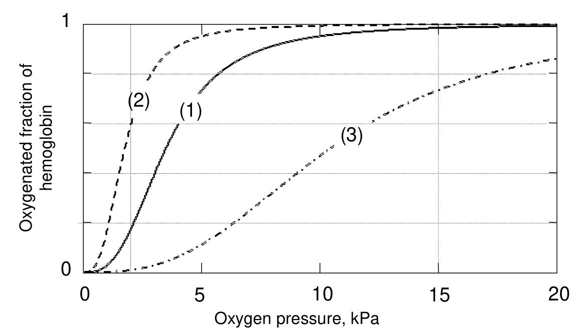
Figure 1
1.1 Using the relation between \(K\) and the standard Gibbs energy \(\Delta G^0\) for a reaction, calculate the difference between the \(\Delta G^0\) values for the heme reactions (1) and (2).
1.2 Estimate from Figure 1 (to 2 significant figures) how many moles of \(\ce{O2}\) are deposited in muscle tissue when one mole of Hb travels from the lungs to the muscles and back again for the three different types of Hb.
1.3 The special S-shaped uptake curve 1 is the result of subtle structural features of Hb. The deficient Hb shown in curve 2 is not optimal because:
- ☐ The binding with \(\ce{O2}\) is too weak.
- ☐ The binding with \(\ce{O2}\) is too strong.
- ☐ The maximum oxygen capacity is too low.
- ☐ The deficiency is caused by carbon monoxide poisoning.
1.4 Calculate how much oxygen (in \(\mathrm{mol\,s^{-1}}\)) can be deposited in tissue by blood with normal Hb (1).
1.5 Calculate the maximum power that the body can produce (assuming it is limited by oxygen transfer).
Official English text of the problem.
Answers
- 1.1: \(\boxed{\Delta G_1^\circ-\Delta G_2^\circ=-22.8\ \mathrm{kJ\,mol^{-1}}}\). The standard Gibbs energy of carbon monoxide binding is lower by \(22.8\ \mathrm{kJ\,mol^{-1}}\).
- 1.2: Approximately \(\boxed{3.2,\ 1.6,\ 2.9\ \mathrm{mol}}\) of \(\ce{O2}\) per mole of Hb for curves (1), (2), and (3), respectively.
- 1.3: Select the second box: “The binding with \(\ce{O2}\) is too strong.”
- 1.4: \(\boxed{2.7\times10^{-3}\ \mathrm{mol\,s^{-1}}}\) of \(\ce{O2}\).
- 1.5: \(\boxed{1.1\times10^3\ \mathrm{W}=1.1\ \mathrm{kW}}\).
Solution
The small molecules involved in the binding comparison are molecular oxygen (dioxygen), \(\ce{O2}\), SMILES O=O, and carbon monoxide, \(\ce{CO}\), SMILES [C-]#[O+]. Hm denotes the heme binding site defined in the problem; its detailed structure is unnecessary for these calculations.
1.1
For the binding equilibria
\[ \ce{Hm + CO <=> Hm.CO} \tag{1} \]
and
\[ \ce{Hm + O2 <=> Hm.O2}, \tag{2} \]
the standard Gibbs energy is related to the equilibrium constant by
\[ \Delta G^\circ=-RT\ln K. \]
Subtracting the values for reactions (1) and (2),
\[ \begin{aligned} \Delta G_1^\circ-\Delta G_2^\circ &=-RT\ln\left(\frac{K_1}{K_2}\right)\\ &=-(8.314\ \mathrm{J\,mol^{-1}\,K^{-1}}) (298\ \mathrm{K})\ln(10\,000)\\ &=-2.2819\times10^4\ \mathrm{J\,mol^{-1}}\\ &=\boxed{-22.8\ \mathrm{kJ\,mol^{-1}}}. \end{aligned} \]
The negative sign means that carbon monoxide binding has the more favorable standard Gibbs energy.
1.2
Let \(f(p)\) be the oxygenated fraction shown in Figure 1. Since each Hb molecule can bind four oxygen molecules, one mole of Hb carries \(4f(p)\) moles of \(\ce{O2}\).
Thus, the amount released in the muscles during one complete circulation is
\[ n_{\mathrm{released}} =4\left[f(15\ \mathrm{kPa})-f(2\ \mathrm{kPa})\right] \quad\text{mol of \(\ce{O2}\) per mol of Hb}. \]
Approximate readings from the graph give:
| Hb curve | Fraction in lungs, \(f(15)\) | Fraction in muscles, \(f(2)\) | Oxygen deposited per mole of Hb |
|---|---|---|---|
| (1), normal | \(0.98\) | \(0.18\) | \(4(0.98-0.18)=\boxed{3.2\ \mathrm{mol}}\) |
| (2), deficient | \(1.00\) | \(0.60\) | \(4(1.00-0.60)=\boxed{1.6\ \mathrm{mol}}\) |
| (3), deficient | \(0.74\) | \(0.01\) | \(4(0.74-0.01)=2.92\approx\boxed{2.9\ \mathrm{mol}}\) |
Returning to the lungs restores the oxygen loading for the next circulation.
1.3
Curve (2) reaches high oxygenation at a lower oxygen pressure than normal Hb, indicating stronger oxygen binding. At the muscle oxygen pressure of \(2\ \mathrm{kPa}\), it remains about \(60\%\) oxygenated, so much of its oxygen remains bound instead of being delivered to the tissue.
Therefore, select:
☑ The binding with \(\ce{O2}\) is too strong.
Its oxygenation in the lungs is essentially complete; the deficiency concerns oxygen release.
1.4
The concentration of Hb given in the problem applies to the red cells, which occupy \(40\%\) of the blood volume. The Hb concentration in whole blood is therefore
\[ c_{\mathrm{Hb}} =\frac{0.40(340\ \mathrm{kg\,m^{-3}})} {64\ \mathrm{kg\,mol^{-1}}} =2.125\ \mathrm{mol\,m^{-3}}. \]
At the maximum blood flow, the molar flow of Hb is
\[ \begin{aligned} \dot n_{\mathrm{Hb}} &=(4\times10^{-4}\ \mathrm{m^3\,s^{-1}}) (2.125\ \mathrm{mol\,m^{-3}})\\ &=8.50\times10^{-4}\ \mathrm{mol\,s^{-1}}. \end{aligned} \]
Normal Hb delivers approximately \(3.2\) moles of oxygen per mole of Hb, so
\[ \begin{aligned} \dot n_{\ce{O2}} &=(8.50\times10^{-4}\ \mathrm{mol\,Hb\,s^{-1}}) \left(3.2\ \frac{\mathrm{mol}\ \ce{O2}}{\mathrm{mol\,Hb}}\right)\\ &=2.72\times10^{-3}\ \mathrm{mol\,s^{-1}}\\ &\approx\boxed{2.7\times10^{-3}\ \mathrm{mol\,s^{-1}}}. \end{aligned} \]
1.5
The energy released is \(400\ \mathrm{kJ}\) per mole of oxygen consumed. Using the unrounded intermediate oxygen-delivery rate,
\[ \begin{aligned} P_{\max} &=\dot n_{\ce{O2}}\left(400\ \mathrm{kJ\,mol^{-1}}\right)\\ &=(2.72\times10^{-3}\ \mathrm{mol\,s^{-1}}) (400\ \mathrm{kJ\,mol^{-1}})\\ &=1.088\ \mathrm{kJ\,s^{-1}}\\ &\approx\boxed{1.1\ \mathrm{kW}}. \end{aligned} \]
This is the maximum chemical-energy release rate under the stated oxygen-transfer limitation.
The solution was checked against the official solution.
IChO 2002 · Theorieaufgabe 1
Sauerstoff in Ihrem Leben
Thema 1 - Chemie des Lebens
Das Leben beruht auf Chemie. Dem Verständnis und der Überwachung von Lebensprozessen wird in der Chemie große Aufmerksamkeit gewidmet.
AUFGABE 1
SAUERSTOFF IN DEINEM LEBEN
Sauerstoff ist für uns alle lebenswichtig. Sauerstoff gelangt über die Lunge in den Körper und wird vom Blut zu den Geweben unseres Körpers transportiert. Dort kann er durch die Oxidation von Zuckern Energie liefern:
Diese Reaktion setzt 400 kJ Energie pro mol Sauerstoff frei. Die Aufnahme von \(\ce{O2}\) durch das Blut erfolgt an vier Hämgruppen (Hm) im Protein Hämoglobin (Hb).
Freies Hm besteht aus einem \(\ce{Fe^2+}\)-Ion, das an vier \(\ce{N}\)-Atome eines \(\ce{{porphyrin}^{2-}}\)-Liganden gebunden ist. Sauerstoff kann an der Koordinationsstelle von \(\ce{Fe^2+}\) binden, wobei ein \(\ce{Hm.O2}\)-Komplex entsteht. Kohlenstoffmonoxid kann auf ähnliche Weise komplexiert werden, wobei ein \(\ce{Hm.CO}\)-Komplex entsteht. \(\ce{CO}\) ist ein Gift, da es stärker an Hm bindet als \(\ce{O2}\). Die Gleichgewichtskonstante \(K_1\) für die Reaktion:
ist 10 000-mal größer als die Gleichgewichtskonstante \(K_2\) für die Reaktion:
Jedes Hb-Molekül kann vier Moleküle \(\ce{O2}\) aufnehmen. Blut, das mit \(\ce{O2}\) in Kontakt steht, nimmt abhängig vom Sauerstoffdruck einen Bruchteil dieser Menge auf, wie in Abbildung 1 (Kurve 1) dargestellt. Ebenfalls dargestellt sind die Kurven (2) und (3) für Blut mit zwei Arten von funktionsgestörtem Hb. Diese kommen bei Patienten mit bestimmten Erbkrankheiten vor.
Relevante Daten: Der \(\ce{O2}\)-Druck in der Lunge beträgt 15 kPa; in den Muskeln beträgt er 2 kPa. Der maximale Blutvolumenstrom durch Herz und Lunge beträgt \(4\times10^{-4}\ \mathrm{m^3\,s^{-1}}\). Die roten Blutkörperchen nehmen 40 % des Blutvolumens ein; innerhalb der Zellen beträgt die Konzentration von Hb \(340\ \mathrm{kg\,m^{-3}}\); Hb hat eine molare Masse von \(64\ \mathrm{kg\,mol^{-1}}\). \(R=8.314\ \mathrm{J\,mol^{-1}\,K^{-1}}\). \(T=298\ \mathrm{K}\).
Abbildung 1
1.1 Berechnen Sie mithilfe der Beziehung zwischen \(K\) und der Standard-Gibbs-Energie \(\Delta G^0\) einer Reaktion die Differenz zwischen den \(\Delta G^0\)-Werten für die Hämreaktionen (1) und (2).
1.2 Schätzen Sie anhand von Abbildung 1 (auf 2 signifikante Stellen), wie viele Mol \(\ce{O2}\) bei den drei verschiedenen Hb-Typen an das Muskelgewebe abgegeben werden, wenn ein Mol Hb von der Lunge zu den Muskeln und wieder zurück gelangt.
1.3 Die besondere S-förmige Aufnahmekurve 1 ist das Ergebnis subtiler Strukturmerkmale von Hb. Das in Kurve 2 dargestellte funktionsgestörte Hb ist nicht optimal, weil:
- ☐ Die Bindung an \(\ce{O2}\) zu schwach ist.
- ☐ Die Bindung an \(\ce{O2}\) zu stark ist.
- ☐ Die maximale Sauerstoffkapazität zu gering ist.
- ☐ Die Funktionsstörung durch eine Kohlenstoffmonoxidvergiftung verursacht wird.
1.4 Berechnen Sie, wie viel Sauerstoff (in \(\mathrm{mol\,s^{-1}}\)) durch Blut mit normalem Hb (1) an das Gewebe abgegeben werden kann.
1.5 Berechnen Sie die maximale Leistung, die der Körper erbringen kann (unter der Annahme, dass sie durch den Sauerstofftransport begrenzt ist).
Aufgabentext aus dem offiziellen englischen Text übersetzt.
Antworten
- 1.1: \(\boxed{\Delta G_1^\circ-\Delta G_2^\circ=-22.8\ \mathrm{kJ\,mol^{-1}}}\). Die Standard-Gibbs-Energie der Bindung von Kohlenstoffmonoxid ist um \(22.8\ \mathrm{kJ\,mol^{-1}}\) niedriger.
- 1.2: Ungefähr \(\boxed{3.2,\ 1.6,\ 2.9\ \mathrm{mol}}\) \(\ce{O2}\) pro Mol Hb für die Kurven (1), (2) bzw. (3).
- 1.3: Kreuzen Sie das zweite Kästchen an: „Die Bindung an \(\ce{O2}\) ist zu stark.“
- 1.4: \(\boxed{2.7\times10^{-3}\ \mathrm{mol\,s^{-1}}}\) \(\ce{O2}\).
- 1.5: \(\boxed{1.1\times10^3\ \mathrm{W}=1.1\ \mathrm{kW}}\).
Lösung
Die kleinen Moleküle, deren Bindung verglichen wird, sind molekularer Sauerstoff (Disauerstoff), \(\ce{O2}\), SMILES O=O, und Kohlenstoffmonoxid, \(\ce{CO}\), SMILES [C-]#[O+]. Hm bezeichnet die in der Aufgabe definierte Häm-Bindungsstelle; ihre genaue Struktur ist für diese Berechnungen nicht erforderlich.
1.1
Für die Bindungsgleichgewichte
\[ \ce{Hm + CO <=> Hm.CO} \tag{1} \]
und
\[ \ce{Hm + O2 <=> Hm.O2}, \tag{2} \]
ist die Standard-Gibbs-Energie mit der Gleichgewichtskonstanten durch folgende Beziehung verknüpft:
\[ \Delta G^\circ=-RT\ln K. \]
Durch Subtraktion der Werte für die Reaktionen (1) und (2) erhält man:
\[ \begin{aligned} \Delta G_1^\circ-\Delta G_2^\circ &=-RT\ln\left(\frac{K_1}{K_2}\right)\\ &=-(8.314\ \mathrm{J\,mol^{-1}\,K^{-1}}) (298\ \mathrm{K})\ln(10\,000)\\ &=-2.2819\times10^4\ \mathrm{J\,mol^{-1}}\\ &=\boxed{-22.8\ \mathrm{kJ\,mol^{-1}}}. \end{aligned} \]
Das negative Vorzeichen bedeutet, dass die Bindung von Kohlenstoffmonoxid die günstigere Standard-Gibbs-Energie aufweist.
1.2
Es sei \(f(p)\) der in Abbildung 1 dargestellte mit Sauerstoff beladene Anteil. Da jedes Hb-Molekül vier Sauerstoffmoleküle binden kann, transportiert ein Mol Hb \(4f(p)\) Mol \(\ce{O2}\).
Somit beträgt die während eines vollständigen Kreislaufdurchgangs in den Muskeln freigesetzte Stoffmenge
\[ n_{\mathrm{released}} =4\left[f(15\ \mathrm{kPa})-f(2\ \mathrm{kPa})\right] \quad\text{mol \(\ce{O2}\) pro mol Hb}. \]
Näherungsweises Ablesen aus dem Diagramm ergibt:
| Hb-Kurve | Anteil in der Lunge, \(f(15)\) | Anteil in den Muskeln, \(f(2)\) | Abgegebener Sauerstoff pro Mol Hb |
|---|---|---|---|
| (1), normal | \(0.98\) | \(0.18\) | \(4(0.98-0.18)=\boxed{3.2\ \mathrm{mol}}\) |
| (2), funktionsgestört | \(1.00\) | \(0.60\) | \(4(1.00-0.60)=\boxed{1.6\ \mathrm{mol}}\) |
| (3), funktionsgestört | \(0.74\) | \(0.01\) | \(4(0.74-0.01)=2.92\approx\boxed{2.9\ \mathrm{mol}}\) |
Bei der Rückkehr in die Lunge wird die Sauerstoffbeladung für den nächsten Kreislaufdurchgang wiederhergestellt.
1.3
Kurve (2) erreicht bereits bei einem niedrigeren Sauerstoffdruck als normales Hb eine hohe Sauerstoffsättigung, was auf eine stärkere Sauerstoffbindung hinweist. Beim Sauerstoffdruck in den Muskeln von \(2\ \mathrm{kPa}\) bleibt es zu etwa \(60\%\) mit Sauerstoff beladen, sodass ein großer Teil seines Sauerstoffs gebunden bleibt, statt an das Gewebe abgegeben zu werden.
Daher ist anzukreuzen:
☑ Die Bindung an \(\ce{O2}\) ist zu stark.
Seine Sauerstoffsättigung in der Lunge ist praktisch vollständig; die Funktionsstörung betrifft die Sauerstoffabgabe.
1.4
Die in der Aufgabe angegebene Hb-Konzentration gilt für die roten Blutkörperchen, die \(40\%\) des Blutvolumens einnehmen. Die Hb-Konzentration im Vollblut beträgt daher
\[ c_{\mathrm{Hb}} =\frac{0.40(340\ \mathrm{kg\,m^{-3}})} {64\ \mathrm{kg\,mol^{-1}}} =2.125\ \mathrm{mol\,m^{-3}}. \]
Beim maximalen Blutvolumenstrom beträgt der Stoffmengenstrom von Hb
\[ \begin{aligned} \dot n_{\mathrm{Hb}} &=(4\times10^{-4}\ \mathrm{m^3\,s^{-1}}) (2.125\ \mathrm{mol\,m^{-3}})\\ &=8.50\times10^{-4}\ \mathrm{mol\,s^{-1}}. \end{aligned} \]
Normales Hb gibt ungefähr \(3.2\) Mol Sauerstoff pro Mol Hb ab, sodass
\[ \begin{aligned} \dot n_{\ce{O2}} &=(8.50\times10^{-4}\ \mathrm{mol\,Hb\,s^{-1}}) \left(3.2\ \frac{\mathrm{mol}\ \ce{O2}}{\mathrm{mol\,Hb}}\right)\\ &=2.72\times10^{-3}\ \mathrm{mol\,s^{-1}}\\ &\approx\boxed{2.7\times10^{-3}\ \mathrm{mol\,s^{-1}}}. \end{aligned} \]
1.5
Die freigesetzte Energie beträgt \(400\ \mathrm{kJ}\) pro Mol verbrauchten Sauerstoffs. Mit dem ungerundeten Zwischenwert für die Sauerstoffabgaberate ergibt sich
\[ \begin{aligned} P_{\max} &=\dot n_{\ce{O2}}\left(400\ \mathrm{kJ\,mol^{-1}}\right)\\ &=(2.72\times10^{-3}\ \mathrm{mol\,s^{-1}}) (400\ \mathrm{kJ\,mol^{-1}})\\ &=1.088\ \mathrm{kJ\,s^{-1}}\\ &\approx\boxed{1.1\ \mathrm{kW}}. \end{aligned} \]
Dies ist die maximale Freisetzungsrate chemischer Energie unter der angegebenen Begrenzung durch den Sauerstofftransport.
Die Lösung wurde mit der offiziellen Lösung abgeglichen.
IChO 2002 · Teori sorusu 1
Yaşamınızda Oksijen
Tema 1 - Yaşamın Kimyası
Yaşam, kimyasal süreçlerle sürer. Yaşamsal süreçlerin anlaşılması ve izlenmesi kimyada büyük ilgi görmektedir.
SORU 1
YAŞAMINIZDA OKSİJEN
Oksijen hepimiz için yaşamsal öneme sahiptir. Oksijen vücuda akciğerler yoluyla girer ve kan tarafından vücudumuzdaki dokulara taşınır. Burada şekerlerin yükseltgenmesi yoluyla enerji sağlayabilir:
Bu tepkime, mol oksijen başına 400 kJ enerji açığa çıkarır. Kanın \(\ce{O2}\) alımı, hemoglobin (Hb) proteinindeki dört hem (Hm) grubunda gerçekleşir.
Serbest Hm, bir \(\ce{{porphyrin}^{2-}}\) ligandının dört \(\ce{N}\) atomuna bağlı bir \(\ce{Fe^2+}\) iyonundan oluşur. Oksijen, \(\ce{Fe^2+}\) iyonunun koordinasyon yerine bağlanarak bir \(\ce{Hm.O2}\) kompleksi oluşturabilir. Karbon monoksit de benzer şekilde kompleksleşerek bir \(\ce{Hm.CO}\) kompleksi oluşturabilir. \(\ce{CO}\), Hm'ye \(\ce{O2}\)'den daha kuvvetli bağlandığı için zehirlidir. Şu tepkimenin \(K_1\) denge sabiti:
aşağıdaki tepkimenin \(K_2\) denge sabitinden 10 000 kat büyüktür:
Her Hb molekülü dört \(\ce{O2}\) molekülü alabilir. \(\ce{O2}\) ile temas hâlindeki kan, Şekil 1'de (eğri 1) gösterildiği gibi oksijen basıncına bağlı olarak bu miktarın bir kesrini soğurur. İki farklı işlev bozukluğu olan Hb türünü içeren kan için (2) ve (3) eğrileri de gösterilmiştir. Bunlar, belirli kalıtsal hastalıkları olan hastalarda görülür.
İlgili veriler: Akciğerlerdeki \(\ce{O2}\) basıncı 15 kPa, kaslardaki ise 2 kPa'dır. Kalp ve akciğerlerden geçen kanın en yüksek hacimsel debisi \(4\times10^{-4}\ \mathrm{m^3\,s^{-1}}\) değerindedir. Kandaki alyuvarlar kan hacminin %40'ını kaplar; hücrelerin içindeki Hb derişimi \(340\ \mathrm{kg\,m^{-3}}\) değerindedir; Hb'nin molar kütlesi \(64\ \mathrm{kg\,mol^{-1}}\) değerindedir. \(R=8.314\ \mathrm{J\,mol^{-1}\,K^{-1}}\). \(T=298\ \mathrm{K}\).
Şekil 1
1.1 Bir tepkimenin \(K\) değeri ile standart Gibbs enerjisi \(\Delta G^0\) arasındaki bağıntıyı kullanarak (1) ve (2) hem tepkimelerinin \(\Delta G^0\) değerleri arasındaki farkı hesaplayınız.
1.2 Üç farklı Hb türü için, bir mol Hb akciğerlerden kaslara gidip tekrar geri döndüğünde kas dokusuna kaç mol \(\ce{O2}\) bırakıldığını Şekil 1'den (2 anlamlı rakamla) tahmin ediniz.
1.3 S biçimindeki özel alım eğrisi 1, Hb'nin ince yapısal özelliklerinin sonucudur. Eğri 2'de gösterilen işlev bozukluğu olan Hb en uygun durumda değildir, çünkü:
- ☐ \(\ce{O2}\) ile bağlanma çok zayıftır.
- ☐ \(\ce{O2}\) ile bağlanma çok kuvvetlidir.
- ☐ En yüksek oksijen kapasitesi çok düşüktür.
- ☐ İşlev bozukluğuna karbon monoksit zehirlenmesi neden olmaktadır.
1.4 Normal Hb (1) içeren kanın dokuya ne kadar oksijen (\(\mathrm{mol\,s^{-1}}\) cinsinden) bırakabileceğini hesaplayınız.
1.5 Vücudun üretebileceği en yüksek gücü (oksijen aktarımıyla sınırlı olduğunu varsayarak) hesaplayınız.
Soru metni resmi İngilizce metinden çevrilmiştir.
Cevaplar
- 1.1: \(\boxed{\Delta G_1^\circ-\Delta G_2^\circ=-22.8\ \mathrm{kJ\,mol^{-1}}}\). Karbon monoksit bağlanmasının standart Gibbs enerjisi \(22.8\ \mathrm{kJ\,mol^{-1}}\) daha düşüktür.
- 1.2: Sırasıyla (1), (2) ve (3) eğrileri için mol Hb başına yaklaşık \(\boxed{3.2,\ 1.6,\ 2.9\ \mathrm{mol}}\) \(\ce{O2}\).
- 1.3: İkinci kutucuğu seçiniz: “\(\ce{O2}\) ile bağlanma çok kuvvetlidir.”
- 1.4: \(\boxed{2.7\times10^{-3}\ \mathrm{mol\,s^{-1}}}\) \(\ce{O2}\).
- 1.5: \(\boxed{1.1\times10^3\ \mathrm{W}=1.1\ \mathrm{kW}}\).
Çözüm
Bağlanma karşılaştırmasında yer alan küçük moleküller, moleküler oksijen (dioksijen), \(\ce{O2}\), SMILES O=O, ve karbon monoksit, \(\ce{CO}\), SMILES [C-]#[O+], molekülleridir. Hm, soruda tanımlanan hem bağlanma yerini belirtir; ayrıntılı yapısı bu hesaplamalar için gerekli değildir.
1.1
Şu bağlanma dengeleri için:
\[ \ce{Hm + CO <=> Hm.CO} \tag{1} \]
ve
\[ \ce{Hm + O2 <=> Hm.O2}, \tag{2} \]
standart Gibbs enerjisi, denge sabitiyle şu bağıntı üzerinden ilişkilidir:
\[ \Delta G^\circ=-RT\ln K. \]
(1) ve (2) tepkimelerinin değerleri birbirinden çıkarıldığında,
\[ \begin{aligned} \Delta G_1^\circ-\Delta G_2^\circ &=-RT\ln\left(\frac{K_1}{K_2}\right)\\ &=-(8.314\ \mathrm{J\,mol^{-1}\,K^{-1}}) (298\ \mathrm{K})\ln(10\,000)\\ &=-2.2819\times10^4\ \mathrm{J\,mol^{-1}}\\ &=\boxed{-22.8\ \mathrm{kJ\,mol^{-1}}}. \end{aligned} \]
Negatif işaret, karbon monoksit bağlanmasının standart Gibbs enerjisi açısından daha elverişli olduğunu gösterir.
1.2
\(f(p)\), Şekil 1'de gösterilen oksijenlenmiş kesir olsun. Her Hb molekülü dört oksijen molekülü bağlayabildiğinden, bir mol Hb, \(4f(p)\) mol \(\ce{O2}\) taşır.
Dolayısıyla, bir tam dolaşım sırasında kaslarda serbest bırakılan miktar şöyledir:
\[ n_{\mathrm{released}} =4\left[f(15\ \mathrm{kPa})-f(2\ \mathrm{kPa})\right] \quad\text{mol Hb başına mol \(\ce{O2}\)}. \]
Grafikten okunan yaklaşık değerler şunları verir:
| Hb eğrisi | Akciğerlerdeki kesir, \(f(15)\) | Kaslardaki kesir, \(f(2)\) | Mol Hb başına bırakılan oksijen |
|---|---|---|---|
| (1), normal | \(0.98\) | \(0.18\) | \(4(0.98-0.18)=\boxed{3.2\ \mathrm{mol}}\) |
| (2), işlev bozukluğu olan | \(1.00\) | \(0.60\) | \(4(1.00-0.60)=\boxed{1.6\ \mathrm{mol}}\) |
| (3), işlev bozukluğu olan | \(0.74\) | \(0.01\) | \(4(0.74-0.01)=2.92\approx\boxed{2.9\ \mathrm{mol}}\) |
Akciğerlere dönüş, bir sonraki dolaşım için oksijen yükünün yeniden kazanılmasını sağlar.
1.3
Eğri (2), normal Hb'ye göre daha düşük oksijen basıncında yüksek oksijenlenmeye ulaşır; bu, oksijenin daha kuvvetli bağlandığını gösterir. Kaslardaki \(2\ \mathrm{kPa}\) oksijen basıncında yaklaşık \(60\%\) oranında oksijenlenmiş durumda kalır; bu nedenle oksijeninin büyük bölümü dokuya verilmek yerine bağlı kalır.
Bu nedenle şu seçeneği işaretleyiniz:
☑ \(\ce{O2}\) ile bağlanma çok kuvvetlidir.
Akciğerlerdeki oksijenlenmesi neredeyse tamdır; işlev bozukluğu oksijenin serbest bırakılmasıyla ilgilidir.
1.4
Soruda verilen Hb derişimi, kan hacminin \(40\%\)'ını kaplayan alyuvarlar için geçerlidir. Dolayısıyla tam kandaki Hb derişimi şöyledir:
\[ c_{\mathrm{Hb}} =\frac{0.40(340\ \mathrm{kg\,m^{-3}})} {64\ \mathrm{kg\,mol^{-1}}} =2.125\ \mathrm{mol\,m^{-3}}. \]
En yüksek kan debisinde Hb'nin molar debisi şöyledir:
\[ \begin{aligned} \dot n_{\mathrm{Hb}} &=(4\times10^{-4}\ \mathrm{m^3\,s^{-1}}) (2.125\ \mathrm{mol\,m^{-3}})\\ &=8.50\times10^{-4}\ \mathrm{mol\,s^{-1}}. \end{aligned} \]
Normal Hb, mol Hb başına yaklaşık \(3.2\) mol oksijen verir; dolayısıyla
\[ \begin{aligned} \dot n_{\ce{O2}} &=(8.50\times10^{-4}\ \mathrm{mol\,Hb\,s^{-1}}) \left(3.2\ \frac{\mathrm{mol}\ \ce{O2}}{\mathrm{mol\,Hb}}\right)\\ &=2.72\times10^{-3}\ \mathrm{mol\,s^{-1}}\\ &\approx\boxed{2.7\times10^{-3}\ \mathrm{mol\,s^{-1}}}. \end{aligned} \]
1.5
Açığa çıkan enerji, tüketilen mol oksijen başına \(400\ \mathrm{kJ}\) değerindedir. Oksijen aktarım hızının yuvarlanmamış ara değeri kullanıldığında,
\[ \begin{aligned} P_{\max} &=\dot n_{\ce{O2}}\left(400\ \mathrm{kJ\,mol^{-1}}\right)\\ &=(2.72\times10^{-3}\ \mathrm{mol\,s^{-1}}) (400\ \mathrm{kJ\,mol^{-1}})\\ &=1.088\ \mathrm{kJ\,s^{-1}}\\ &\approx\boxed{1.1\ \mathrm{kW}}. \end{aligned} \]
Bu, belirtilen oksijen aktarımı sınırlaması altında kimyasal enerjinin açığa çıkabileceği en yüksek hızdır.
Çözüm, resmi çözümle karşılaştırılarak kontrol edildi.
IChO 2002 · Problema teórico 1
El oxígeno en su vida
Tema 1 - Química de la vida
La vida funciona gracias a la química. La comprensión y el seguimiento de los procesos vitales reciben mucha atención en química.
PROBLEMA 1
EL OXÍGENO EN SU VIDA
El oxígeno es de vital importancia para todos nosotros. El oxígeno entra en el organismo a través de los pulmones y la sangre lo transporta a los tejidos de nuestro cuerpo. Allí puede proporcionar energía mediante la oxidación de los azúcares:
Esta reacción libera 400 kJ de energía por mol de oxígeno. La captación de \(\ce{O2}\) por la sangre tiene lugar en cuatro grupos hemo (Hm) de la proteína hemoglobina (Hb).
El Hm libre consta de un ion \(\ce{Fe^2+}\) unido a cuatro átomos de \(\ce{N}\) de un ligando \(\ce{{porphyrin}^{2-}}\). El oxígeno puede unirse al sitio de coordinación del \(\ce{Fe^2+}\), formando un complejo \(\ce{Hm.O2}\). El monóxido de carbono puede formar un complejo de manera similar, dando lugar a un complejo \(\ce{Hm.CO}\). El \(\ce{CO}\) es un veneno, ya que se une al Hm con mayor fuerza que el \(\ce{O2}\). La constante de equilibrio \(K_1\) de la reacción:
es 10 000 veces mayor que la constante de equilibrio \(K_2\) de la reacción:
Cada molécula de Hb puede captar cuatro moléculas de \(\ce{O2}\). La sangre en contacto con \(\ce{O2}\) absorbe una fracción de esta cantidad, que depende de la presión de oxígeno, como se muestra en la Figura 1 (curva 1). También se muestran las curvas (2) y (3) correspondientes a sangre con dos tipos de Hb defectuosa. Estos se encuentran en pacientes con determinadas enfermedades hereditarias.
Datos relevantes: la presión de \(\ce{O2}\) en los pulmones es de 15 kPa; en los músculos es de 2 kPa. El caudal máximo de sangre a través del corazón y los pulmones es de \(4\times10^{-4}\ \mathrm{m^3\,s^{-1}}\). Los glóbulos rojos ocupan el 40 % del volumen de la sangre; en el interior de estas células, la concentración de Hb es de \(340\ \mathrm{kg\,m^{-3}}\); la Hb tiene una masa molar de \(64\ \mathrm{kg\,mol^{-1}}\). \(R=8.314\ \mathrm{J\,mol^{-1}\,K^{-1}}\). \(T=298\ \mathrm{K}\).
Figura 1
1.1 Utilizando la relación entre \(K\) y la energía de Gibbs estándar \(\Delta G^0\) de una reacción, calcule la diferencia entre los valores de \(\Delta G^0\) de las reacciones del hemo (1) y (2).
1.2 Estime a partir de la Figura 1 (con 2 cifras significativas) cuántos moles de \(\ce{O2}\) se depositan en el tejido muscular cuando un mol de Hb viaja de los pulmones a los músculos y regresa, para los tres tipos distintos de Hb.
1.3 La peculiar forma de S de la curva de captación 1 se debe a características estructurales sutiles de la Hb. La Hb defectuosa representada en la curva 2 no es óptima porque:
- ☐ La unión con el \(\ce{O2}\) es demasiado débil.
- ☐ La unión con el \(\ce{O2}\) es demasiado fuerte.
- ☐ La capacidad máxima de oxígeno es demasiado baja.
- ☐ La deficiencia se debe a una intoxicación por monóxido de carbono.
1.4 Calcule cuánto oxígeno (en \(\mathrm{mol\,s^{-1}}\)) puede depositar en los tejidos la sangre con Hb normal (1).
1.5 Calcule la potencia máxima que puede producir el organismo (suponiendo que está limitada por la transferencia de oxígeno).
Enunciado traducido del texto oficial en inglés.
Respuestas
- 1.1: \(\boxed{\Delta G_1^\circ-\Delta G_2^\circ=-22.8\ \mathrm{kJ\,mol^{-1}}}\). La energía de Gibbs estándar de la unión del monóxido de carbono es menor en \(22.8\ \mathrm{kJ\,mol^{-1}}\).
- 1.2: Aproximadamente \(\boxed{3.2,\ 1.6,\ 2.9\ \mathrm{mol}}\) de \(\ce{O2}\) por mol de Hb para las curvas (1), (2) y (3), respectivamente.
- 1.3: Seleccione la segunda casilla: «La unión con el \(\ce{O2}\) es demasiado fuerte».
- 1.4: \(\boxed{2.7\times10^{-3}\ \mathrm{mol\,s^{-1}}}\) de \(\ce{O2}\).
- 1.5: \(\boxed{1.1\times10^3\ \mathrm{W}=1.1\ \mathrm{kW}}\).
Solución
Las moléculas pequeñas que intervienen en la comparación de la unión son el oxígeno molecular (dioxígeno), \(\ce{O2}\), SMILES O=O, y el monóxido de carbono, \(\ce{CO}\), SMILES [C-]#[O+]. Hm designa el sitio de unión del hemo definido en el problema; no es necesario conocer su estructura detallada para estos cálculos.
1.1
Para los equilibrios de unión
\[ \ce{Hm + CO <=> Hm.CO} \tag{1} \]
y
\[ \ce{Hm + O2 <=> Hm.O2}, \tag{2} \]
la energía de Gibbs estándar se relaciona con la constante de equilibrio mediante
\[ \Delta G^\circ=-RT\ln K. \]
Al restar los valores de las reacciones (1) y (2),
\[ \begin{aligned} \Delta G_1^\circ-\Delta G_2^\circ &=-RT\ln\left(\frac{K_1}{K_2}\right)\\ &=-(8.314\ \mathrm{J\,mol^{-1}\,K^{-1}}) (298\ \mathrm{K})\ln(10\,000)\\ &=-2.2819\times10^4\ \mathrm{J\,mol^{-1}}\\ &=\boxed{-22.8\ \mathrm{kJ\,mol^{-1}}}. \end{aligned} \]
El signo negativo significa que la unión del monóxido de carbono tiene la energía de Gibbs estándar más favorable.
1.2
Sea \(f(p)\) la fracción oxigenada que se muestra en la Figura 1. Como cada molécula de Hb puede unirse a cuatro moléculas de oxígeno, un mol de Hb transporta \(4f(p)\) moles de \(\ce{O2}\).
Por tanto, la cantidad liberada en los músculos durante un recorrido circulatorio completo es
\[ n_{\mathrm{released}} =4\left[f(15\ \mathrm{kPa})-f(2\ \mathrm{kPa})\right] \quad\text{mol de \(\ce{O2}\) por mol de Hb}. \]
Las lecturas aproximadas de la gráfica dan:
| Curva de Hb | Fracción en los pulmones, \(f(15)\) | Fracción en los músculos, \(f(2)\) | Oxígeno depositado por mol de Hb |
|---|---|---|---|
| (1), normal | \(0.98\) | \(0.18\) | \(4(0.98-0.18)=\boxed{3.2\ \mathrm{mol}}\) |
| (2), defectuosa | \(1.00\) | \(0.60\) | \(4(1.00-0.60)=\boxed{1.6\ \mathrm{mol}}\) |
| (3), defectuosa | \(0.74\) | \(0.01\) | \(4(0.74-0.01)=2.92\approx\boxed{2.9\ \mathrm{mol}}\) |
El regreso a los pulmones restablece la carga de oxígeno para el siguiente recorrido circulatorio.
1.3
La curva (2) alcanza una oxigenación elevada a una presión de oxígeno menor que la Hb normal, lo que indica una unión más fuerte al oxígeno. A la presión de oxígeno muscular de \(2\ \mathrm{kPa}\), permanece oxigenada aproximadamente en un \(60\%\), por lo que gran parte de su oxígeno sigue unido en lugar de ser entregado al tejido.
Por tanto, seleccione:
☑ La unión con el \(\ce{O2}\) es demasiado fuerte.
Su oxigenación en los pulmones es prácticamente completa; la deficiencia afecta a la liberación de oxígeno.
1.4
La concentración de Hb indicada en el problema corresponde a los glóbulos rojos, que ocupan el \(40\%\) del volumen sanguíneo. Por tanto, la concentración de Hb en la sangre total es
\[ c_{\mathrm{Hb}} =\frac{0.40(340\ \mathrm{kg\,m^{-3}})} {64\ \mathrm{kg\,mol^{-1}}} =2.125\ \mathrm{mol\,m^{-3}}. \]
Con el caudal sanguíneo máximo, el caudal molar de Hb es
\[ \begin{aligned} \dot n_{\mathrm{Hb}} &=(4\times10^{-4}\ \mathrm{m^3\,s^{-1}}) (2.125\ \mathrm{mol\,m^{-3}})\\ &=8.50\times10^{-4}\ \mathrm{mol\,s^{-1}}. \end{aligned} \]
La Hb normal entrega aproximadamente \(3.2\) moles de oxígeno por mol de Hb, de modo que
\[ \begin{aligned} \dot n_{\ce{O2}} &=(8.50\times10^{-4}\ \mathrm{mol\,Hb\,s^{-1}}) \left(3.2\ \frac{\mathrm{mol}\ \ce{O2}}{\mathrm{mol\,Hb}}\right)\\ &=2.72\times10^{-3}\ \mathrm{mol\,s^{-1}}\\ &\approx\boxed{2.7\times10^{-3}\ \mathrm{mol\,s^{-1}}}. \end{aligned} \]
1.5
La energía liberada es de \(400\ \mathrm{kJ}\) por mol de oxígeno consumido. Utilizando el valor intermedio sin redondear de la tasa de suministro de oxígeno,
\[ \begin{aligned} P_{\max} &=\dot n_{\ce{O2}}\left(400\ \mathrm{kJ\,mol^{-1}}\right)\\ &=(2.72\times10^{-3}\ \mathrm{mol\,s^{-1}}) (400\ \mathrm{kJ\,mol^{-1}})\\ &=1.088\ \mathrm{kJ\,s^{-1}}\\ &\approx\boxed{1.1\ \mathrm{kW}}. \end{aligned} \]
Esta es la tasa máxima de liberación de energía química bajo la limitación de transferencia de oxígeno indicada.
La solución se comprobó con la solución oficial.
IChO 2002 · 理论题 1
你生命中的氧
主题 1 - 生命化学
生命的运转依赖于化学。理解和监测生命过程在化学领域备受关注。
题目 1
你生命中的氧
氧对我们每个人都至关重要。氧经肺进入人体,并由血液输送到体内各组织。在那里,氧可以通过氧化糖类来提供能量:
该反应每消耗一摩尔氧气释放 400 kJ 的能量。血液对 \(\ce{O2}\) 的摄取发生在血红蛋白(Hb)中的四个血红素(Hm)基团上。
游离 Hm 由一个 \(\ce{Fe^2+}\) 离子与一个 \(\ce{{porphyrin}^{2-}}\) 配体的四个 \(\ce{N}\) 原子配位而成。氧气可以结合在 \(\ce{Fe^2+}\) 的配位位点上,形成 \(\ce{Hm.O2}\) 配合物。一氧化碳也可以类似地发生配位,形成 \(\ce{Hm.CO}\) 配合物。\(\ce{CO}\) 是一种毒物,因为它与 Hm 的结合比 \(\ce{O2}\) 更强。下列反应的平衡常数 \(K_1\):
是下列反应的平衡常数 \(K_2\) 的 10 000 倍:
每个 Hb 分子可以摄取四个 \(\ce{O2}\) 分子。血液与 \(\ce{O2}\) 接触时,所吸收的氧量占此最大量的比例取决于氧分压,如图 1(曲线 1)所示。图中还给出了含有两种缺陷型 Hb 的血液所对应的曲线(2)和(3)。这两种缺陷型 Hb 存在于某些遗传病患者中。
相关数据:肺中的 \(\ce{O2}\) 分压为 15 kPa;肌肉中的氧分压为 2 kPa。通过心脏和肺的最大血液流量为 \(4\times10^{-4}\ \mathrm{m^3\,s^{-1}}\)。血液中的红细胞占血液体积的 40 %;细胞内 Hb 的浓度为 \(340\ \mathrm{kg\,m^{-3}}\);Hb 的摩尔质量为 \(64\ \mathrm{kg\,mol^{-1}}\)。\(R=8.314\ \mathrm{J\,mol^{-1}\,K^{-1}}\)。\(T=298\ \mathrm{K}\)。
图 1
1.1 利用反应的 \(K\) 与标准吉布斯能 \(\Delta G^0\) 之间的关系,计算血红素反应(1)和(2)的 \(\Delta G^0\) 值之差。
1.2 对于这三种不同类型的 Hb,根据图 1 估算一摩尔 Hb 从肺到肌肉再返回肺的过程中向肌肉组织释放的 \(\ce{O2}\) 的物质的量(保留 2 位有效数字)。
1.3 特殊的 S 形摄氧曲线 1 源于 Hb 的精细结构特征。曲线 2 所示的缺陷型 Hb 并非最优,原因是:
- ☐ 与 \(\ce{O2}\) 的结合太弱。
- ☐ 与 \(\ce{O2}\) 的结合太强。
- ☐ 最大携氧容量太低。
- ☐ 这种缺陷由一氧化碳中毒引起。
1.4 计算含有正常 Hb(1)的血液能够向组织释放多少氧气(以 \(\mathrm{mol\,s^{-1}}\) 为单位)。
1.5 计算人体能够产生的最大功率(假设其受氧输送的限制)。
题目译自官方英文原文。
答案
- 1.1: \(\boxed{\Delta G_1^\circ-\Delta G_2^\circ=-22.8\ \mathrm{kJ\,mol^{-1}}}\)。一氧化碳结合反应的标准吉布斯能低 \(22.8\ \mathrm{kJ\,mol^{-1}}\)。
- 1.2: 对于曲线 (1)、(2) 和 (3),每摩尔 Hb 释放的 \(\ce{O2}\) 分别约为 \(\boxed{3.2,\ 1.6,\ 2.9\ \mathrm{mol}}\)。
- 1.3: 选择第二个方框:“与 \(\ce{O2}\) 的结合太强。”
- 1.4: \(\boxed{2.7\times10^{-3}\ \mathrm{mol\,s^{-1}}}\) 的 \(\ce{O2}\)。
- 1.5: \(\boxed{1.1\times10^3\ \mathrm{W}=1.1\ \mathrm{kW}}\)。
解答
结合能力比较所涉及的小分子为分子氧(双原子氧)\(\ce{O2}\),其 SMILES 为 O=O,以及一氧化碳 \(\ce{CO}\),其 SMILES 为 [C-]#[O+]。Hm 表示题目中定义的血红素结合位点;这些计算不需要其详细结构。
1.1
对于结合平衡
\[ \ce{Hm + CO <=> Hm.CO} \tag{1} \]
和
\[ \ce{Hm + O2 <=> Hm.O2}, \tag{2} \]
标准吉布斯能与平衡常数之间的关系为
\[ \Delta G^\circ=-RT\ln K. \]
将反应(1)和(2)的相应值相减,得
\[ \begin{aligned} \Delta G_1^\circ-\Delta G_2^\circ &=-RT\ln\left(\frac{K_1}{K_2}\right)\\ &=-(8.314\ \mathrm{J\,mol^{-1}\,K^{-1}}) (298\ \mathrm{K})\ln(10\,000)\\ &=-2.2819\times10^4\ \mathrm{J\,mol^{-1}}\\ &=\boxed{-22.8\ \mathrm{kJ\,mol^{-1}}}. \end{aligned} \]
负号表示一氧化碳结合反应的标准吉布斯能更有利。
1.2
令 \(f(p)\) 为图 1 所示的氧合比例。由于每个 Hb 分子可以结合四个氧分子,一摩尔 Hb 携带 \(4f(p)\) 摩尔 \(\ce{O2}\)。
因此,在一次完整循环中向肌肉释放的量为
\[ n_{\mathrm{released}} =4\left[f(15\ \mathrm{kPa})-f(2\ \mathrm{kPa})\right] \quad\text{每 mol Hb 释放的 \(\ce{O2}\) 的 mol 数}. \]
从图中读取的近似值如下:
| Hb 曲线 | 肺中的氧合比例,\(f(15)\) | 肌肉中的氧合比例,\(f(2)\) | 每摩尔 Hb 释放的氧量 |
|---|---|---|---|
| (1),正常型 | \(0.98\) | \(0.18\) | \(4(0.98-0.18)=\boxed{3.2\ \mathrm{mol}}\) |
| (2),缺陷型 | \(1.00\) | \(0.60\) | \(4(1.00-0.60)=\boxed{1.6\ \mathrm{mol}}\) |
| (3),缺陷型 | \(0.74\) | \(0.01\) | \(4(0.74-0.01)=2.92\approx\boxed{2.9\ \mathrm{mol}}\) |
返回肺后,Hb 的载氧量恢复,为下一次循环做好准备。
1.3
曲线(2)在比正常 Hb 更低的氧分压下就达到较高的氧合程度,表明其与氧的结合更强。在肌肉中的氧分压 \(2\ \mathrm{kPa}\) 下,其氧合比例仍约为 \(60\%\),因此它所携带的大部分氧仍处于结合状态,而没有被输送给组织。
因此,应选择:
☑ 与 \(\ce{O2}\) 的结合太强。
它在肺中基本上已完全氧合;其缺陷在于氧的释放。
1.4
题目给出的 Hb 浓度是红细胞内的浓度,而红细胞占血液体积的 \(40\%\)。因此,全血中的 Hb 浓度为
\[ c_{\mathrm{Hb}} =\frac{0.40(340\ \mathrm{kg\,m^{-3}})} {64\ \mathrm{kg\,mol^{-1}}} =2.125\ \mathrm{mol\,m^{-3}}. \]
在最大血液流量下,Hb 的摩尔流量为
\[ \begin{aligned} \dot n_{\mathrm{Hb}} &=(4\times10^{-4}\ \mathrm{m^3\,s^{-1}}) (2.125\ \mathrm{mol\,m^{-3}})\\ &=8.50\times10^{-4}\ \mathrm{mol\,s^{-1}}. \end{aligned} \]
正常 Hb 每摩尔可输送约 \(3.2\) 摩尔氧气,因此
\[ \begin{aligned} \dot n_{\ce{O2}} &=(8.50\times10^{-4}\ \mathrm{mol\,Hb\,s^{-1}}) \left(3.2\ \frac{\mathrm{mol}\ \ce{O2}}{\mathrm{mol\,Hb}}\right)\\ &=2.72\times10^{-3}\ \mathrm{mol\,s^{-1}}\\ &\approx\boxed{2.7\times10^{-3}\ \mathrm{mol\,s^{-1}}}. \end{aligned} \]
1.5
每消耗一摩尔氧气释放的能量为 \(400\ \mathrm{kJ}\)。使用未经舍入的氧输送速率中间值,得
\[ \begin{aligned} P_{\max} &=\dot n_{\ce{O2}}\left(400\ \mathrm{kJ\,mol^{-1}}\right)\\ &=(2.72\times10^{-3}\ \mathrm{mol\,s^{-1}}) (400\ \mathrm{kJ\,mol^{-1}})\\ &=1.088\ \mathrm{kJ\,s^{-1}}\\ &\approx\boxed{1.1\ \mathrm{kW}}. \end{aligned} \]
这就是在题设氧输送限制下的最大化学能释放速率。
解答已与官方解答核对。
IChO 2002 · Теоретическая задача 1
Кислород в вашей жизни
Тема 1 - Химия жизни
В основе жизни лежат химические процессы. Изучению и мониторингу жизненных процессов в химии уделяется большое внимание.
ЗАДАЧА 1
КИСЛОРОД В ВАШЕЙ ЖИЗНИ
Кислород жизненно необходим каждому из нас. Кислород поступает в организм через лёгкие и переносится кровью к тканям нашего тела. Там он может обеспечивать выделение энергии при окислении сахаров:
В этой реакции выделяется 400 кДж энергии на моль кислорода. Поглощение \(\ce{O2}\) кровью происходит на четырёх гемовых (Hm) группах белка гемоглобина (Hb).
Свободный Hm состоит из иона \(\ce{Fe^2+}\), связанного с четырьмя атомами \(\ce{N}\) лиганда \(\ce{{porphyrin}^{2-}}\). Кислород может занимать координационное место у \(\ce{Fe^2+}\), образуя комплекс \(\ce{Hm.O2}\). Монооксид углерода может аналогичным образом образовывать комплекс \(\ce{Hm.CO}\). \(\ce{CO}\) ядовит, поскольку связывается с Hm прочнее, чем \(\ce{O2}\). Константа равновесия \(K_1\) реакции:
в 10 000 раз больше константы равновесия \(K_2\) реакции:
Каждая молекула Hb может связывать четыре молекулы \(\ce{O2}\). Кровь, контактирующая с \(\ce{O2}\), поглощает определённую долю этого количества в зависимости от давления кислорода, как показано на рисунке 1 (кривая 1). Также приведены кривые (2) и (3) для крови с двумя разновидностями дефектного Hb. Они встречаются у пациентов с некоторыми наследственными заболеваниями.
Необходимые данные: давление \(\ce{O2}\) в лёгких составляет 15 кПа; в мышцах — 2 кПа. Максимальный объёмный расход крови через сердце и лёгкие составляет \(4\times10^{-4}\ \mathrm{m^3\,s^{-1}}\). Эритроциты занимают 40 % объёма крови; концентрация Hb внутри клеток составляет \(340\ \mathrm{kg\,m^{-3}}\); молярная масса Hb равна \(64\ \mathrm{kg\,mol^{-1}}\). \(R=8.314\ \mathrm{J\,mol^{-1}\,K^{-1}}\). \(T=298\ \mathrm{K}\).
Рисунок 1
1.1 Используя связь между \(K\) и стандартной энергией Гиббса \(\Delta G^0\) реакции, рассчитайте разность значений \(\Delta G^0\) для реакций гема (1) и (2).
1.2 По рисунку 1 оцените (с точностью до 2 значащих цифр), сколько молей \(\ce{O2}\) отдаётся мышечной ткани при переносе одного моля Hb из лёгких в мышцы и обратно для трёх различных типов Hb.
1.3 Характерная S-образная кривая поглощения 1 обусловлена тонкими особенностями структуры Hb. Дефектный Hb, которому соответствует кривая 2, не оптимален, поскольку:
- ☐ Связывание с \(\ce{O2}\) слишком слабое.
- ☐ Связывание с \(\ce{O2}\) слишком сильное.
- ☐ Максимальная кислородная ёмкость слишком мала.
- ☐ Дефект вызван отравлением монооксидом углерода.
1.4 Рассчитайте, сколько кислорода (в \(\mathrm{mol\,s^{-1}}\)) может отдавать тканям кровь с нормальным Hb (1).
1.5 Рассчитайте максимальную мощность, которую может развивать организм (считая, что она ограничена переносом кислорода).
Условие переведено с официального английского текста.
Ответы
- 1.1: \(\boxed{\Delta G_1^\circ-\Delta G_2^\circ=-22.8\ \mathrm{kJ\,mol^{-1}}}\). Стандартная энергия Гиббса связывания монооксида углерода ниже на \(22.8\ \mathrm{kJ\,mol^{-1}}\).
- 1.2: Приблизительно \(\boxed{3.2,\ 1.6,\ 2.9\ \mathrm{mol}}\) \(\ce{O2}\) на моль Hb для кривых (1), (2) и (3) соответственно.
- 1.3: Отметьте второй вариант: «Связывание с \(\ce{O2}\) слишком сильное».
- 1.4: \(\boxed{2.7\times10^{-3}\ \mathrm{mol\,s^{-1}}}\) \(\ce{O2}\).
- 1.5: \(\boxed{1.1\times10^3\ \mathrm{W}=1.1\ \mathrm{kW}}\).
Решение
Малые молекулы, связывание которых сравнивается, — это молекулярный кислород (дикислород), \(\ce{O2}\), SMILES O=O, и монооксид углерода, \(\ce{CO}\), SMILES [C-]#[O+]. Hm обозначает гемовый центр связывания, определённый в условии задачи; для этих расчётов его подробное строение не требуется.
1.1
Для равновесий связывания
\[ \ce{Hm + CO <=> Hm.CO} \tag{1} \]
и
\[ \ce{Hm + O2 <=> Hm.O2}, \tag{2} \]
стандартная энергия Гиббса связана с константой равновесия соотношением
\[ \Delta G^\circ=-RT\ln K. \]
Вычитая значения для реакций (1) и (2), получаем
\[ \begin{aligned} \Delta G_1^\circ-\Delta G_2^\circ &=-RT\ln\left(\frac{K_1}{K_2}\right)\\ &=-(8.314\ \mathrm{J\,mol^{-1}\,K^{-1}}) (298\ \mathrm{K})\ln(10\,000)\\ &=-2.2819\times10^4\ \mathrm{J\,mol^{-1}}\\ &=\boxed{-22.8\ \mathrm{kJ\,mol^{-1}}}. \end{aligned} \]
Отрицательный знак означает, что связыванию монооксида углерода соответствует более выгодное значение стандартной энергии Гиббса.
1.2
Пусть \(f(p)\) — доля насыщения кислородом, показанная на рисунке 1. Поскольку каждая молекула Hb может связывать четыре молекулы кислорода, один моль Hb переносит \(4f(p)\) молей \(\ce{O2}\).
Таким образом, количество кислорода, высвобождаемого в мышцах за один полный круг кровообращения, составляет
\[ n_{\mathrm{released}} =4\left[f(15\ \mathrm{kPa})-f(2\ \mathrm{kPa})\right] \quad\text{моль \(\ce{O2}\) на моль Hb}. \]
Приблизительные значения, считанные с графика, дают:
| Кривая для Hb | Доля насыщения в лёгких, \(f(15)\) | Доля насыщения в мышцах, \(f(2)\) | Количество кислорода, отдаваемого на моль Hb |
|---|---|---|---|
| (1), нормальный | \(0.98\) | \(0.18\) | \(4(0.98-0.18)=\boxed{3.2\ \mathrm{mol}}\) |
| (2), дефектный | \(1.00\) | \(0.60\) | \(4(1.00-0.60)=\boxed{1.6\ \mathrm{mol}}\) |
| (3), дефектный | \(0.74\) | \(0.01\) | \(4(0.74-0.01)=2.92\approx\boxed{2.9\ \mathrm{mol}}\) |
При возвращении в лёгкие насыщение кислородом восстанавливается для следующего круга кровообращения.
1.3
Кривая (2) достигает высокой степени насыщения кислородом при более низком давлении кислорода, чем нормальный Hb, что указывает на более сильное связывание кислорода. При давлении кислорода в мышцах \(2\ \mathrm{kPa}\) степень его насыщения кислородом остаётся около \(60\%\), поэтому значительная часть кислорода остаётся связанной, вместо того чтобы поступать в ткань.
Следовательно, следует выбрать:
☑ Связывание с \(\ce{O2}\) слишком сильное.
Его насыщение кислородом в лёгких практически полное; нарушение связано с высвобождением кислорода.
1.4
Приведённая в условии концентрация Hb относится к эритроцитам, которые занимают \(40\%\) объёма крови. Поэтому концентрация Hb в цельной крови равна
\[ c_{\mathrm{Hb}} =\frac{0.40(340\ \mathrm{kg\,m^{-3}})} {64\ \mathrm{kg\,mol^{-1}}} =2.125\ \mathrm{mol\,m^{-3}}. \]
При максимальном объёмном расходе крови молярный расход Hb составляет
\[ \begin{aligned} \dot n_{\mathrm{Hb}} &=(4\times10^{-4}\ \mathrm{m^3\,s^{-1}}) (2.125\ \mathrm{mol\,m^{-3}})\\ &=8.50\times10^{-4}\ \mathrm{mol\,s^{-1}}. \end{aligned} \]
Нормальный Hb отдаёт приблизительно \(3.2\) моля кислорода на моль Hb, поэтому
\[ \begin{aligned} \dot n_{\ce{O2}} &=(8.50\times10^{-4}\ \mathrm{mol\,Hb\,s^{-1}}) \left(3.2\ \frac{\mathrm{mol}\ \ce{O2}}{\mathrm{mol\,Hb}}\right)\\ &=2.72\times10^{-3}\ \mathrm{mol\,s^{-1}}\\ &\approx\boxed{2.7\times10^{-3}\ \mathrm{mol\,s^{-1}}}. \end{aligned} \]
1.5
На моль потреблённого кислорода выделяется \(400\ \mathrm{kJ}\) энергии. Используя неокруглённое промежуточное значение скорости доставки кислорода, получаем
\[ \begin{aligned} P_{\max} &=\dot n_{\ce{O2}}\left(400\ \mathrm{kJ\,mol^{-1}}\right)\\ &=(2.72\times10^{-3}\ \mathrm{mol\,s^{-1}}) (400\ \mathrm{kJ\,mol^{-1}})\\ &=1.088\ \mathrm{kJ\,s^{-1}}\\ &\approx\boxed{1.1\ \mathrm{kW}}. \end{aligned} \]
Это максимальная скорость высвобождения химической энергии при указанном ограничении, обусловленном переносом кислорода.
Решение проверено по официальному решению.
IChO 2002 · مسألة نظرية 1
الأكسجين في حياتك
المحور 1 - كيمياء الحياة
تقوم الحياة على الكيمياء. ويحظى فهم العمليات الحيوية ومراقبتها باهتمام كبير في الكيمياء.
المسألة 1
الأكسجين في حياتك
للأكسجين أهمية حيوية لنا جميعًا. يدخل الأكسجين الجسم عبر الرئتين، وينقله الدم إلى أنسجة أجسامنا. وهناك يمكنه توفير الطاقة من خلال أكسدة السكريات:
يحرّر هذا التفاعل 400 kJ من الطاقة لكل mol من الأكسجين. ويحدث امتصاص الدم لـ \(\ce{O2}\) عند أربع مجموعات هيم (Hm) في بروتين الهيموغلوبين (Hb).
يتكوّن Hm الحر من أيون \(\ce{Fe^2+}\) مرتبط بأربع ذرات \(\ce{N}\) تابعة لربيطة \(\ce{{porphyrin}^{2-}}\). ويمكن للأكسجين الارتباط بموقع التناسق في \(\ce{Fe^2+}\)، مكوّنًا معقّد \(\ce{Hm.O2}\). ويمكن لأول أكسيد الكربون تكوين معقّد بطريقة مماثلة، فينتج معقّد \(\ce{Hm.CO}\). ويُعدّ \(\ce{CO}\) سمًّا لأنه يرتبط بـ Hm بقوة أكبر من \(\ce{O2}\). وثابت الاتزان \(K_1\) للتفاعل:
أكبر بمقدار 10 000 مرة من ثابت الاتزان \(K_2\) للتفاعل:
يمكن لكل جزيء Hb أن يستوعب أربعة جزيئات من \(\ce{O2}\). ويمتص الدم الملامس لـ \(\ce{O2}\) جزءًا من هذه الكمية يعتمد على ضغط الأكسجين، كما هو موضح في الشكل 1 (المنحنى 1). ويُظهر الشكل أيضًا المنحنيين (2) و(3) لدم يحتوي على نوعين من Hb المختلّ وظيفيًا. ويوجد هذان النوعان لدى مرضى مصابين بأمراض وراثية معيّنة.
المعطيات ذات الصلة: ضغط \(\ce{O2}\) في الرئتين هو 15 kPa؛ وفي العضلات هو 2 kPa. وأقصى معدل لتدفق الدم عبر القلب والرئتين هو \(4\times10^{-4}\ \mathrm{m^3\,s^{-1}}\). تشغل كريات الدم الحمراء 40 % من حجم الدم؛ ويبلغ تركيز Hb داخل هذه الخلايا \(340\ \mathrm{kg\,m^{-3}}\)؛ وتبلغ الكتلة المولية لـ Hb مقدار \(64\ \mathrm{kg\,mol^{-1}}\). \(R=8.314\ \mathrm{J\,mol^{-1}\,K^{-1}}\). \(T=298\ \mathrm{K}\).
الشكل 1
1.1 باستخدام العلاقة بين \(K\) وطاقة غيبس القياسية \(\Delta G^0\) لتفاعل، احسب الفرق بين قيمتي \(\Delta G^0\) لتفاعلي الهيم (1) و(2).
1.2 قدّر من الشكل 1 (برقمين معنويين) عدد مولات \(\ce{O2}\) التي تُسلَّم إلى النسيج العضلي عندما ينتقل مول واحد من Hb من الرئتين إلى العضلات ثم يعود، وذلك للأنواع الثلاثة المختلفة من Hb.
1.3 ينتج منحنى الامتصاص 1 المميّز، الذي يتخذ شكل حرف S، عن سمات بنيوية دقيقة في Hb. ولا يكون Hb المختلّ وظيفيًا الممثَّل بالمنحنى 2 أمثلَ لأن:
- ☐ الارتباط بـ \(\ce{O2}\) أضعف مما ينبغي.
- ☐ الارتباط بـ \(\ce{O2}\) أقوى مما ينبغي.
- ☐ السعة القصوى للأكسجين أقل مما ينبغي.
- ☐ الخلل ناجم عن التسمم بأول أكسيد الكربون.
1.4 احسب كمية الأكسجين (بوحدة \(\mathrm{mol\,s^{-1}}\)) التي يمكن أن يسلّمها الدم المحتوي على Hb الطبيعي (1) إلى الأنسجة.
1.5 احسب القدرة القصوى التي يمكن للجسم إنتاجها (بافتراض أن انتقال الأكسجين هو العامل المحدِّد لها).
نص المسألة مترجم عن النص الإنجليزي الرسمي.
الإجابات
- 1.1: \(\boxed{\Delta G_1^\circ-\Delta G_2^\circ=-22.8\ \mathrm{kJ\,mol^{-1}}}\). طاقة غيبس القياسية لارتباط أول أكسيد الكربون أقل بمقدار \(22.8\ \mathrm{kJ\,mol^{-1}}\).
- 1.2: نحو \(\boxed{3.2,\ 1.6,\ 2.9\ \mathrm{mol}}\) من \(\ce{O2}\) لكل مول من Hb للمنحنيات (1)، (2)، و(3)، على الترتيب.
- 1.3: اختر المربع الثاني: «الارتباط بـ \(\ce{O2}\) أقوى مما ينبغي.»
- 1.4: \(\boxed{2.7\times10^{-3}\ \mathrm{mol\,s^{-1}}}\) من \(\ce{O2}\).
- 1.5: \(\boxed{1.1\times10^3\ \mathrm{W}=1.1\ \mathrm{kW}}\).
الحل
الجزيئان الصغيران المعنيّان بمقارنة الارتباط هما الأكسجين الجزيئي (ثنائي الأكسجين)، \(\ce{O2}\)، بترميز SMILES O=O، وأول أكسيد الكربون، \(\ce{CO}\)، بترميز SMILES [C-]#[O+]. ويرمز Hm إلى موقع ارتباط الهيم المعرَّف في المسألة؛ ولا تلزم معرفة بنيته التفصيلية لإجراء هذه الحسابات.
1.1
لاتزاني الارتباط
\[ \ce{Hm + CO <=> Hm.CO} \tag{1} \]
و
\[ \ce{Hm + O2 <=> Hm.O2}, \tag{2} \]
ترتبط طاقة غيبس القياسية بثابت الاتزان بالعلاقة
\[ \Delta G^\circ=-RT\ln K. \]
بطرح القيمتين للتفاعلين (1) و(2)،
\[ \begin{aligned} \Delta G_1^\circ-\Delta G_2^\circ &=-RT\ln\left(\frac{K_1}{K_2}\right)\\ &=-(8.314\ \mathrm{J\,mol^{-1}\,K^{-1}}) (298\ \mathrm{K})\ln(10\,000)\\ &=-2.2819\times10^4\ \mathrm{J\,mol^{-1}}\\ &=\boxed{-22.8\ \mathrm{kJ\,mol^{-1}}}. \end{aligned} \]
تعني الإشارة السالبة أن ارتباط أول أكسيد الكربون له طاقة غيبس قياسية أكثر ملاءمة.
1.2
لتكن \(f(p)\) نسبة الإشباع بالأكسجين الموضّحة في الشكل 1. وبما أن كل جزيء Hb يمكن أن يرتبط بأربعة جزيئات أكسجين، فإن مولًا واحدًا من Hb يحمل \(4f(p)\) مولًا من \(\ce{O2}\).
وعليه، فإن الكمية المحرَّرة في العضلات خلال دورة دموية كاملة واحدة هي
\[ n_{\mathrm{released}} =4\left[f(15\ \mathrm{kPa})-f(2\ \mathrm{kPa})\right] \quad\text{mol من \(\ce{O2}\) لكل mol من Hb}. \]
تعطي القراءات التقريبية من الرسم البياني ما يأتي:
| منحنى Hb | نسبة الإشباع في الرئتين، \(f(15)\) | نسبة الإشباع في العضلات، \(f(2)\) | الأكسجين المسلَّم لكل مول من Hb |
|---|---|---|---|
| (1)، طبيعي | \(0.98\) | \(0.18\) | \(4(0.98-0.18)=\boxed{3.2\ \mathrm{mol}}\) |
| (2)، مختلّ وظيفيًا | \(1.00\) | \(0.60\) | \(4(1.00-0.60)=\boxed{1.6\ \mathrm{mol}}\) |
| (3)، مختلّ وظيفيًا | \(0.74\) | \(0.01\) | \(4(0.74-0.01)=2.92\approx\boxed{2.9\ \mathrm{mol}}\) |
تُعيد العودة إلى الرئتين تحميل الأكسجين استعدادًا للدورة الدموية التالية.
1.3
يصل المنحنى (2) إلى درجة عالية من الإشباع بالأكسجين عند ضغط أكسجين أقل من اللازم لـ Hb الطبيعي، مما يشير إلى ارتباط أقوى بالأكسجين. وعند ضغط الأكسجين في العضلات، البالغ \(2\ \mathrm{kPa}\)، تبقى نسبة إشباعه بالأكسجين نحو \(60\%\)، ولذلك يظل جزء كبير من الأكسجين مرتبطًا به بدلًا من أن يُسلَّم إلى النسيج.
لذلك، اختر:
☑ الارتباط بـ \(\ce{O2}\) أقوى مما ينبغي.
يكون إشباعه بالأكسجين في الرئتين كاملًا تقريبًا؛ ويتعلق الخلل بتحرير الأكسجين.
1.4
ينطبق تركيز Hb المعطى في المسألة على كريات الدم الحمراء، التي تشغل \(40\%\) من حجم الدم. ومن ثم فإن تركيز Hb في الدم الكامل هو
\[ c_{\mathrm{Hb}} =\frac{0.40(340\ \mathrm{kg\,m^{-3}})} {64\ \mathrm{kg\,mol^{-1}}} =2.125\ \mathrm{mol\,m^{-3}}. \]
عند أقصى معدل لتدفق الدم، يكون معدل التدفق المولي لـ Hb
\[ \begin{aligned} \dot n_{\mathrm{Hb}} &=(4\times10^{-4}\ \mathrm{m^3\,s^{-1}}) (2.125\ \mathrm{mol\,m^{-3}})\\ &=8.50\times10^{-4}\ \mathrm{mol\,s^{-1}}. \end{aligned} \]
يسلّم Hb الطبيعي نحو \(3.2\) مولًا من الأكسجين لكل مول من Hb، لذا
\[ \begin{aligned} \dot n_{\ce{O2}} &=(8.50\times10^{-4}\ \mathrm{mol\,Hb\,s^{-1}}) \left(3.2\ \frac{\mathrm{mol}\ \ce{O2}}{\mathrm{mol\,Hb}}\right)\\ &=2.72\times10^{-3}\ \mathrm{mol\,s^{-1}}\\ &\approx\boxed{2.7\times10^{-3}\ \mathrm{mol\,s^{-1}}}. \end{aligned} \]
1.5
تبلغ الطاقة المحرَّرة \(400\ \mathrm{kJ}\) لكل مول من الأكسجين المستهلَك. وباستخدام القيمة الوسيطة غير المقرَّبة لمعدل إيصال الأكسجين،
\[ \begin{aligned} P_{\max} &=\dot n_{\ce{O2}}\left(400\ \mathrm{kJ\,mol^{-1}}\right)\\ &=(2.72\times10^{-3}\ \mathrm{mol\,s^{-1}}) (400\ \mathrm{kJ\,mol^{-1}})\\ &=1.088\ \mathrm{kJ\,s^{-1}}\\ &\approx\boxed{1.1\ \mathrm{kW}}. \end{aligned} \]
هذا هو أقصى معدل لتحرير الطاقة الكيميائية في ظل القيد المذكور المتعلق بانتقال الأكسجين.
تمت مراجعة الحل بمقارنته بالحل الرسمي.