IChO 2000 · Theory problem 5
Protein and DNA
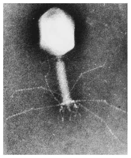
DNA is composed of 2’-deoxy-nucleotides carrying the bases adenine (A), guanine (G), cytosine (C) and thymine (T). The molar mass of the 2’-deoxy-nucleotide-5’-triphosphates is given in table 2:
Table 2
| dNTP | Molar mass /g mol\(^{-1}\) |
|---|---|
| dATP | 487 |
| dGTP | 503 |
| dCTP | 464 |
| dTTP | 478 |
5.1 Calculate the molar mass of a double stranded DNA fragment consisting of 1000 base pairs with a uniform distribution of the four bases.
This DNA fragment can be isolated and cloned by using the PCR method (polymerase chain reaction), in which a heat stable DNA polymerase enzyme multiplies the number of molecules of a specific piece of DNA in a cyclic process. Under optimal conditions the number of double-stranded DNA copies doubles in each cycle. Using the PCR method you perform 30 cycles starting from a single double stranded DNA molecule.
5.2 Calculate the approximate mass of the DNA you obtain from this experiment.
The bacteria-virus T4 enzyme - polynucleotide kinase (PNK) catalyzes the transfer of the terminal phosphate of ATP (\(\gamma\)-orthophosphate) to the 5'-hydroxyl termini of ribo- and deoxyribonucleotides:
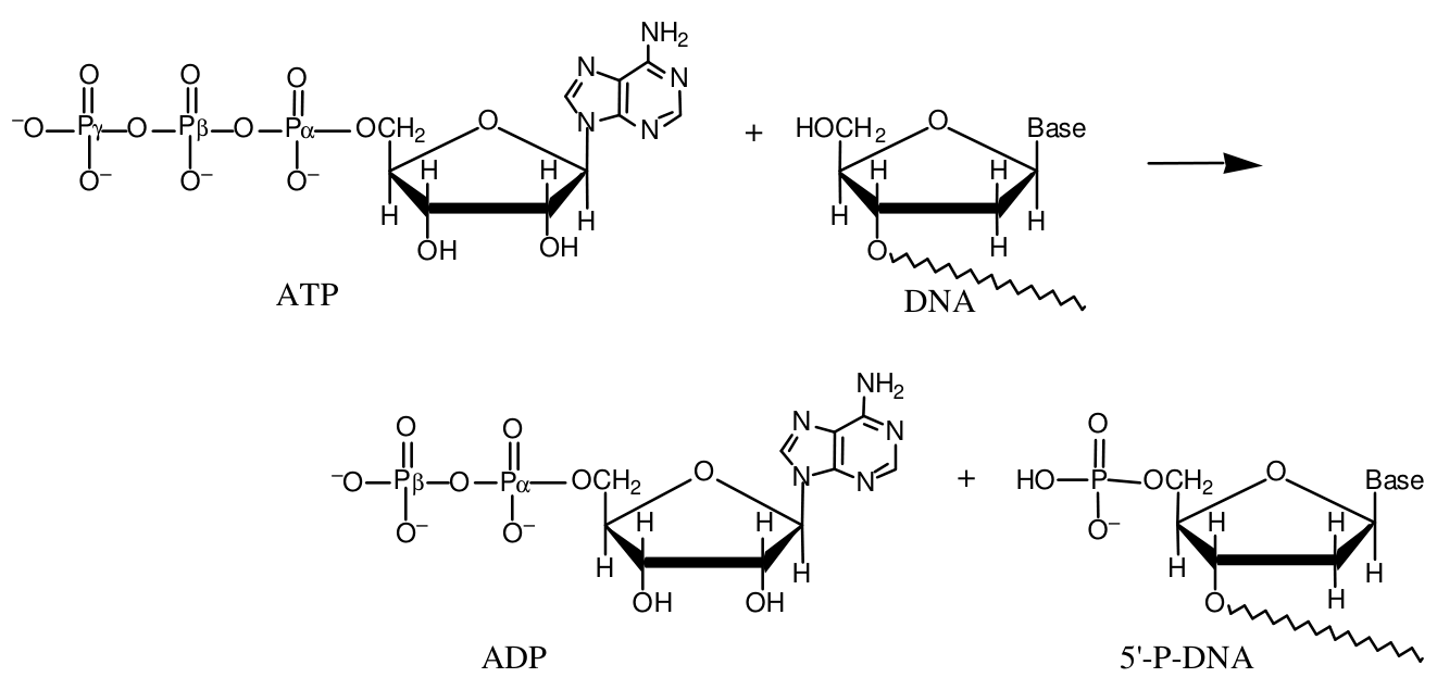
PNK is commonly used to label DNA at the 5’-end with the radioactive phosphorus isotope \(\ce{^{32}P}\) using ATP in which the \(\gamma\)-P (the outermost of the phosphorus atoms) is replaced with \(\ce{^{32}P}\). The amount of \(\ce{^{32}P}\) and thus the amount of labeled DNA can be measured.
A 10 µL solution containing double stranded DNA is labelled 100 % with \([\gamma\text{-}\ce{^{32}P}]\mathrm{ATP}\) by PNK. 37 days ago, the specific activity of \([\gamma\text{-}\ce{^{32}P}]\mathrm{ATP}\) was 10 Ci/mmol or \(370\cdot10^9\) Bq/mmol. \(\ce{^{32}P}\) has a half-life of 14.2 days, and during the decay a \(\beta\)-particle is emitted. Now the labelled DNA emits 40000 \(\beta\)-particles/s.
5-3 Calculate the concentration of the DNA solution.
In an experiment in which PNK is incubated with \([\gamma\text{-}\ce{^{32}P}]\mathrm{ATP}\) and single stranded DNA, the reaction can be monitored by isolating labeled DNA and measuring the \(\beta\)-particle emission. Using this kind of measurements in a 1 cm\(^{3}\) experimental mixture, a labeling of 9 nmol DNA/min was calculated. PNK has a catalytic rate constant (turnover number) of 0.05 s\(^{-1}\) and molar mass of 34620 g mol\(^{-1}\).
5.4 Calculate the concentration (in mg/cm\(^{3}\)) of PNK in the experimental mixture.
Aromatic amino acids, tryptophan, tyrosine and phenylalanine absorb UV light of a wavelength between 240 nm and 300 nm. In a protein containing several aromatic amino acids, the sum of the molar absorptivity per amino acid \(\Sigma\varepsilon_{\text{amino acid}}\), is approximately equal to the molar absorptivity, \(\varepsilon_{\text{protein}}\), for the protein. The molar absorptivity, \(\varepsilon_{\text{amino acid}}\), at 280 nm for tyrosine, tryptophan and phenylalanine is 1400 M\(^{-1}\) cm\(^{-1}\), 5600 M\(^{-1}\) cm\(^{-1}\) and 5 M\(^{-1}\) cm\(^{-1}\), respectively. The absorbance of a 10 µM solution of PNK is 0.644 at 280 nm and with 1.00 cm light path. The amino acid sequence of PNK contains 14 tyrosines and 9 phenylalanines.
\((\mathrm{M}=\mathrm{mol\,dm^{-3}})\)
5.5 Calculate the number of tryptophan residues in a PNK molecule.
Official English text of the problem.
Answers
- 5.1: \(M_{\mathrm{DNA}}\approx6.18\times10^5\ \mathrm{g\,mol^{-1}}\).
- 5.2: \(m_{\mathrm{DNA}}\approx1.10\times10^{-9}\ \mathrm{g}=1.10\ \mathrm{ng}\).
- 5.3: \(c_{\mathrm{DNA}}\approx6.58\times10^{-5}\ \mathrm{mol\,dm^{-3}}=65.8\ \mu\mathrm{M}\), counting each labelled 5′ end as one DNA molecule (amount of labelled DNA = amount of 32P, as the statement says).
- 5.4: \(c_m(\mathrm{PNK})\approx0.104\ \mathrm{mg\,cm^{-3}}\), or about \(0.10\ \mathrm{mg\,cm^{-3}}\).
- 5.5: 8 tryptophan residues per PNK molecule.
Solution
5.1 — Molar mass of the DNA fragment
There are two nucleotides per base pair, so the fragment contains
\[ N_{\mathrm{nt}}=2(1000)=2000 \]
nucleotide residues. A uniform distribution means 500 residues of each base.
During incorporation into DNA, the two terminal phosphate groups of a nucleotide triphosphate leave as pyrophosphate. Using the ionic mass convention of the table, the removed pyrophosphate group is \(\ce{P2O7^{4-}}\), with
\[ M_{\mathrm{PP_i}} =2(31)+7(16) =174\ \mathrm{g\,mol^{-1}}. \]
Pyrophosphate ion has SMILES [O-]P(=O)([O-])OP(=O)([O-])[O-].
The average molar mass of an incorporated nucleotide residue is therefore
\[ \overline M_{\mathrm{residue}} =\frac{487+503+464+478}{4}-174 =309\ \mathrm{g\,mol^{-1}}. \]
Neglecting the small contribution from terminal groups,
\[ \boxed{ M_{\mathrm{DNA}}\approx2000(309) =6.18\times10^5\ \mathrm{g\,mol^{-1}} }. \]
5.2 — Mass after 30 PCR cycles
Each cycle doubles the number of double-stranded DNA molecules. Starting from one molecule,
\[ N=2^{30}=1\,073\,741\,824. \]
Using \(N_{\mathrm A}=6.022\times10^{23}\ \mathrm{mol^{-1}}\),
\[ n_{\mathrm{DNA}} =\frac{2^{30}}{N_{\mathrm A}} =1.783\times10^{-15}\ \mathrm{mol}. \]
Thus,
\[ m_{\mathrm{DNA}} =n_{\mathrm{DNA}}M_{\mathrm{DNA}} =(1.783\times10^{-15})(6.18\times10^5)\ \mathrm{g}, \]
\[ \boxed{ m_{\mathrm{DNA}}\approx1.10\times10^{-9}\ \mathrm{g} =1.10\ \mathrm{ng} }. \]
5.3 — DNA concentration from radioactive labelling
The specific activity decreases according to
\[ a(t)=a_0\,2^{-t/t_{1/2}}. \]
After 37 days,
\[ a(37) =(370\times10^9)\,2^{-37/14.2} =6.079\times10^{10}\ \mathrm{Bq\,mmol^{-1}}. \]
One emitted \(\beta\)-particle corresponds to one decay, so the measured activity is
\[ A=40000\ \mathrm{Bq}. \]
Since the amount of labelled DNA equals the amount of 32P (each labelled 5′ end counts as one DNA molecule),
\[ n_{\mathrm{DNA}} =\frac{A}{a(37)} =\frac{40000}{6.079\times10^{10}} =6.580\times10^{-7}\ \mathrm{mmol} =6.580\times10^{-10}\ \mathrm{mol}. \]
The sample volume is
\[ V=10\ \mu\mathrm{L}=1.0\times10^{-5}\ \mathrm{dm^3}. \]
Consequently,
\[ \boxed{ c_{\mathrm{DNA}} =\frac{6.580\times10^{-10}}{1.0\times10^{-5}} =6.58\times10^{-5}\ \mathrm{mol\,dm^{-3}} =65.8\ \mu\mathrm{M} }. \]
No factor of two is applied, because each labelled 5′ end counts as one DNA molecule.
5.4 — PNK mass concentration
The observed labelling rate is
\[ \dot n_{\mathrm{DNA}} =\frac{9\ \mathrm{nmol}}{60\ \mathrm{s}} =0.15\ \mathrm{nmol\,s^{-1}}. \]
Using the stated turnover number as the reaction rate per enzyme molecule,
\[ \dot n_{\mathrm{DNA}} =k_{\mathrm{cat}}n_{\mathrm{PNK}}. \]
Therefore,
\[ n_{\mathrm{PNK}} =\frac{0.15\ \mathrm{nmol\,s^{-1}}}{0.05\ \mathrm{s^{-1}}} =3.0\ \mathrm{nmol}. \]
The corresponding enzyme mass is
\[ m_{\mathrm{PNK}} =(3.0\times10^{-9}\ \mathrm{mol}) (34620\ \mathrm{g\,mol^{-1}}) =1.0386\times10^{-4}\ \mathrm{g} =0.10386\ \mathrm{mg}. \]
Since the experimental volume is \(1\ \mathrm{cm^3}\),
\[ \boxed{ c_m(\mathrm{PNK}) =0.10386\ \mathrm{mg\,cm^{-3}} \approx0.10\ \mathrm{mg\,cm^{-3}} }. \]
5.5 — Number of tryptophan residues
The Beer–Lambert law gives
\[ A=\varepsilon_{\mathrm{PNK}}cl. \]
Hence,
\[ \varepsilon_{\mathrm{PNK}} =\frac{0.644} {(10\times10^{-6}\ \mathrm{mol\,dm^{-3}})(1.00\ \mathrm{cm})} =64400\ \mathrm{M^{-1}\,cm^{-1}}. \]
Let \(x\) be the number of tryptophan residues. Adding the contributions from the three aromatic amino acids,
\[ 64400=14(1400)+x(5600)+9(5). \]
Thus,
\[ x=\frac{64400-19600-45}{5600} =7.99196. \]
The residue count must be an integer, and the absorptivity relation is approximate:
\[ \boxed{x=8}. \]
The residue is L-tryptophan; its neutral free-amino-acid SMILES is N[C@@H](Cc1c[nH]c2ccccc12)C(=O)O. Within PNK, its amino and carboxyl groups participate in the peptide backbone.
The solution was checked against the official solution.
IChO 2000 · Theorieaufgabe 5
Protein und DNA
DNA besteht aus 2’-Desoxynukleotiden mit den Basen Adenin (A), Guanin (G), Cytosin (C) und Thymin (T). Die molaren Massen der 2’-Desoxynukleotid-5’-triphosphate sind in Tabelle 2 angegeben:
Tabelle 2
| dNTP | Molare Masse /g mol\(^{-1}\) |
|---|---|
| dATP | 487 |
| dGTP | 503 |
| dCTP | 464 |
| dTTP | 478 |
5.1 Berechnen Sie die molare Masse eines doppelsträngigen DNA-Fragments aus 1000 Basenpaaren mit einer gleichmäßigen Verteilung der vier Basen.
Dieses DNA-Fragment kann mithilfe der PCR-Methode (Polymerase-Kettenreaktion) isoliert und kloniert werden. Dabei vervielfältigt eine thermostabile DNA-Polymerase die Anzahl der Moleküle eines bestimmten DNA-Abschnitts in einem zyklischen Prozess. Unter optimalen Bedingungen verdoppelt sich die Anzahl der doppelsträngigen DNA-Kopien in jedem Zyklus. Ausgehend von einem einzigen doppelsträngigen DNA-Molekül führen Sie mit der PCR-Methode 30 Zyklen durch.
5.2 Berechnen Sie die ungefähre Masse der DNA, die Sie bei diesem Experiment erhalten.
Das Enzym Polynukleotidkinase (PNK) des Bakteriophagen T4 katalysiert die Übertragung der endständigen Phosphatgruppe von ATP (\(\gamma\)-Orthophosphat) auf die 5'-Hydroxylenden von Ribo- und Desoxyribonukleotiden:
PNK wird häufig verwendet, um DNA am 5’-Ende mit dem radioaktiven Phosphorisotop \(\ce{^{32}P}\) zu markieren. Dazu wird ATP eingesetzt, in dem das \(\gamma\)-P (das äußerste der Phosphoratome) durch \(\ce{^{32}P}\) ersetzt ist. Die Menge an \(\ce{^{32}P}\) und damit die Menge an markierter DNA kann gemessen werden.
Eine 10 µL große Lösung, die doppelsträngige DNA enthält, wird durch PNK mit \([\gamma\text{-}\ce{^{32}P}]\mathrm{ATP}\) zu 100 % markiert. Vor 37 Tagen betrug die spezifische Aktivität von \([\gamma\text{-}\ce{^{32}P}]\mathrm{ATP}\) 10 Ci/mmol oder \(370\cdot10^9\) Bq/mmol. \(\ce{^{32}P}\) hat eine Halbwertszeit von 14.2 Tagen, und beim Zerfall wird ein \(\beta\)-Teilchen emittiert. Jetzt emittiert die markierte DNA 40000 \(\beta\)-Teilchen/s.
5-3 Berechnen Sie die Konzentration der DNA-Lösung.
In einem Experiment, bei dem PNK mit \([\gamma\text{-}\ce{^{32}P}]\mathrm{ATP}\) und einzelsträngiger DNA inkubiert wird, kann die Reaktion verfolgt werden, indem die markierte DNA isoliert und die Emission von \(\beta\)-Teilchen gemessen wird. Aus solchen Messungen in einem Reaktionsansatz von 1 cm\(^{3}\) wurde eine Markierungsrate von 9 nmol DNA/min berechnet. PNK hat eine katalytische Geschwindigkeitskonstante (Wechselzahl) von 0.05 s\(^{-1}\) und eine molare Masse von 34620 g mol\(^{-1}\).
5.4 Berechnen Sie die Konzentration (in mg/cm\(^{3}\)) von PNK im Reaktionsansatz.
Die aromatischen Aminosäuren Tryptophan, Tyrosin und Phenylalanin absorbieren UV-Licht mit einer Wellenlänge zwischen 240 nm und 300 nm. Bei einem Protein, das mehrere aromatische Aminosäuren enthält, ist die Summe der molaren Absorptionskoeffizienten der einzelnen Aminosäuren, \(\Sigma\varepsilon_{\text{Aminosäure}}\), näherungsweise gleich dem molaren Absorptionskoeffizienten \(\varepsilon_{\text{Protein}}\) des Proteins. Der molare Absorptionskoeffizient \(\varepsilon_{\text{Aminosäure}}\) bei 280 nm beträgt für Tyrosin, Tryptophan und Phenylalanin 1400 M\(^{-1}\) cm\(^{-1}\), 5600 M\(^{-1}\) cm\(^{-1}\) bzw. 5 M\(^{-1}\) cm\(^{-1}\). Die Extinktion einer 10 µM PNK-Lösung beträgt bei 280 nm und einer Schichtdicke von 1.00 cm 0.644. Die Aminosäuresequenz von PNK enthält 14 Tyrosinreste und 9 Phenylalaninreste.
\((\mathrm{M}=\mathrm{mol\,dm^{-3}})\)
5.5 Berechnen Sie die Anzahl der Tryptophanreste in einem PNK-Molekül.
Aufgabentext aus dem offiziellen englischen Text übersetzt.
Antworten
- 5.1: \(M_{\mathrm{DNA}}\approx6.18\times10^5\ \mathrm{g\,mol^{-1}}\).
- 5.2: \(m_{\mathrm{DNA}}\approx1.10\times10^{-9}\ \mathrm{g}=1.10\ \mathrm{ng}\).
- 5.3: \(c_{\mathrm{DNA}}\approx6.58\times10^{-5}\ \mathrm{mol\,dm^{-3}}=65.8\ \mu\mathrm{M}\), wobei jedes markierte 5′-Ende als ein DNA-Molekül gezählt wird (Stoffmenge der markierten DNA = Stoffmenge von 32P, wie in der Aufgabenstellung angegeben).
- 5.4: \(c_m(\mathrm{PNK})\approx0.104\ \mathrm{mg\,cm^{-3}}\), also etwa \(0.10\ \mathrm{mg\,cm^{-3}}\).
- 5.5: 8 Tryptophanreste pro PNK-Molekül.
Lösung
5.1 — Molare Masse des DNA-Fragments
Auf jedes Basenpaar entfallen zwei Nukleotide, sodass das Fragment
\[ N_{\mathrm{nt}}=2(1000)=2000 \]
Nukleotidreste enthält. Eine gleichmäßige Verteilung bedeutet 500 Reste jeder Base.
Beim Einbau in die DNA werden die beiden endständigen Phosphatgruppen eines Nukleotidtriphosphats als Pyrophosphat abgespalten. Nach der in der Tabelle verwendeten Konvention für die Ionenmassen ist die abgespaltene Pyrophosphatgruppe \(\ce{P2O7^{4-}}\), mit
\[ M_{\mathrm{PP_i}} =2(31)+7(16) =174\ \mathrm{g\,mol^{-1}}. \]
Das Pyrophosphat-Ion hat die SMILES-Darstellung [O-]P(=O)([O-])OP(=O)([O-])[O-].
Die mittlere molare Masse eines eingebauten Nukleotidrests beträgt daher
\[ \overline M_{\mathrm{residue}} =\frac{487+503+464+478}{4}-174 =309\ \mathrm{g\,mol^{-1}}. \]
Unter Vernachlässigung des kleinen Beitrags der Endgruppen ergibt sich
\[ \boxed{ M_{\mathrm{DNA}}\approx2000(309) =6.18\times10^5\ \mathrm{g\,mol^{-1}} }. \]
5.2 — Masse nach 30 PCR-Zyklen
Jeder Zyklus verdoppelt die Anzahl der doppelsträngigen DNA-Moleküle. Ausgehend von einem Molekül gilt
\[ N=2^{30}=1\,073\,741\,824. \]
Mit \(N_{\mathrm A}=6.022\times10^{23}\ \mathrm{mol^{-1}}\) ergibt sich
\[ n_{\mathrm{DNA}} =\frac{2^{30}}{N_{\mathrm A}} =1.783\times10^{-15}\ \mathrm{mol}. \]
Damit gilt
\[ m_{\mathrm{DNA}} =n_{\mathrm{DNA}}M_{\mathrm{DNA}} =(1.783\times10^{-15})(6.18\times10^5)\ \mathrm{g}, \]
\[ \boxed{ m_{\mathrm{DNA}}\approx1.10\times10^{-9}\ \mathrm{g} =1.10\ \mathrm{ng} }. \]
5.3 — DNA-Konzentration aus der radioaktiven Markierung
Die Abnahme der spezifischen Aktivität folgt der Beziehung
\[ a(t)=a_0\,2^{-t/t_{1/2}}. \]
Nach 37 Tagen gilt
\[ a(37) =(370\times10^9)\,2^{-37/14.2} =6.079\times10^{10}\ \mathrm{Bq\,mmol^{-1}}. \]
Ein emittiertes \(\beta\)-Teilchen entspricht einem Zerfall, sodass für die gemessene Aktivität gilt:
\[ A=40000\ \mathrm{Bq}. \]
Da die Stoffmenge der markierten DNA gleich der Stoffmenge von 32P ist (jedes markierte 5′-Ende zählt als ein DNA-Molekül), gilt
\[ n_{\mathrm{DNA}} =\frac{A}{a(37)} =\frac{40000}{6.079\times10^{10}} =6.580\times10^{-7}\ \mathrm{mmol} =6.580\times10^{-10}\ \mathrm{mol}. \]
Das Probenvolumen beträgt
\[ V=10\ \mu\mathrm{L}=1.0\times10^{-5}\ \mathrm{dm^3}. \]
Folglich gilt
\[ \boxed{ c_{\mathrm{DNA}} =\frac{6.580\times10^{-10}}{1.0\times10^{-5}} =6.58\times10^{-5}\ \mathrm{mol\,dm^{-3}} =65.8\ \mu\mathrm{M} }. \]
Es wird kein Faktor zwei angewendet, da jedes markierte 5′-Ende als ein DNA-Molekül zählt.
5.4 — Massenkonzentration von PNK
Die beobachtete Markierungsrate beträgt
\[ \dot n_{\mathrm{DNA}} =\frac{9\ \mathrm{nmol}}{60\ \mathrm{s}} =0.15\ \mathrm{nmol\,s^{-1}}. \]
Verwendet man die angegebene Wechselzahl als Reaktionsrate pro Enzymmolekül, gilt
\[ \dot n_{\mathrm{DNA}} =k_{\mathrm{cat}}n_{\mathrm{PNK}}. \]
Daher ergibt sich
\[ n_{\mathrm{PNK}} =\frac{0.15\ \mathrm{nmol\,s^{-1}}}{0.05\ \mathrm{s^{-1}}} =3.0\ \mathrm{nmol}. \]
Die entsprechende Enzymmasse beträgt
\[ m_{\mathrm{PNK}} =(3.0\times10^{-9}\ \mathrm{mol}) (34620\ \mathrm{g\,mol^{-1}}) =1.0386\times10^{-4}\ \mathrm{g} =0.10386\ \mathrm{mg}. \]
Da das Volumen des Reaktionsansatzes \(1\ \mathrm{cm^3}\) beträgt, gilt
\[ \boxed{ c_m(\mathrm{PNK}) =0.10386\ \mathrm{mg\,cm^{-3}} \approx0.10\ \mathrm{mg\,cm^{-3}} }. \]
5.5 — Anzahl der Tryptophanreste
Das Lambert-Beer-Gesetz liefert
\[ A=\varepsilon_{\mathrm{PNK}}cl. \]
Daraus folgt
\[ \varepsilon_{\mathrm{PNK}} =\frac{0.644} {(10\times10^{-6}\ \mathrm{mol\,dm^{-3}})(1.00\ \mathrm{cm})} =64400\ \mathrm{M^{-1}\,cm^{-1}}. \]
Sei \(x\) die Anzahl der Tryptophanreste. Durch Addition der Beiträge der drei aromatischen Aminosäuren erhält man
\[ 64400=14(1400)+x(5600)+9(5). \]
Somit gilt
\[ x=\frac{64400-19600-45}{5600} =7.99196. \]
Die Anzahl der Reste muss ganzzahlig sein, und die Beziehung für die Absorptionskoeffizienten gilt nur näherungsweise:
\[ \boxed{x=8}. \]
Bei dem Rest handelt es sich um L-Tryptophan; die SMILES-Darstellung der neutralen freien Aminosäure lautet N[C@@H](Cc1c[nH]c2ccccc12)C(=O)O. Innerhalb von PNK sind seine Amino- und Carboxylgruppen am Peptidrückgrat beteiligt.
Die Lösung wurde mit der offiziellen Lösung abgeglichen.
IChO 2000 · Teori sorusu 5
Protein ve DNA
DNA, adenin (A), guanin (G), sitozin (C) ve timin (T) bazlarını taşıyan 2’-deoksinükleotitlerden oluşur. 2’-deoksinükleotit-5’-trifosfatların molar kütleleri tablo 2’de verilmiştir:
Tablo 2
| dNTP | Molar kütle /g mol\(^{-1}\) |
|---|---|
| dATP | 487 |
| dGTP | 503 |
| dCTP | 464 |
| dTTP | 478 |
5.1 Dört bazın eşit dağıldığı, 1000 baz çiftinden oluşan çift zincirli bir DNA parçasının molar kütlesini hesaplayınız.
Bu DNA parçası, ısıya dayanıklı bir DNA polimeraz enziminin döngüsel bir süreçte belirli bir DNA parçasına ait moleküllerin sayısını artırdığı PCR (polimeraz zincir reaksiyonu) yöntemi kullanılarak izole edilebilir ve klonlanabilir. En uygun koşullarda çift zincirli DNA kopyalarının sayısı her döngüde iki katına çıkar. Tek bir çift zincirli DNA molekülünden başlayarak PCR yöntemiyle 30 döngü gerçekleştiriyorsunuz.
5.2 Bu deneyden elde ettiğiniz DNA’nın yaklaşık kütlesini hesaplayınız.
Bakteriyofaj T4’ün polinükleotit kinaz (PNK) enzimi, ATP’nin uç fosfatının (\(\gamma\)-ortofosfat) ribonükleotitlerin ve deoksiribonükleotitlerin 5'-hidroksil uçlarına aktarılmasını katalizler:
PNK, DNA’yı 5’ ucunda radyoaktif fosfor izotopu \(\ce{^{32}P}\) ile işaretlemek için yaygın olarak kullanılır; bunun için \(\gamma\)-P’nin (fosfor atomlarının en dışta olanının) \(\ce{^{32}P}\) ile değiştirildiği ATP kullanılır. \(\ce{^{32}P}\) miktarı ve dolayısıyla işaretli DNA miktarı ölçülebilir.
Çift zincirli DNA içeren 10 µL’lik bir çözelti, PNK aracılığıyla \([\gamma\text{-}\ce{^{32}P}]\mathrm{ATP}\) kullanılarak %100 oranında işaretlenmiştir. 37 gün önce \([\gamma\text{-}\ce{^{32}P}]\mathrm{ATP}\)’nin özgül aktivitesi 10 Ci/mmol veya \(370\cdot10^9\) Bq/mmol idi. \(\ce{^{32}P}\)’nin yarı ömrü 14.2 gündür ve bozunma sırasında bir \(\beta\) parçacığı yayılır. Şu anda işaretli DNA saniyede 40000 \(\beta\) parçacığı yaymaktadır.
5-3 DNA çözeltisinin derişimini hesaplayınız.
PNK’nin \([\gamma\text{-}\ce{^{32}P}]\mathrm{ATP}\) ve tek zincirli DNA ile inkübe edildiği bir deneyde, işaretli DNA izole edilip \(\beta\) parçacığı yayımı ölçülerek tepkime izlenebilir. 1 cm\(^{3}\)’lük bir deney karışımında bu tür ölçümler kullanılarak işaretleme hızı 9 nmol DNA/min olarak hesaplanmıştır. PNK’nin katalitik hız sabiti (çevrim sayısı) 0.05 s\(^{-1}\), molar kütlesi ise 34620 g mol\(^{-1}\)’dir.
5.4 Deney karışımındaki PNK derişimini (mg/cm\(^{3}\) cinsinden) hesaplayınız.
Aromatik amino asitler olan triptofan, tirozin ve fenilalanin, dalga boyu 240 nm ile 300 nm arasında olan UV ışığını soğurur. Birden fazla aromatik amino asit içeren bir proteinde, her bir amino asidin molar soğurma katsayılarının toplamı olan \(\Sigma\varepsilon_{\text{amino asit}}\), proteinin molar soğurma katsayısı \(\varepsilon_{\text{protein}}\)’e yaklaşık olarak eşittir. Tirozin, triptofan ve fenilalaninin 280 nm’deki molar soğurma katsayıları \(\varepsilon_{\text{amino asit}}\), sırasıyla 1400 M\(^{-1}\) cm\(^{-1}\), 5600 M\(^{-1}\) cm\(^{-1}\) ve 5 M\(^{-1}\) cm\(^{-1}\)’dir. 10 µM’lık bir PNK çözeltisinin absorbansı, 280 nm’de ve 1.00 cm optik yol uzunluğunda 0.644’tür. PNK’nin amino asit dizisi 14 tirozin ve 9 fenilalanin içerir.
\((\mathrm{M}=\mathrm{mol\,dm^{-3}})\)
5.5 Bir PNK molekülündeki triptofan kalıntılarının sayısını hesaplayınız.
Soru metni resmi İngilizce metinden çevrilmiştir.
Cevaplar
- 5.1: \(M_{\mathrm{DNA}}\approx6.18\times10^5\ \mathrm{g\,mol^{-1}}\).
- 5.2: \(m_{\mathrm{DNA}}\approx1.10\times10^{-9}\ \mathrm{g}=1.10\ \mathrm{ng}\).
- 5.3: \(c_{\mathrm{DNA}}\approx6.58\times10^{-5}\ \mathrm{mol\,dm^{-3}}=65.8\ \mu\mathrm{M}\); işaretli her 5′ ucu bir DNA molekülü olarak sayılır (soru metninde belirtildiği gibi, işaretli DNA miktarı = 32P miktarı).
- 5.4: \(c_m(\mathrm{PNK})\approx0.104\ \mathrm{mg\,cm^{-3}}\) veya yaklaşık \(0.10\ \mathrm{mg\,cm^{-3}}\).
- 5.5: PNK molekülü başına 8 triptofan kalıntısı.
Çözüm
5.1 — DNA parçasının molar kütlesi
Her baz çiftinde iki nükleotit bulunduğundan parça,
\[ N_{\mathrm{nt}}=2(1000)=2000 \]
nükleotit kalıntısı içerir. Eşit dağılım, her bazdan 500 kalıntı bulunması anlamına gelir.
DNA’ya katılma sırasında bir nükleotit trifosfatın uçtaki iki fosfat grubu pirofosfat olarak ayrılır. Tabloda kullanılan iyonik kütle kabulüne göre, ayrılan pirofosfat grubu \(\ce{P2O7^{4-}}\) olup molar kütlesi şöyledir:
\[ M_{\mathrm{PP_i}} =2(31)+7(16) =174\ \mathrm{g\,mol^{-1}}. \]
Pirofosfat iyonunun SMILES gösterimi [O-]P(=O)([O-])OP(=O)([O-])[O-] şeklindedir.
Dolayısıyla DNA’ya katılan bir nükleotit kalıntısının ortalama molar kütlesi
\[ \overline M_{\mathrm{residue}} =\frac{487+503+464+478}{4}-174 =309\ \mathrm{g\,mol^{-1}}. \]
Uç grupların küçük katkısı ihmal edilirse,
\[ \boxed{ M_{\mathrm{DNA}}\approx2000(309) =6.18\times10^5\ \mathrm{g\,mol^{-1}} }. \]
5.2 — 30 PCR döngüsünden sonraki kütle
Her döngü, çift zincirli DNA moleküllerinin sayısını iki katına çıkarır. Tek bir molekülden başlandığında,
\[ N=2^{30}=1\,073\,741\,824. \]
\(N_{\mathrm A}=6.022\times10^{23}\ \mathrm{mol^{-1}}\) kullanılarak,
\[ n_{\mathrm{DNA}} =\frac{2^{30}}{N_{\mathrm A}} =1.783\times10^{-15}\ \mathrm{mol}. \]
Böylece,
\[ m_{\mathrm{DNA}} =n_{\mathrm{DNA}}M_{\mathrm{DNA}} =(1.783\times10^{-15})(6.18\times10^5)\ \mathrm{g}, \]
\[ \boxed{ m_{\mathrm{DNA}}\approx1.10\times10^{-9}\ \mathrm{g} =1.10\ \mathrm{ng} }. \]
5.3 — Radyoaktif işaretlemeden DNA derişiminin hesaplanması
Özgül aktivite aşağıdaki bağıntıya göre azalır:
\[ a(t)=a_0\,2^{-t/t_{1/2}}. \]
37 gün sonra,
\[ a(37) =(370\times10^9)\,2^{-37/14.2} =6.079\times10^{10}\ \mathrm{Bq\,mmol^{-1}}. \]
Yayılan her \(\beta\) parçacığı bir bozunmaya karşılık geldiğinden ölçülen aktivite
\[ A=40000\ \mathrm{Bq}. \]
İşaretli DNA miktarı 32P miktarına eşit olduğundan (işaretli her 5′ ucu bir DNA molekülü olarak sayılır),
\[ n_{\mathrm{DNA}} =\frac{A}{a(37)} =\frac{40000}{6.079\times10^{10}} =6.580\times10^{-7}\ \mathrm{mmol} =6.580\times10^{-10}\ \mathrm{mol}. \]
Örneğin hacmi
\[ V=10\ \mu\mathrm{L}=1.0\times10^{-5}\ \mathrm{dm^3}. \]
Sonuç olarak,
\[ \boxed{ c_{\mathrm{DNA}} =\frac{6.580\times10^{-10}}{1.0\times10^{-5}} =6.58\times10^{-5}\ \mathrm{mol\,dm^{-3}} =65.8\ \mu\mathrm{M} }. \]
İşaretli her 5′ ucu bir DNA molekülü olarak sayıldığından iki katsayısı uygulanmaz.
5.4 — PNK’nin kütle derişimi
Gözlenen işaretleme hızı
\[ \dot n_{\mathrm{DNA}} =\frac{9\ \mathrm{nmol}}{60\ \mathrm{s}} =0.15\ \mathrm{nmol\,s^{-1}}. \]
Verilen çevrim sayısı, enzim molekülü başına tepkime hızı olarak kullanıldığında,
\[ \dot n_{\mathrm{DNA}} =k_{\mathrm{cat}}n_{\mathrm{PNK}}. \]
Dolayısıyla,
\[ n_{\mathrm{PNK}} =\frac{0.15\ \mathrm{nmol\,s^{-1}}}{0.05\ \mathrm{s^{-1}}} =3.0\ \mathrm{nmol}. \]
Buna karşılık gelen enzim kütlesi
\[ m_{\mathrm{PNK}} =(3.0\times10^{-9}\ \mathrm{mol}) (34620\ \mathrm{g\,mol^{-1}}) =1.0386\times10^{-4}\ \mathrm{g} =0.10386\ \mathrm{mg}. \]
Deney hacmi \(1\ \mathrm{cm^3}\) olduğundan,
\[ \boxed{ c_m(\mathrm{PNK}) =0.10386\ \mathrm{mg\,cm^{-3}} \approx0.10\ \mathrm{mg\,cm^{-3}} }. \]
5.5 — Triptofan kalıntılarının sayısı
Beer–Lambert yasasına göre,
\[ A=\varepsilon_{\mathrm{PNK}}cl. \]
Buradan,
\[ \varepsilon_{\mathrm{PNK}} =\frac{0.644} {(10\times10^{-6}\ \mathrm{mol\,dm^{-3}})(1.00\ \mathrm{cm})} =64400\ \mathrm{M^{-1}\,cm^{-1}}. \]
Triptofan kalıntılarının sayısı \(x\) olsun. Üç aromatik amino asidin katkıları toplandığında,
\[ 64400=14(1400)+x(5600)+9(5). \]
Böylece,
\[ x=\frac{64400-19600-45}{5600} =7.99196. \]
Kalıntı sayısı bir tam sayı olmalıdır ve soğurma katsayısı bağıntısı yaklaşık bir bağıntıdır:
\[ \boxed{x=8}. \]
Bu kalıntı L-triptofandır; nötr, serbest amino asit hâlinin SMILES gösterimi N[C@@H](Cc1c[nH]c2ccccc12)C(=O)O şeklindedir. PNK içinde amino ve karboksil grupları peptit omurgasına katılır.
Çözüm, resmi çözümle karşılaştırılarak kontrol edildi.
IChO 2000 · Problema teórico 5
Proteína y ADN
El ADN está compuesto por 2’-desoxinucleótidos que contienen las bases adenina (A), guanina (G), citosina (C) y timina (T). La masa molar de los 2’-desoxinucleótidos-5’-trifosfato se indica en la tabla 2:
Tabla 2
| dNTP | Masa molar /g mol\(^{-1}\) |
|---|---|
| dATP | 487 |
| dGTP | 503 |
| dCTP | 464 |
| dTTP | 478 |
5.1 Calcule la masa molar de un fragmento de ADN de doble cadena formado por 1000 pares de bases con una distribución uniforme de las cuatro bases.
Este fragmento de ADN puede aislarse y clonarse mediante el método de PCR (reacción en cadena de la polimerasa), en el que una enzima ADN polimerasa termoestable multiplica el número de moléculas de un fragmento específico de ADN en un proceso cíclico. En condiciones óptimas, el número de copias de ADN de doble cadena se duplica en cada ciclo. Utilizando el método de PCR, usted realiza 30 ciclos a partir de una única molécula de ADN de doble cadena.
5.2 Calcule la masa aproximada de ADN que obtiene en este experimento.
La enzima polinucleótido quinasa (PNK) del bacteriófago T4 cataliza la transferencia del fosfato terminal del ATP (\(\gamma\)-ortofosfato) a los grupos hidroxilo de los extremos 5' de ribonucleótidos y desoxirribonucleótidos:
La PNK se utiliza habitualmente para marcar el ADN en el extremo 5’ con el isótopo radiactivo del fósforo \(\ce{^{32}P}\), utilizando ATP en el que el \(\gamma\)-P (el más externo de los átomos de fósforo) se sustituye por \(\ce{^{32}P}\). Se puede medir la cantidad de \(\ce{^{32}P}\) y, por tanto, la cantidad de ADN marcado.
Una disolución de 10 µL que contiene ADN de doble cadena se marca al 100 % con \([\gamma\text{-}\ce{^{32}P}]\mathrm{ATP}\) mediante PNK. Hace 37 días, la actividad específica del \([\gamma\text{-}\ce{^{32}P}]\mathrm{ATP}\) era de 10 Ci/mmol o \(370\cdot10^9\) Bq/mmol. El \(\ce{^{32}P}\) tiene un período de semidesintegración de 14.2 días, y durante la desintegración se emite una partícula \(\beta\). Ahora, el ADN marcado emite 40000 partículas \(\beta\)/s.
5-3 Calcule la concentración de la disolución de ADN.
En un experimento en el que se incuba PNK con \([\gamma\text{-}\ce{^{32}P}]\mathrm{ATP}\) y ADN de cadena sencilla, se puede seguir la reacción aislando el ADN marcado y midiendo la emisión de partículas \(\beta\). Mediante este tipo de mediciones en una mezcla experimental de 1 cm\(^{3}\), se calculó una velocidad de marcaje de 9 nmol de ADN/min. La PNK tiene una constante de velocidad catalítica (número de recambio) de 0.05 s\(^{-1}\) y una masa molar de 34620 g mol\(^{-1}\).
5.4 Calcule la concentración (en mg/cm\(^{3}\)) de PNK en la mezcla experimental.
Los aminoácidos aromáticos triptófano, tirosina y fenilalanina absorben luz UV de longitudes de onda comprendidas entre 240 nm y 300 nm. En una proteína que contiene varios aminoácidos aromáticos, la suma de las absortividades molares de cada aminoácido, \(\Sigma\varepsilon_{\text{aminoácido}}\), es aproximadamente igual a la absortividad molar, \(\varepsilon_{\text{proteína}}\), de la proteína. La absortividad molar, \(\varepsilon_{\text{aminoácido}}\), a 280 nm de la tirosina, el triptófano y la fenilalanina es de 1400 M\(^{-1}\) cm\(^{-1}\), 5600 M\(^{-1}\) cm\(^{-1}\) y 5 M\(^{-1}\) cm\(^{-1}\), respectivamente. La absorbancia de una disolución de PNK de 10 µM es de 0.644 a 280 nm y con un paso óptico de 1.00 cm. La secuencia de aminoácidos de la PNK contiene 14 tirosinas y 9 fenilalaninas.
\((\mathrm{M}=\mathrm{mol\,dm^{-3}})\)
5.5 Calcule el número de residuos de triptófano en una molécula de PNK.
Enunciado traducido del texto oficial en inglés.
Respuestas
- 5.1: \(M_{\mathrm{DNA}}\approx6.18\times10^5\ \mathrm{g\,mol^{-1}}\).
- 5.2: \(m_{\mathrm{DNA}}\approx1.10\times10^{-9}\ \mathrm{g}=1.10\ \mathrm{ng}\).
- 5.3: \(c_{\mathrm{DNA}}\approx6.58\times10^{-5}\ \mathrm{mol\,dm^{-3}}=65.8\ \mu\mathrm{M}\), contando cada extremo 5′ marcado como una molécula de ADN (cantidad de ADN marcado = cantidad de 32P, como indica el enunciado).
- 5.4: \(c_m(\mathrm{PNK})\approx0.104\ \mathrm{mg\,cm^{-3}}\), o aproximadamente \(0.10\ \mathrm{mg\,cm^{-3}}\).
- 5.5: 8 residuos de triptófano por molécula de PNK.
Solución
5.1 — Masa molar del fragmento de ADN
Hay dos nucleótidos por par de bases, por lo que el fragmento contiene
\[ N_{\mathrm{nt}}=2(1000)=2000 \]
residuos de nucleótidos. Una distribución uniforme significa que hay 500 residuos de cada base.
Durante la incorporación al ADN, los dos grupos fosfato terminales de un nucleótido trifosfato se liberan como pirofosfato. Utilizando la convención de masas iónicas de la tabla, el grupo pirofosfato eliminado es \(\ce{P2O7^{4-}}\), con
\[ M_{\mathrm{PP_i}} =2(31)+7(16) =174\ \mathrm{g\,mol^{-1}}. \]
El ion pirofosfato tiene el SMILES [O-]P(=O)([O-])OP(=O)([O-])[O-].
Por tanto, la masa molar media de un residuo de nucleótido incorporado es
\[ \overline M_{\mathrm{residue}} =\frac{487+503+464+478}{4}-174 =309\ \mathrm{g\,mol^{-1}}. \]
Despreciando la pequeña contribución de los grupos terminales,
\[ \boxed{ M_{\mathrm{DNA}}\approx2000(309) =6.18\times10^5\ \mathrm{g\,mol^{-1}} }. \]
5.2 — Masa después de 30 ciclos de PCR
Cada ciclo duplica el número de moléculas de ADN de doble cadena. Partiendo de una molécula,
\[ N=2^{30}=1\,073\,741\,824. \]
Utilizando \(N_{\mathrm A}=6.022\times10^{23}\ \mathrm{mol^{-1}}\),
\[ n_{\mathrm{DNA}} =\frac{2^{30}}{N_{\mathrm A}} =1.783\times10^{-15}\ \mathrm{mol}. \]
Así,
\[ m_{\mathrm{DNA}} =n_{\mathrm{DNA}}M_{\mathrm{DNA}} =(1.783\times10^{-15})(6.18\times10^5)\ \mathrm{g}, \]
\[ \boxed{ m_{\mathrm{DNA}}\approx1.10\times10^{-9}\ \mathrm{g} =1.10\ \mathrm{ng} }. \]
5.3 — Concentración de ADN a partir del marcaje radiactivo
La actividad específica disminuye según
\[ a(t)=a_0\,2^{-t/t_{1/2}}. \]
Después de 37 días,
\[ a(37) =(370\times10^9)\,2^{-37/14.2} =6.079\times10^{10}\ \mathrm{Bq\,mmol^{-1}}. \]
Cada partícula \(\beta\) emitida corresponde a una desintegración, por lo que la actividad medida es
\[ A=40000\ \mathrm{Bq}. \]
Como la cantidad de ADN marcado es igual a la cantidad de 32P (cada extremo 5′ marcado cuenta como una molécula de ADN),
\[ n_{\mathrm{DNA}} =\frac{A}{a(37)} =\frac{40000}{6.079\times10^{10}} =6.580\times10^{-7}\ \mathrm{mmol} =6.580\times10^{-10}\ \mathrm{mol}. \]
El volumen de la muestra es
\[ V=10\ \mu\mathrm{L}=1.0\times10^{-5}\ \mathrm{dm^3}. \]
En consecuencia,
\[ \boxed{ c_{\mathrm{DNA}} =\frac{6.580\times10^{-10}}{1.0\times10^{-5}} =6.58\times10^{-5}\ \mathrm{mol\,dm^{-3}} =65.8\ \mu\mathrm{M} }. \]
No se aplica ningún factor de dos, porque cada extremo 5′ marcado cuenta como una molécula de ADN.
5.4 — Concentración másica de PNK
La velocidad de marcaje observada es
\[ \dot n_{\mathrm{DNA}} =\frac{9\ \mathrm{nmol}}{60\ \mathrm{s}} =0.15\ \mathrm{nmol\,s^{-1}}. \]
Utilizando el número de recambio indicado como velocidad de reacción por molécula de enzima,
\[ \dot n_{\mathrm{DNA}} =k_{\mathrm{cat}}n_{\mathrm{PNK}}. \]
Por tanto,
\[ n_{\mathrm{PNK}} =\frac{0.15\ \mathrm{nmol\,s^{-1}}}{0.05\ \mathrm{s^{-1}}} =3.0\ \mathrm{nmol}. \]
La masa de enzima correspondiente es
\[ m_{\mathrm{PNK}} =(3.0\times10^{-9}\ \mathrm{mol}) (34620\ \mathrm{g\,mol^{-1}}) =1.0386\times10^{-4}\ \mathrm{g} =0.10386\ \mathrm{mg}. \]
Como el volumen experimental es de \(1\ \mathrm{cm^3}\),
\[ \boxed{ c_m(\mathrm{PNK}) =0.10386\ \mathrm{mg\,cm^{-3}} \approx0.10\ \mathrm{mg\,cm^{-3}} }. \]
5.5 — Número de residuos de triptófano
La ley de Beer–Lambert da
\[ A=\varepsilon_{\mathrm{PNK}}cl. \]
Por consiguiente,
\[ \varepsilon_{\mathrm{PNK}} =\frac{0.644} {(10\times10^{-6}\ \mathrm{mol\,dm^{-3}})(1.00\ \mathrm{cm})} =64400\ \mathrm{M^{-1}\,cm^{-1}}. \]
Sea \(x\) el número de residuos de triptófano. Sumando las contribuciones de los tres aminoácidos aromáticos,
\[ 64400=14(1400)+x(5600)+9(5). \]
Así,
\[ x=\frac{64400-19600-45}{5600} =7.99196. \]
El número de residuos debe ser entero, y la relación de absortividades es aproximada:
\[ \boxed{x=8}. \]
El residuo es L-triptófano; su SMILES como aminoácido libre neutro es N[C@@H](Cc1c[nH]c2ccccc12)C(=O)O. En la PNK, sus grupos amino y carboxilo forman parte del esqueleto peptídico.
La solución se comprobó con la solución oficial.
IChO 2000 · 理论题 5
蛋白质与 DNA
DNA 由含有腺嘌呤(A)、鸟嘌呤(G)、胞嘧啶(C)和胸腺嘧啶(T)这些碱基的 2′-脱氧核苷酸组成。2′-脱氧核苷-5′-三磷酸的摩尔质量列于表 2:
表 2
| dNTP | 摩尔质量 /g mol\(^{-1}\) |
|---|---|
| dATP | 487 |
| dGTP | 503 |
| dCTP | 464 |
| dTTP | 478 |
5.1 计算一个由 1000 个碱基对组成、四种碱基均匀分布的双链 DNA 片段的摩尔质量。
该 DNA 片段可以利用 PCR 法(聚合酶链式反应)进行分离和克隆。在这一方法中,耐热的 DNA 聚合酶通过循环过程增加特定 DNA 片段的分子数。在最佳条件下,双链 DNA 的拷贝数在每个循环中增加一倍。你从一个双链 DNA 分子开始,利用 PCR 法进行 30 个循环。
5.2 计算通过该实验获得的 DNA 的近似质量。
T4 噬菌体的多核苷酸激酶(PNK)催化 ATP 的末端磷酸基团(\(\gamma\)-正磷酸基团)转移至核糖核苷酸和脱氧核糖核苷酸的 5′-羟基末端:
PNK 常用于利用放射性磷同位素 \(\ce{^{32}P}\) 标记 DNA 的 5′ 端,所用的 ATP 中,\(\gamma\)-P(最外侧的磷原子)被 \(\ce{^{32}P}\) 取代。可以测量 \(\ce{^{32}P}\) 的量,从而测定被标记的 DNA 的量。
用 PNK 和 \([\gamma\text{-}\ce{^{32}P}]\mathrm{ATP}\) 对 10 µL 含双链 DNA 的溶液进行 100 % 标记。37 天前,\([\gamma\text{-}\ce{^{32}P}]\mathrm{ATP}\) 的比活度为 10 Ci/mmol,即 \(370\cdot10^9\) Bq/mmol。\(\ce{^{32}P}\) 的半衰期为 14.2 天,衰变时会发射一个 \(\beta\) 粒子。现在,被标记的 DNA 每秒发射 40000 个 \(\beta\) 粒子。
5-3 计算该 DNA 溶液的浓度。
在将 PNK 与 \([\gamma\text{-}\ce{^{32}P}]\mathrm{ATP}\) 及单链 DNA 一同孵育的实验中,可以通过分离被标记的 DNA 并测量其 \(\beta\) 粒子发射速率来监测反应。对体积为 1 cm\(^{3}\) 的实验混合物进行这类测量,计算得到 DNA 的标记速率为 9 nmol DNA/min。PNK 的催化速率常数(转换数)为 0.05 s\(^{-1}\),摩尔质量为 34620 g mol\(^{-1}\)。
5.4 计算实验混合物中 PNK 的浓度(以 mg/cm\(^{3}\) 表示)。
芳香族氨基酸色氨酸、酪氨酸和苯丙氨酸可吸收波长介于 240 nm 和 300 nm 之间的紫外光。对于含有多个芳香族氨基酸的蛋白质,各氨基酸的摩尔吸光系数之和 \(\Sigma\varepsilon_{\text{氨基酸}}\) 近似等于该蛋白质的摩尔吸光系数 \(\varepsilon_{\text{蛋白质}}\)。酪氨酸、色氨酸和苯丙氨酸在 280 nm 处的摩尔吸光系数 \(\varepsilon_{\text{氨基酸}}\) 分别为 1400 M\(^{-1}\) cm\(^{-1}\)、5600 M\(^{-1}\) cm\(^{-1}\) 和 5 M\(^{-1}\) cm\(^{-1}\)。浓度为 10 µM 的 PNK 溶液在 280 nm 处、光程为 1.00 cm 时的吸光度为 0.644。PNK 的氨基酸序列含有 14 个酪氨酸残基和 9 个苯丙氨酸残基。
\((\mathrm{M}=\mathrm{mol\,dm^{-3}})\)
5.5 计算一个 PNK 分子中色氨酸残基的数目。
题目译自官方英文原文。
答案
- 5.1: \(M_{\mathrm{DNA}}\approx6.18\times10^5\ \mathrm{g\,mol^{-1}}\)。
- 5.2: \(m_{\mathrm{DNA}}\approx1.10\times10^{-9}\ \mathrm{g}=1.10\ \mathrm{ng}\)。
- 5.3: \(c_{\mathrm{DNA}}\approx6.58\times10^{-5}\ \mathrm{mol\,dm^{-3}}=65.8\ \mu\mathrm{M}\),将每个被标记的 5′ 端计为一个 DNA 分子(如题目所述,被标记的 DNA 的物质的量 = 32P 的物质的量)。
- 5.4: \(c_m(\mathrm{PNK})\approx0.104\ \mathrm{mg\,cm^{-3}}\),即约为 \(0.10\ \mathrm{mg\,cm^{-3}}\)。
- 5.5: 每个 PNK 分子含有 8 个色氨酸残基。
解答
5.1 — DNA 片段的摩尔质量
每个碱基对含有两个核苷酸,因此该片段含有
\[ N_{\mathrm{nt}}=2(1000)=2000 \]
个核苷酸残基。均匀分布意味着每种碱基对应的残基各有 500 个。
核苷三磷酸掺入 DNA 时,其末端的两个磷酸基团以焦磷酸根的形式离去。按照表中采用的离子质量约定,脱去的焦磷酸根基团为 \(\ce{P2O7^{4-}}\),其摩尔质量为
\[ M_{\mathrm{PP_i}} =2(31)+7(16) =174\ \mathrm{g\,mol^{-1}}. \]
焦磷酸根离子的 SMILES 为 [O-]P(=O)([O-])OP(=O)([O-])[O-]。
因此,掺入 DNA 的核苷酸残基的平均摩尔质量为
\[ \overline M_{\mathrm{residue}} =\frac{487+503+464+478}{4}-174 =309\ \mathrm{g\,mol^{-1}}. \]
忽略末端基团的微小贡献,
\[ \boxed{ M_{\mathrm{DNA}}\approx2000(309) =6.18\times10^5\ \mathrm{g\,mol^{-1}} }. \]
5.2 — 30 个 PCR 循环后的质量
每个循环都使双链 DNA 分子数增加一倍。从一个分子开始,
\[ N=2^{30}=1\,073\,741\,824. \]
取 \(N_{\mathrm A}=6.022\times10^{23}\ \mathrm{mol^{-1}}\),
\[ n_{\mathrm{DNA}} =\frac{2^{30}}{N_{\mathrm A}} =1.783\times10^{-15}\ \mathrm{mol}. \]
因此,
\[ m_{\mathrm{DNA}} =n_{\mathrm{DNA}}M_{\mathrm{DNA}} =(1.783\times10^{-15})(6.18\times10^5)\ \mathrm{g}, \]
\[ \boxed{ m_{\mathrm{DNA}}\approx1.10\times10^{-9}\ \mathrm{g} =1.10\ \mathrm{ng} }. \]
5.3 — 由放射性标记求 DNA 浓度
比活度按照下式降低:
\[ a(t)=a_0\,2^{-t/t_{1/2}}. \]
37 天后,
\[ a(37) =(370\times10^9)\,2^{-37/14.2} =6.079\times10^{10}\ \mathrm{Bq\,mmol^{-1}}. \]
每发射一个 \(\beta\) 粒子对应一次衰变,因此测得的放射性活度为
\[ A=40000\ \mathrm{Bq}. \]
由于被标记的 DNA 的物质的量等于 32P 的物质的量(每个被标记的 5′ 端计为一个 DNA 分子),
\[ n_{\mathrm{DNA}} =\frac{A}{a(37)} =\frac{40000}{6.079\times10^{10}} =6.580\times10^{-7}\ \mathrm{mmol} =6.580\times10^{-10}\ \mathrm{mol}. \]
样品体积为
\[ V=10\ \mu\mathrm{L}=1.0\times10^{-5}\ \mathrm{dm^3}. \]
所以,
\[ \boxed{ c_{\mathrm{DNA}} =\frac{6.580\times10^{-10}}{1.0\times10^{-5}} =6.58\times10^{-5}\ \mathrm{mol\,dm^{-3}} =65.8\ \mu\mathrm{M} }. \]
不引入因子 2,因为每个被标记的 5′ 端都计为一个 DNA 分子。
5.4 — PNK 的质量浓度
测得的标记速率为
\[ \dot n_{\mathrm{DNA}} =\frac{9\ \mathrm{nmol}}{60\ \mathrm{s}} =0.15\ \mathrm{nmol\,s^{-1}}. \]
将给定的转换数作为每个酶分子的反应速率,
\[ \dot n_{\mathrm{DNA}} =k_{\mathrm{cat}}n_{\mathrm{PNK}}. \]
因此,
\[ n_{\mathrm{PNK}} =\frac{0.15\ \mathrm{nmol\,s^{-1}}}{0.05\ \mathrm{s^{-1}}} =3.0\ \mathrm{nmol}. \]
相应的酶质量为
\[ m_{\mathrm{PNK}} =(3.0\times10^{-9}\ \mathrm{mol}) (34620\ \mathrm{g\,mol^{-1}}) =1.0386\times10^{-4}\ \mathrm{g} =0.10386\ \mathrm{mg}. \]
由于实验体积为 \(1\ \mathrm{cm^3}\),
\[ \boxed{ c_m(\mathrm{PNK}) =0.10386\ \mathrm{mg\,cm^{-3}} \approx0.10\ \mathrm{mg\,cm^{-3}} }. \]
5.5 — 色氨酸残基的数目
根据朗伯–比尔定律,
\[ A=\varepsilon_{\mathrm{PNK}}cl. \]
所以,
\[ \varepsilon_{\mathrm{PNK}} =\frac{0.644} {(10\times10^{-6}\ \mathrm{mol\,dm^{-3}})(1.00\ \mathrm{cm})} =64400\ \mathrm{M^{-1}\,cm^{-1}}. \]
设 \(x\) 为色氨酸残基的数目。将三种芳香族氨基酸的贡献相加,
\[ 64400=14(1400)+x(5600)+9(5). \]
因此,
\[ x=\frac{64400-19600-45}{5600} =7.99196. \]
残基数目必须为整数,且吸光系数的加和关系是近似关系:
\[ \boxed{x=8}. \]
该残基为 L-色氨酸残基;其中性游离氨基酸的 SMILES 为 N[C@@H](Cc1c[nH]c2ccccc12)C(=O)O。在 PNK 中,其氨基和羧基参与构成肽链骨架。
解答已与官方解答核对。
IChO 2000 · Теоретическая задача 5
Белок и ДНК
ДНК состоит из 2’-дезоксинуклеотидов, содержащих основания аденин (A), гуанин (G), цитозин (C) и тимин (T). Молярные массы 5’-трифосфатов 2’-дезоксинуклеотидов приведены в таблице 2:
Таблица 2
| dNTP | Молярная масса /г моль\(^{-1}\) |
|---|---|
| dATP | 487 |
| dGTP | 503 |
| dCTP | 464 |
| dTTP | 478 |
5.1 Рассчитайте молярную массу двухцепочечного фрагмента ДНК, состоящего из 1000 пар оснований, при равномерном распределении четырёх оснований.
Этот фрагмент ДНК можно выделить и клонировать с помощью метода ПЦР (полимеразной цепной реакции), в котором термостабильный фермент ДНК-полимераза увеличивает число молекул определённого участка ДНК в ходе циклического процесса. В оптимальных условиях число двухцепочечных копий ДНК удваивается в каждом цикле. Используя метод ПЦР, вы проводите 30 циклов, начиная с одной двухцепочечной молекулы ДНК.
5.2 Рассчитайте приблизительную массу ДНК, полученной в этом эксперименте.
Фермент бактериофага T4 — полинуклеотидкиназа (PNK) — катализирует перенос концевой фосфатной группы АТФ (\(\gamma\)-ортофосфата) на концевые 5'-гидроксильные группы рибо- и дезоксирибонуклеотидов:
PNK обычно используют для мечения ДНК по 5’-концу радиоактивным изотопом фосфора \(\ce{^{32}P}\), применяя АТФ, в которой \(\gamma\)-P (самый удалённый от рибозы атом фосфора) заменён на \(\ce{^{32}P}\). Количество \(\ce{^{32}P}\) и, следовательно, количество меченой ДНК можно измерить.
ДНК в растворе объёмом 10 мкл, содержащем двухцепочечную ДНК, метят на 100 % с помощью \([\gamma\text{-}\ce{^{32}P}]\mathrm{ATP}\) и PNK. 37 дней назад удельная активность \([\gamma\text{-}\ce{^{32}P}]\mathrm{ATP}\) составляла 10 Ки/ммоль, или \(370\cdot10^9\) Бк/ммоль. Период полураспада \(\ce{^{32}P}\) составляет 14.2 суток, и при распаде испускается \(\beta\)-частица. Сейчас меченая ДНК испускает 40000 \(\beta\)-частиц/с.
5-3 Рассчитайте концентрацию раствора ДНК.
В эксперименте, в котором PNK инкубируют с \([\gamma\text{-}\ce{^{32}P}]\mathrm{ATP}\) и одноцепочечной ДНК, за ходом реакции можно следить, выделяя меченую ДНК и измеряя испускание \(\beta\)-частиц. На основе таких измерений для реакционной смеси объёмом 1 см\(^{3}\) была рассчитана скорость мечения ДНК, равная 9 нмоль/мин. Каталитическая константа скорости (число оборотов) PNK составляет 0.05 с\(^{-1}\), а молярная масса — 34620 г моль\(^{-1}\).
5.4 Рассчитайте концентрацию PNK (в мг/см\(^{3}\)) в реакционной смеси.
Ароматические аминокислоты триптофан, тирозин и фенилаланин поглощают УФ-излучение с длиной волны от 240 нм до 300 нм. В белке, содержащем несколько ароматических аминокислот, сумма молярных коэффициентов поглощения отдельных аминокислот \(\Sigma\varepsilon_{\text{аминокислота}}\) приблизительно равна молярному коэффициенту поглощения белка \(\varepsilon_{\text{белок}}\). Молярный коэффициент поглощения \(\varepsilon_{\text{аминокислота}}\) при 280 нм для тирозина, триптофана и фенилаланина составляет соответственно 1400 М\(^{-1}\) см\(^{-1}\), 5600 М\(^{-1}\) см\(^{-1}\) и 5 М\(^{-1}\) см\(^{-1}\). Оптическая плотность раствора PNK с концентрацией 10 мкМ при 280 нм и длине оптического пути 1.00 см равна 0.644. Аминокислотная последовательность PNK содержит 14 остатков тирозина и 9 остатков фенилаланина.
\((\mathrm{M}=\mathrm{mol\,dm^{-3}})\)
5.5 Рассчитайте число остатков триптофана в молекуле PNK.
Условие переведено с официального английского текста.
Ответы
- 5.1: \(M_{\mathrm{DNA}}\approx6.18\times10^5\ \mathrm{g\,mol^{-1}}\).
- 5.2: \(m_{\mathrm{DNA}}\approx1.10\times10^{-9}\ \mathrm{g}=1.10\ \mathrm{ng}\).
- 5.3: \(c_{\mathrm{DNA}}\approx6.58\times10^{-5}\ \mathrm{mol\,dm^{-3}}=65.8\ \mu\mathrm{M}\), если считать каждый меченый 5′-конец одной молекулой ДНК (количество меченой ДНК = количество 32P, как указано в условии).
- 5.4: \(c_m(\mathrm{PNK})\approx0.104\ \mathrm{mg\,cm^{-3}}\), или около \(0.10\ \mathrm{mg\,cm^{-3}}\).
- 5.5: 8 остатков триптофана на молекулу PNK.
Решение
5.1 — Молярная масса фрагмента ДНК
На каждую пару оснований приходится два нуклеотида, поэтому фрагмент содержит
\[ N_{\mathrm{nt}}=2(1000)=2000 \]
нуклеотидных остатков. Равномерное распределение означает наличие 500 остатков каждого основания.
При включении в ДНК две концевые фосфатные группы нуклеозидтрифосфата отщепляются в виде пирофосфата. В соответствии с принятой в таблице системой расчёта масс ионных форм отщепляющаяся пирофосфатная группа представляет собой \(\ce{P2O7^{4-}}\), причём
\[ M_{\mathrm{PP_i}} =2(31)+7(16) =174\ \mathrm{g\,mol^{-1}}. \]
SMILES пирофосфат-иона: [O-]P(=O)([O-])OP(=O)([O-])[O-].
Следовательно, средняя молярная масса включённого в цепь нуклеотидного остатка равна
\[ \overline M_{\mathrm{residue}} =\frac{487+503+464+478}{4}-174 =309\ \mathrm{g\,mol^{-1}}. \]
Пренебрегая небольшим вкладом концевых групп, получаем
\[ \boxed{ M_{\mathrm{DNA}}\approx2000(309) =6.18\times10^5\ \mathrm{g\,mol^{-1}} }. \]
5.2 — Масса после 30 циклов ПЦР
В каждом цикле число двухцепочечных молекул ДНК удваивается. Если начать с одной молекулы,
\[ N=2^{30}=1\,073\,741\,824. \]
Используя \(N_{\mathrm A}=6.022\times10^{23}\ \mathrm{mol^{-1}}\), получаем
\[ n_{\mathrm{DNA}} =\frac{2^{30}}{N_{\mathrm A}} =1.783\times10^{-15}\ \mathrm{mol}. \]
Таким образом,
\[ m_{\mathrm{DNA}} =n_{\mathrm{DNA}}M_{\mathrm{DNA}} =(1.783\times10^{-15})(6.18\times10^5)\ \mathrm{g}, \]
\[ \boxed{ m_{\mathrm{DNA}}\approx1.10\times10^{-9}\ \mathrm{g} =1.10\ \mathrm{ng} }. \]
5.3 — Концентрация ДНК по данным радиоактивного мечения
Удельная активность уменьшается по закону
\[ a(t)=a_0\,2^{-t/t_{1/2}}. \]
Через 37 суток
\[ a(37) =(370\times10^9)\,2^{-37/14.2} =6.079\times10^{10}\ \mathrm{Bq\,mmol^{-1}}. \]
Одна испущенная \(\beta\)-частица соответствует одному распаду, поэтому измеренная активность равна
\[ A=40000\ \mathrm{Bq}. \]
Поскольку количество меченой ДНК равно количеству 32P (каждый меченый 5′-конец считается одной молекулой ДНК),
\[ n_{\mathrm{DNA}} =\frac{A}{a(37)} =\frac{40000}{6.079\times10^{10}} =6.580\times10^{-7}\ \mathrm{mmol} =6.580\times10^{-10}\ \mathrm{mol}. \]
Объём образца равен
\[ V=10\ \mu\mathrm{L}=1.0\times10^{-5}\ \mathrm{dm^3}. \]
Следовательно,
\[ \boxed{ c_{\mathrm{DNA}} =\frac{6.580\times10^{-10}}{1.0\times10^{-5}} =6.58\times10^{-5}\ \mathrm{mol\,dm^{-3}} =65.8\ \mu\mathrm{M} }. \]
Множитель два не вводится, поскольку каждый меченый 5′-конец считается одной молекулой ДНК.
5.4 — Массовая концентрация PNK
Наблюдаемая скорость мечения равна
\[ \dot n_{\mathrm{DNA}} =\frac{9\ \mathrm{nmol}}{60\ \mathrm{s}} =0.15\ \mathrm{nmol\,s^{-1}}. \]
Используя указанное число оборотов как скорость реакции в расчёте на одну молекулу фермента, получаем
\[ \dot n_{\mathrm{DNA}} =k_{\mathrm{cat}}n_{\mathrm{PNK}}. \]
Следовательно,
\[ n_{\mathrm{PNK}} =\frac{0.15\ \mathrm{nmol\,s^{-1}}}{0.05\ \mathrm{s^{-1}}} =3.0\ \mathrm{nmol}. \]
Соответствующая масса фермента равна
\[ m_{\mathrm{PNK}} =(3.0\times10^{-9}\ \mathrm{mol}) (34620\ \mathrm{g\,mol^{-1}}) =1.0386\times10^{-4}\ \mathrm{g} =0.10386\ \mathrm{mg}. \]
Поскольку объём реакционной смеси составляет \(1\ \mathrm{cm^3}\),
\[ \boxed{ c_m(\mathrm{PNK}) =0.10386\ \mathrm{mg\,cm^{-3}} \approx0.10\ \mathrm{mg\,cm^{-3}} }. \]
5.5 — Число остатков триптофана
Согласно закону Бугера — Ламберта — Бера,
\[ A=\varepsilon_{\mathrm{PNK}}cl. \]
Отсюда
\[ \varepsilon_{\mathrm{PNK}} =\frac{0.644} {(10\times10^{-6}\ \mathrm{mol\,dm^{-3}})(1.00\ \mathrm{cm})} =64400\ \mathrm{M^{-1}\,cm^{-1}}. \]
Пусть \(x\) — число остатков триптофана. Складывая вклады трёх ароматических аминокислот, получаем
\[ 64400=14(1400)+x(5600)+9(5). \]
Таким образом,
\[ x=\frac{64400-19600-45}{5600} =7.99196. \]
Число остатков должно быть целым, а соотношение для коэффициентов поглощения является приближённым:
\[ \boxed{x=8}. \]
Этот остаток представляет собой остаток L-триптофана; SMILES свободной аминокислоты в нейтральной форме: N[C@@H](Cc1c[nH]c2ccccc12)C(=O)O. В составе PNK его амино- и карбоксильная группы участвуют в образовании пептидного остова.
Решение проверено по официальному решению.
IChO 2000 · مسألة نظرية 5
البروتين وDNA
يتكوّن DNA من نيوكليوتيدات منزوعة الأكسجين عند الموضع 2’ تحمل القواعد الأدينين (A) والغوانين (G) والسيتوزين (C) والثايمين (T). ترد الكتل المولية لثلاثيات فوسفات النيوكليوتيدات المنزوعة الأكسجين عند الموضع 2’، المرتبطة عند الموضع 5’، في الجدول 2:
الجدول 2
| dNTP | الكتلة المولية /g mol\(^{-1}\) |
|---|---|
| dATP | 487 |
| dGTP | 503 |
| dCTP | 464 |
| dTTP | 478 |
5.1 احسب الكتلة المولية لقطعة DNA مزدوجة السلسلة تتكوّن من 1000 زوج من القواعد، مع توزّع متساوٍ للقواعد الأربع.
يمكن عزل قطعة DNA هذه واستنساخها باستخدام طريقة PCR (تفاعل البوليميراز المتسلسل)، التي يُضاعف فيها إنزيم بوليميراز DNA المقاوم للحرارة عدد جزيئات قطعة محددة من DNA في عملية دورية. في الظروف المثلى، يتضاعف عدد نسخ DNA المزدوجة السلسلة في كل دورة. باستخدام طريقة PCR، تُجري 30 دورة انطلاقًا من جزيء DNA واحد مزدوج السلسلة.
5.2 احسب الكتلة التقريبية لـDNA الذي تحصل عليه من هذه التجربة.
يحفّز إنزيم كيناز عديد النيوكليوتيد (PNK) الخاص بالعاثية T4 انتقال مجموعة الفوسفات الطرفية في ATP (الأورثوفوسفات \(\gamma\)) إلى نهايات الهيدروكسيل عند الموضع 5' في النيوكليوتيدات الريبية والنيوكليوتيدات الريبية المنزوعة الأكسجين:
يُستخدم PNK عادةً لوسم DNA عند النهاية 5’ بنظير الفوسفور المشع \(\ce{^{32}P}\)، باستخدام ATP استُبدلت فيه ذرة \(\gamma\)-P (أبعد ذرات الفوسفور عن الداخل) بـ\(\ce{^{32}P}\). ويمكن قياس كمية \(\ce{^{32}P}\)، ومن ثم كمية DNA الموسوم.
يُوسم محلول حجمه 10 µL يحتوي على DNA مزدوج السلسلة بنسبة 100 % باستخدام \([\gamma\text{-}\ce{^{32}P}]\mathrm{ATP}\) بوساطة PNK. قبل 37 يومًا، كان النشاط النوعي لـ\([\gamma\text{-}\ce{^{32}P}]\mathrm{ATP}\) يساوي 10 Ci/mmol أو \(370\cdot10^9\) Bq/mmol. يبلغ عمر النصف لـ\(\ce{^{32}P}\) مقدار 14.2 يومًا، وينبعث جسيم \(\beta\) أثناء الاضمحلال. يُصدر DNA الموسوم الآن 40000 جسيم \(\beta\)/s.
5-3 احسب تركيز محلول DNA.
في تجربة يُحضَّن فيها PNK مع \([\gamma\text{-}\ce{^{32}P}]\mathrm{ATP}\) وDNA أحادي السلسلة، يمكن متابعة التفاعل بعزل DNA الموسوم وقياس انبعاث جسيمات \(\beta\). باستخدام هذا النوع من القياسات في مزيج تجريبي حجمه 1 cm\(^{3}\)، حُسب معدل وسم مقداره 9 nmol DNA/min. يبلغ ثابت السرعة التحفيزية (عدد الدوران) لـPNK مقدار 0.05 s\(^{-1}\)، وتبلغ كتلته المولية 34620 g mol\(^{-1}\).
5.4 احسب تركيز PNK (بوحدة mg/cm\(^{3}\)) في المزيج التجريبي.
تمتص الأحماض الأمينية العطرية، التريبتوفان والتيروزين والفينيل ألانين، الأشعة فوق البنفسجية ذات الأطوال الموجية بين 240 nm و300 nm. في بروتين يحتوي على عدة أحماض أمينية عطرية، يكون مجموع معاملات الامتصاص المولية لكل حمض أميني، \(\Sigma\varepsilon_{\text{الحمض الأميني}}\)، مساويًا تقريبًا لمعامل الامتصاص المولي للبروتين، \(\varepsilon_{\text{البروتين}}\). يبلغ معامل الامتصاص المولي، \(\varepsilon_{\text{الحمض الأميني}}\)، عند 280 nm للتيروزين والتريبتوفان والفينيل ألانين 1400 M\(^{-1}\) cm\(^{-1}\) و5600 M\(^{-1}\) cm\(^{-1}\) و5 M\(^{-1}\) cm\(^{-1}\)، على الترتيب. تبلغ امتصاصية محلول PNK بتركيز 10 µM مقدار 0.644 عند 280 nm، وبطول مسار ضوئي 1.00 cm. يحتوي تسلسل الأحماض الأمينية في PNK على 14 وحدة تيروزين و9 وحدات فينيل ألانين.
\((\mathrm{M}=\mathrm{mol\,dm^{-3}})\)
5.5 احسب عدد بقايا التريبتوفان في جزيء PNK.
نص المسألة مترجم عن النص الإنجليزي الرسمي.
الإجابات
- 5.1: \(M_{\mathrm{DNA}}\approx6.18\times10^5\ \mathrm{g\,mol^{-1}}\).
- 5.2: \(m_{\mathrm{DNA}}\approx1.10\times10^{-9}\ \mathrm{g}=1.10\ \mathrm{ng}\).
- 5.3: \(c_{\mathrm{DNA}}\approx6.58\times10^{-5}\ \mathrm{mol\,dm^{-3}}=65.8\ \mu\mathrm{M}\)، مع احتساب كل نهاية 5′ موسومة جزيءَ DNA واحدًا (كمية DNA الموسوم = كمية 32P، كما ينصّ نص المسألة).
- 5.4: \(c_m(\mathrm{PNK})\approx0.104\ \mathrm{mg\,cm^{-3}}\)، أو نحو \(0.10\ \mathrm{mg\,cm^{-3}}\).
- 5.5: 8 بقايا تريبتوفان لكل جزيء PNK.
الحل
5.1 — الكتلة المولية لقطعة DNA
يوجد نيوكليوتيدان لكل زوج من القواعد، لذا تحتوي القطعة على
\[ N_{\mathrm{nt}}=2(1000)=2000 \]
بقية نيوكليوتيدية. ويعني التوزّع المتساوي وجود 500 بقية من كل قاعدة.
عند إدماج النيوكليوتيد ثلاثي الفوسفات في DNA، تغادر مجموعتا الفوسفات الطرفيتان على هيئة بيروفوسفات. وفق اصطلاح الكتل الأيونية المستخدم في الجدول، تكون مجموعة البيروفوسفات المُزالة هي \(\ce{P2O7^{4-}}\)، حيث
\[ M_{\mathrm{PP_i}} =2(31)+7(16) =174\ \mathrm{g\,mol^{-1}}. \]
تمثيل SMILES لأيون البيروفوسفات هو [O-]P(=O)([O-])OP(=O)([O-])[O-].
إذن، متوسط الكتلة المولية للبقية النيوكليوتيدية المُدمجة هو
\[ \overline M_{\mathrm{residue}} =\frac{487+503+464+478}{4}-174 =309\ \mathrm{g\,mol^{-1}}. \]
بإهمال المساهمة الصغيرة للمجموعات الطرفية،
\[ \boxed{ M_{\mathrm{DNA}}\approx2000(309) =6.18\times10^5\ \mathrm{g\,mol^{-1}} }. \]
5.2 — الكتلة بعد 30 دورة PCR
تُضاعف كل دورة عدد جزيئات DNA المزدوجة السلسلة. وبالبدء من جزيء واحد،
\[ N=2^{30}=1\,073\,741\,824. \]
باستخدام \(N_{\mathrm A}=6.022\times10^{23}\ \mathrm{mol^{-1}}\)،
\[ n_{\mathrm{DNA}} =\frac{2^{30}}{N_{\mathrm A}} =1.783\times10^{-15}\ \mathrm{mol}. \]
ومن ثم،
\[ m_{\mathrm{DNA}} =n_{\mathrm{DNA}}M_{\mathrm{DNA}} =(1.783\times10^{-15})(6.18\times10^5)\ \mathrm{g}, \]
\[ \boxed{ m_{\mathrm{DNA}}\approx1.10\times10^{-9}\ \mathrm{g} =1.10\ \mathrm{ng} }. \]
5.3 — تركيز DNA من الوسم الإشعاعي
يتناقص النشاط النوعي وفق العلاقة
\[ a(t)=a_0\,2^{-t/t_{1/2}}. \]
بعد 37 يومًا،
\[ a(37) =(370\times10^9)\,2^{-37/14.2} =6.079\times10^{10}\ \mathrm{Bq\,mmol^{-1}}. \]
يقابل كل جسيم \(\beta\) منبعث اضمحلالًا واحدًا، لذا فإن النشاط المقاس هو
\[ A=40000\ \mathrm{Bq}. \]
بما أن كمية DNA الموسوم تساوي كمية 32P (تُحتسب كل نهاية 5′ موسومة جزيءَ DNA واحدًا)،
\[ n_{\mathrm{DNA}} =\frac{A}{a(37)} =\frac{40000}{6.079\times10^{10}} =6.580\times10^{-7}\ \mathrm{mmol} =6.580\times10^{-10}\ \mathrm{mol}. \]
حجم العينة هو
\[ V=10\ \mu\mathrm{L}=1.0\times10^{-5}\ \mathrm{dm^3}. \]
وبناءً على ذلك،
\[ \boxed{ c_{\mathrm{DNA}} =\frac{6.580\times10^{-10}}{1.0\times10^{-5}} =6.58\times10^{-5}\ \mathrm{mol\,dm^{-3}} =65.8\ \mu\mathrm{M} }. \]
لا يُطبَّق عامل مقداره اثنان، لأن كل نهاية 5′ موسومة تُحتسب جزيءَ DNA واحدًا.
5.4 — التركيز الكتلي لـPNK
معدل الوسم المرصود هو
\[ \dot n_{\mathrm{DNA}} =\frac{9\ \mathrm{nmol}}{60\ \mathrm{s}} =0.15\ \mathrm{nmol\,s^{-1}}. \]
باستخدام عدد الدوران المعطى بوصفه معدل التفاعل لكل جزيء إنزيم،
\[ \dot n_{\mathrm{DNA}} =k_{\mathrm{cat}}n_{\mathrm{PNK}}. \]
إذن،
\[ n_{\mathrm{PNK}} =\frac{0.15\ \mathrm{nmol\,s^{-1}}}{0.05\ \mathrm{s^{-1}}} =3.0\ \mathrm{nmol}. \]
كتلة الإنزيم المقابلة هي
\[ m_{\mathrm{PNK}} =(3.0\times10^{-9}\ \mathrm{mol}) (34620\ \mathrm{g\,mol^{-1}}) =1.0386\times10^{-4}\ \mathrm{g} =0.10386\ \mathrm{mg}. \]
بما أن حجم المزيج التجريبي هو \(1\ \mathrm{cm^3}\)،
\[ \boxed{ c_m(\mathrm{PNK}) =0.10386\ \mathrm{mg\,cm^{-3}} \approx0.10\ \mathrm{mg\,cm^{-3}} }. \]
5.5 — عدد بقايا التريبتوفان
يعطي قانون بير–لامبرت
\[ A=\varepsilon_{\mathrm{PNK}}cl. \]
وعليه،
\[ \varepsilon_{\mathrm{PNK}} =\frac{0.644} {(10\times10^{-6}\ \mathrm{mol\,dm^{-3}})(1.00\ \mathrm{cm})} =64400\ \mathrm{M^{-1}\,cm^{-1}}. \]
ليكن \(x\) عدد بقايا التريبتوفان. بجمع مساهمات الأحماض الأمينية العطرية الثلاثة،
\[ 64400=14(1400)+x(5600)+9(5). \]
ومن ثم،
\[ x=\frac{64400-19600-45}{5600} =7.99196. \]
يجب أن يكون عدد البقايا عددًا صحيحًا، وعلاقة معاملات الامتصاص تقريبية:
\[ \boxed{x=8}. \]
البقية هي L-تريبتوفان؛ وتمثيل SMILES للحمض الأميني الحر المتعادل هو N[C@@H](Cc1c[nH]c2ccccc12)C(=O)O. وتشارك مجموعتا الأمين والكربوكسيل فيه ضمن PNK في تكوين السلسلة الرئيسية الببتيدية.
تمت مراجعة الحل بمقارنته بالحل الرسمي.